Download as PPSX, PPTX
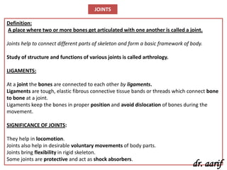
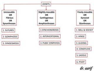
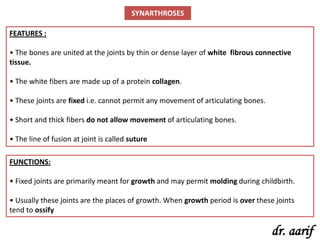


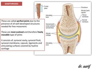


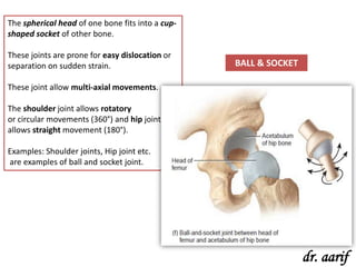
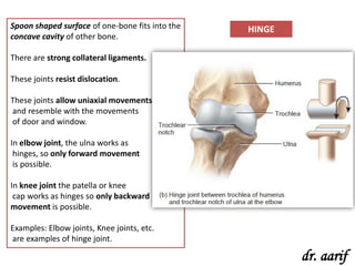
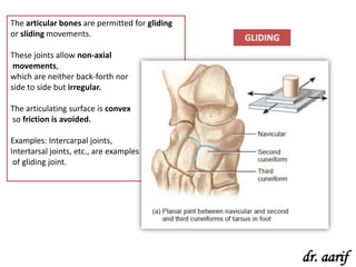
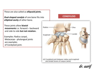
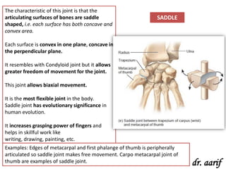
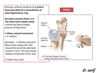


Joints connect bones and allow movement. There are three main types of joints - immovable joints which do not allow movement (e.g. sutures in the skull), slightly movable joints which allow some movement (e.g. symphysis pubis joint), and freely movable joints which allow extensive movement (e.g. ball and socket joint in the shoulder). Freely movable joints have structures like synovial fluid and membranes that reduce friction and allow movement. Examples of different freely movable joints are ball and socket, hinge, gliding, condyloid, saddle, and pivot joints.


