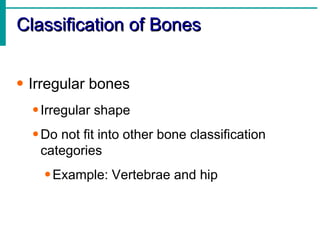
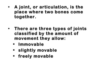
The Skeletal System: Structure, Function, and Diseases of the bones and joints
The Skeletal System Parts of the skeletal system Bones (skeleton) Joints Cartilages Ligaments (bone to bone)(tendon=bone to muscle) Divided into two divisions Axial skeleton Appendicular skeleton – limbs and girdle
Functions of Bones Support of the body Protection of soft organs Movement due to attached skeletal muscles Storage of minerals and fats Blood cell formation
Bones of the Human Body The skeleton has 206 bones Two basic types of bone tissue Compact bone Homogeneous Spongy bone Small needle-like  pieces of bone Many open spaces
Microscopic Anatomy of Bone
Bones are classified by their  shape :   long  short  flat  irregular
Classification of Bones on the Basis of Shape
Classification of Bones Long bones Typically longer than wide Have a shaft with heads at both ends Contain mostly compact bone Examples: Femur, humerus
Gross Anatomy of a Long Bone Diaphysis Shaft Composed of compact bone Epiphysis   Ends of the bone Composed mostly of spongy bone
Structures of a Long Bone Periosteum Outside covering of the diaphysis Fibrous connective tissue membrane Sharpey’s fibers Secure periosteum to underlying bone Arteries Supply bone cells with nutrients
 
Classification of Bones Short bones Generally cube-shape Contain mostly spongy bone Examples: Carpals, tarsals
Classification of Bones Flat bones Thin and flattened Usually curved Thin layers of compact bone around a layer of spongy bone Examples: Skull, ribs, sternum
Classification of Bones Irregular bones Irregular shape Do not fit into other bone classification categories Example: Vertebrae and hip
Surface features of bones Sites of attachments for muscles, tendons, and ligaments Passages for nerves and blood vessels Categories of bone markings Projections and processes – grow out from the bone surface Depressions or cavities – indentations
Types of Bone Cells Osteocytes Mature bone cells Osteoblasts Bone-forming cells Osteoclasts Bone-destroying cells Break down bone matrix for remodeling and release of calcium Bone remodeling is a process by both osteoblasts and osteoclasts
Changes in the Human Skeleton In embryos, the skeleton is primarily hyaline cartilage During development, much of this cartilage is replaced by bone Cartilage remains in isolated areas Bridge of the nose Parts of ribs Joints
Bone Growth Epiphyseal plates allow for growth of long bone during childhood New cartilage is continuously formed Older cartilage becomes ossified Cartilage is broken down Bone replaces cartilage
 
Bone Fractures A break in a bone Types of bone fractures Closed (simple) fracture – break that does not penetrate the skin Open (compound) fracture – broken bone penetrates through the skin Bone fractures are treated  by reduction and immobilization Realignment of the bone
Common Types of Fractures
Stages in the Healing of a Bone Fracture
Axial skeleton supports and protects organs of head, neck and trunk   Axial skeleton: skull (cranium and facial bones)   hyoid bone (anchors tongue and muscles associated with swallowing)   vertebral column (vertebrae and disks)   bony thorax (ribs and sternum)  Appendicular   skeleton includes bones of limbs and  bones that anchor them to the axial skeleton Appendicular skeleton: pectoral girdle (clavicles and scapulae) upper limbs (arms) pelvic girdle (sacrum, coccyx) lower limbs (legs) Articulation-  where joints meet, connect, and are formed.
22 bones in skull 6 in middle ears 1 hyoid bone 26 in vertebral column 25 in thoracic cage 4 in pectoral girdle 60 in upper limbs 60 in lower limbs 2 in pelvic girdle 206 bones in all
 
The Axial Skeleton Forms the longitudinal part of the body Divided into three parts Skull Vertebral column Bony thorax
The Axial Skeleton
The skull 8 sutured bones in cranium Facial bones: 13 sutured bones, 1 mandible Cranium encases brain attachments for muscles sinuses
 
Bones of the Skull
Allows for growth
Human Skull, Superior View
Human Skull, Inferior View Figure 5.9
Paranasal Sinuses Hollow portions of bones surrounding the nasal cavity
The Hyoid Bone The only bone that does not articulate with another bone Serves as a moveable base for the tongue
The Vertebral Column Vertebrae separated by intervertebral discs The spine has a normal curvature Each vertebrae is given a name according to its location
Vertebral column  7 cervial vertebrae 12 thoracic 5 lumbar 1 sacrum (5 fused  1 coccyx (4 fused) Vertebrae vary in size and morphology
 
Structure of a Typical Vertebrae
Thoracic cage ribs thoracic vertebrae sternum costal cartilages True ribs are directly attached to the sternum (first seven pairs) Three false ribs are joined to the 7 th  rib Two pairs of floating ribs
 
Joints Fibrous - Fibrous joints connect bones without allowing any movement. The bones of your skull and pelvis are held together by fibrous joints.  Cartilaginous - Cartilaginous joints are joints in which the bones are attached by cartilage. These joints allow for only a little movement, such as in the spine or  ribs. Synovial - Synovial joints allow for much more movement than cartilaginous joints. Cavities between bones in synovial joints are filled with synovial fluid. This fluid helps lubricate and protect the bones. Bursa sacks contain the synovial fluid.  within fixed limits
A joint, or articulation, is the place where two bones come together.  There are three types of joints classified by the amount of movement they allow:  Immovable slightly movable freely movable
Types of Joints Hinge-  
A hinge joint allows extension and retraction of an appendage. (Elbow, Knee)
Ball and Socket-  
A ball and socket joint allows for radial movement in almost any direction. They are found in   the hips and shoulders. (Hip, Shoulder)
Gliding-  
In a gliding or plane joint bones slide past each other. Mid-carpal and mid-tarsal joints are gliding joints. (Hands, Feet)
Saddle-  This type of joint occurs when the touching surfaces of two bones have both concave and convex regions with the shapes of the two bones complementing one other and allowing a wide range of movement. (Thumb)
Structures Associated with the Synovial Joint Bursae – flattened fibrous sacs Lined with synovial membranes Filled with synovial fluid Not actually part of the joint Tendon sheath Elongated bursa that wraps around a tendon
The Synovial Joint
Types of Synovial Joints Based on Shape
Diseases and Conditions   of the Skeletal System
Arthritis


Bursitis Inflammation of the Bursa sac can become inflamed from injury or infection localized pain or swelling
 
 
Tendonitis inflammation of the tendon. movement becomes painful.  most common cause of tendonitis is overuse.
 
Carpal Tunnel Syndrome swelling or change in position of the tissue within the carpal tunnel squeezes and irritates the median nerve.  causes tingling and numbness of the thumb, index, and the middle fingers
 
 
Osteoporosis means "porous bones."  bones have lost minerals  ( especially calcium)   making them weak, brittle, and susceptible to fractures  most common places where fractures occur are the back, hips, and wrists.


Scoliosis abnormal curvature of the spine.  one or more abnormal  More girls than boys have severe scoliosis.


Kyphosis spine may develop a hump.  can occur as a result of developmental problems or degenerative diseases
 
 
Lordosis A curvature of the vertebrae in the lower back area, giving a "swayback" appearance.
 
 
Rickets softening and weakening of bones in children usually because of an extreme and prolonged vitamin D deficiency.


Gout results from an overload of uric acid in the body.  leads to the formation of urate crystals that deposit in the joints.  crystals in the joints causes recurring attacks of joint inflammation (arthritis).  Chronic gout may cause joint destruction, decreased kidney function, and kidney stones.


Acromegaly occurs when the body produces too much of the hormones that control growth.  tissues grow larger than normal.  excessive growth can cause serious disease and even premature death.


Spina Bifida birth defect that involves the incomplete development of the spinal cord or its coverings.  spina bifida literally means "split" or "open" spine. The condition usually is   detected before a baby is born and treated right away.


Talipes Equinovarus- “Clubfoot” deformity of the whole foot that is present at birth.  the child is born with the foot pointing down and twisted inwards at the ankle.


Sarcoma Osteosarcoma most common type of bone cancer. most commonly found in children and adolescents


Myeloma cancer in which abnormal cells collect in the bone marrow and form tumors
 
 
Leukemia cancer of the blood cells starts in the bone marrow where blood cells are  the bone marrow starts to make a lot of abnormal white blood cells, called leukemia cells.  leukemia cells don't do the work of normal WBC’s and they don't stop growing when they should.
 
 
Bone Marrow Biopsy

Skeletal system

  • 1.
    The Skeletal System:Structure, Function, and Diseases of the bones and joints
  • 2.
    The Skeletal SystemParts of the skeletal system Bones (skeleton) Joints Cartilages Ligaments (bone to bone)(tendon=bone to muscle) Divided into two divisions Axial skeleton Appendicular skeleton – limbs and girdle
  • 3.
    Functions of BonesSupport of the body Protection of soft organs Movement due to attached skeletal muscles Storage of minerals and fats Blood cell formation
  • 4.
    Bones of theHuman Body The skeleton has 206 bones Two basic types of bone tissue Compact bone Homogeneous Spongy bone Small needle-like pieces of bone Many open spaces
  • 5.
  • 6.
    Bones are classifiedby their shape : long short flat irregular
  • 7.
    Classification of Boneson the Basis of Shape
  • 8.
    Classification of BonesLong bones Typically longer than wide Have a shaft with heads at both ends Contain mostly compact bone Examples: Femur, humerus
  • 9.
    Gross Anatomy ofa Long Bone Diaphysis Shaft Composed of compact bone Epiphysis Ends of the bone Composed mostly of spongy bone
  • 10.
    Structures of aLong Bone Periosteum Outside covering of the diaphysis Fibrous connective tissue membrane Sharpey’s fibers Secure periosteum to underlying bone Arteries Supply bone cells with nutrients
  • 11.
  • 12.
    Classification of BonesShort bones Generally cube-shape Contain mostly spongy bone Examples: Carpals, tarsals
  • 13.
    Classification of BonesFlat bones Thin and flattened Usually curved Thin layers of compact bone around a layer of spongy bone Examples: Skull, ribs, sternum
  • 14.
    Classification of BonesIrregular bones Irregular shape Do not fit into other bone classification categories Example: Vertebrae and hip
  • 15.
    Surface features ofbones Sites of attachments for muscles, tendons, and ligaments Passages for nerves and blood vessels Categories of bone markings Projections and processes – grow out from the bone surface Depressions or cavities – indentations
  • 16.
    Types of BoneCells Osteocytes Mature bone cells Osteoblasts Bone-forming cells Osteoclasts Bone-destroying cells Break down bone matrix for remodeling and release of calcium Bone remodeling is a process by both osteoblasts and osteoclasts
  • 17.
    Changes in theHuman Skeleton In embryos, the skeleton is primarily hyaline cartilage During development, much of this cartilage is replaced by bone Cartilage remains in isolated areas Bridge of the nose Parts of ribs Joints
  • 18.
    Bone Growth Epiphysealplates allow for growth of long bone during childhood New cartilage is continuously formed Older cartilage becomes ossified Cartilage is broken down Bone replaces cartilage
  • 19.
  • 20.
    Bone Fractures Abreak in a bone Types of bone fractures Closed (simple) fracture – break that does not penetrate the skin Open (compound) fracture – broken bone penetrates through the skin Bone fractures are treated by reduction and immobilization Realignment of the bone
  • 21.
    Common Types ofFractures
  • 22.
    Stages in theHealing of a Bone Fracture
  • 23.
    Axial skeleton supportsand protects organs of head, neck and trunk Axial skeleton: skull (cranium and facial bones) hyoid bone (anchors tongue and muscles associated with swallowing) vertebral column (vertebrae and disks) bony thorax (ribs and sternum) Appendicular skeleton includes bones of limbs and bones that anchor them to the axial skeleton Appendicular skeleton: pectoral girdle (clavicles and scapulae) upper limbs (arms) pelvic girdle (sacrum, coccyx) lower limbs (legs) Articulation- where joints meet, connect, and are formed.
  • 24.
    22 bones inskull 6 in middle ears 1 hyoid bone 26 in vertebral column 25 in thoracic cage 4 in pectoral girdle 60 in upper limbs 60 in lower limbs 2 in pelvic girdle 206 bones in all
  • 25.
  • 26.
    The Axial SkeletonForms the longitudinal part of the body Divided into three parts Skull Vertebral column Bony thorax
  • 27.
  • 28.
    The skull 8sutured bones in cranium Facial bones: 13 sutured bones, 1 mandible Cranium encases brain attachments for muscles sinuses
  • 29.
  • 30.
  • 31.
  • 32.
  • 33.
    Human Skull, InferiorView Figure 5.9
  • 34.
    Paranasal Sinuses Hollowportions of bones surrounding the nasal cavity
  • 35.
    The Hyoid BoneThe only bone that does not articulate with another bone Serves as a moveable base for the tongue
  • 36.
    The Vertebral ColumnVertebrae separated by intervertebral discs The spine has a normal curvature Each vertebrae is given a name according to its location
  • 37.
    Vertebral column 7 cervial vertebrae 12 thoracic 5 lumbar 1 sacrum (5 fused 1 coccyx (4 fused) Vertebrae vary in size and morphology
  • 38.
  • 39.
    Structure of aTypical Vertebrae
  • 40.
    Thoracic cage ribsthoracic vertebrae sternum costal cartilages True ribs are directly attached to the sternum (first seven pairs) Three false ribs are joined to the 7 th rib Two pairs of floating ribs
  • 41.
  • 42.
    Joints Fibrous -Fibrous joints connect bones without allowing any movement. The bones of your skull and pelvis are held together by fibrous joints. Cartilaginous - Cartilaginous joints are joints in which the bones are attached by cartilage. These joints allow for only a little movement, such as in the spine or ribs. Synovial - Synovial joints allow for much more movement than cartilaginous joints. Cavities between bones in synovial joints are filled with synovial fluid. This fluid helps lubricate and protect the bones. Bursa sacks contain the synovial fluid. within fixed limits
  • 43.
    A joint, orarticulation, is the place where two bones come together. There are three types of joints classified by the amount of movement they allow: Immovable slightly movable freely movable
  • 44.
    Types of JointsHinge- 
A hinge joint allows extension and retraction of an appendage. (Elbow, Knee)
  • 45.
    Ball and Socket- 
A ball and socket joint allows for radial movement in almost any direction. They are found in the hips and shoulders. (Hip, Shoulder)
  • 46.
    Gliding- 
Ina gliding or plane joint bones slide past each other. Mid-carpal and mid-tarsal joints are gliding joints. (Hands, Feet)
  • 47.
    Saddle- Thistype of joint occurs when the touching surfaces of two bones have both concave and convex regions with the shapes of the two bones complementing one other and allowing a wide range of movement. (Thumb)
  • 48.
    Structures Associated withthe Synovial Joint Bursae – flattened fibrous sacs Lined with synovial membranes Filled with synovial fluid Not actually part of the joint Tendon sheath Elongated bursa that wraps around a tendon
  • 49.
  • 50.
    Types of SynovialJoints Based on Shape
  • 51.
    Types of SynovialJoints Based on Shape
  • 52.
    Diseases and Conditions of the Skeletal System
  • 53.
  • 54.
  • 55.
  • 56.
  • 57.
    Bursitis Inflammation ofthe Bursa sac can become inflamed from injury or infection localized pain or swelling
  • 58.
  • 59.
  • 60.
    Tendonitis inflammation ofthe tendon. movement becomes painful. most common cause of tendonitis is overuse.
  • 61.
  • 62.
    Carpal Tunnel Syndromeswelling or change in position of the tissue within the carpal tunnel squeezes and irritates the median nerve. causes tingling and numbness of the thumb, index, and the middle fingers
  • 63.
  • 64.
  • 65.
    Osteoporosis means "porousbones." bones have lost minerals ( especially calcium) making them weak, brittle, and susceptible to fractures most common places where fractures occur are the back, hips, and wrists.
  • 66.
  • 67.
  • 68.
  • 69.
    Scoliosis abnormal curvatureof the spine. one or more abnormal More girls than boys have severe scoliosis.
  • 70.
  • 71.
  • 72.
  • 73.
  • 74.
    Kyphosis spine maydevelop a hump. can occur as a result of developmental problems or degenerative diseases
  • 75.
  • 76.
  • 77.
    Lordosis A curvatureof the vertebrae in the lower back area, giving a "swayback" appearance.
  • 78.
  • 79.
  • 80.
    Rickets softening andweakening of bones in children usually because of an extreme and prolonged vitamin D deficiency.
  • 81.
  • 82.
  • 83.
  • 84.
    Gout results froman overload of uric acid in the body. leads to the formation of urate crystals that deposit in the joints. crystals in the joints causes recurring attacks of joint inflammation (arthritis). Chronic gout may cause joint destruction, decreased kidney function, and kidney stones.
  • 85.
  • 86.
  • 87.
  • 88.
    Acromegaly occurs whenthe body produces too much of the hormones that control growth. tissues grow larger than normal. excessive growth can cause serious disease and even premature death.
  • 89.
  • 90.
  • 91.
  • 92.
  • 93.
    Spina Bifida birthdefect that involves the incomplete development of the spinal cord or its coverings. spina bifida literally means "split" or "open" spine. The condition usually is detected before a baby is born and treated right away.
  • 94.
  • 95.
  • 96.
  • 97.
    Talipes Equinovarus- “Clubfoot”deformity of the whole foot that is present at birth. the child is born with the foot pointing down and twisted inwards at the ankle.
  • 98.
  • 99.
  • 100.
  • 101.
    Sarcoma Osteosarcoma mostcommon type of bone cancer. most commonly found in children and adolescents
  • 102.
  • 103.
  • 104.
  • 105.
  • 106.
  • 107.
    Myeloma cancer inwhich abnormal cells collect in the bone marrow and form tumors
  • 108.
  • 109.
  • 110.
    Leukemia cancer ofthe blood cells starts in the bone marrow where blood cells are the bone marrow starts to make a lot of abnormal white blood cells, called leukemia cells. leukemia cells don't do the work of normal WBC’s and they don't stop growing when they should.
  • 111.
  • 112.
  • 113.