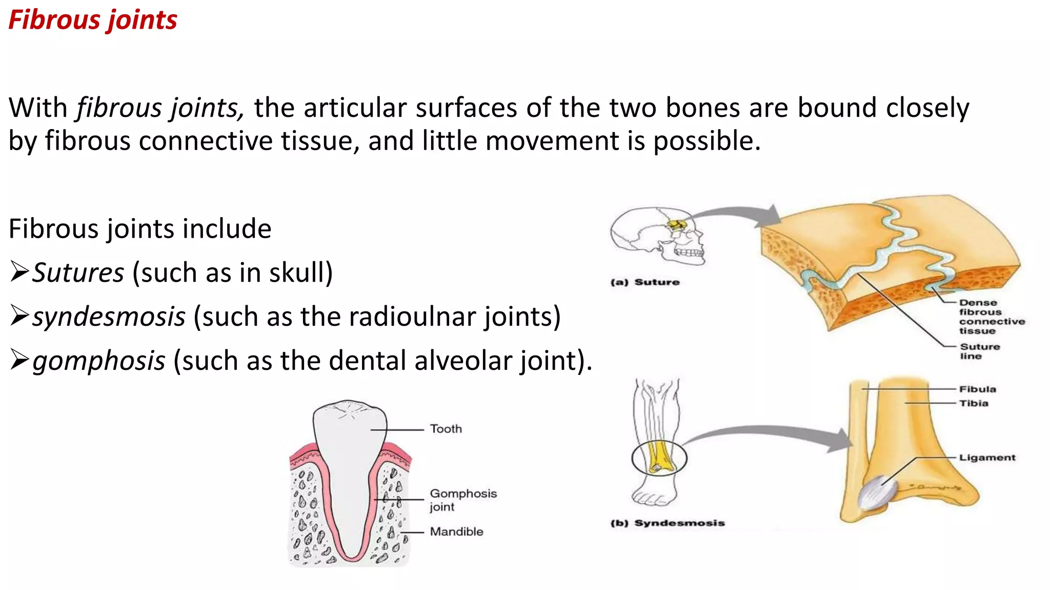
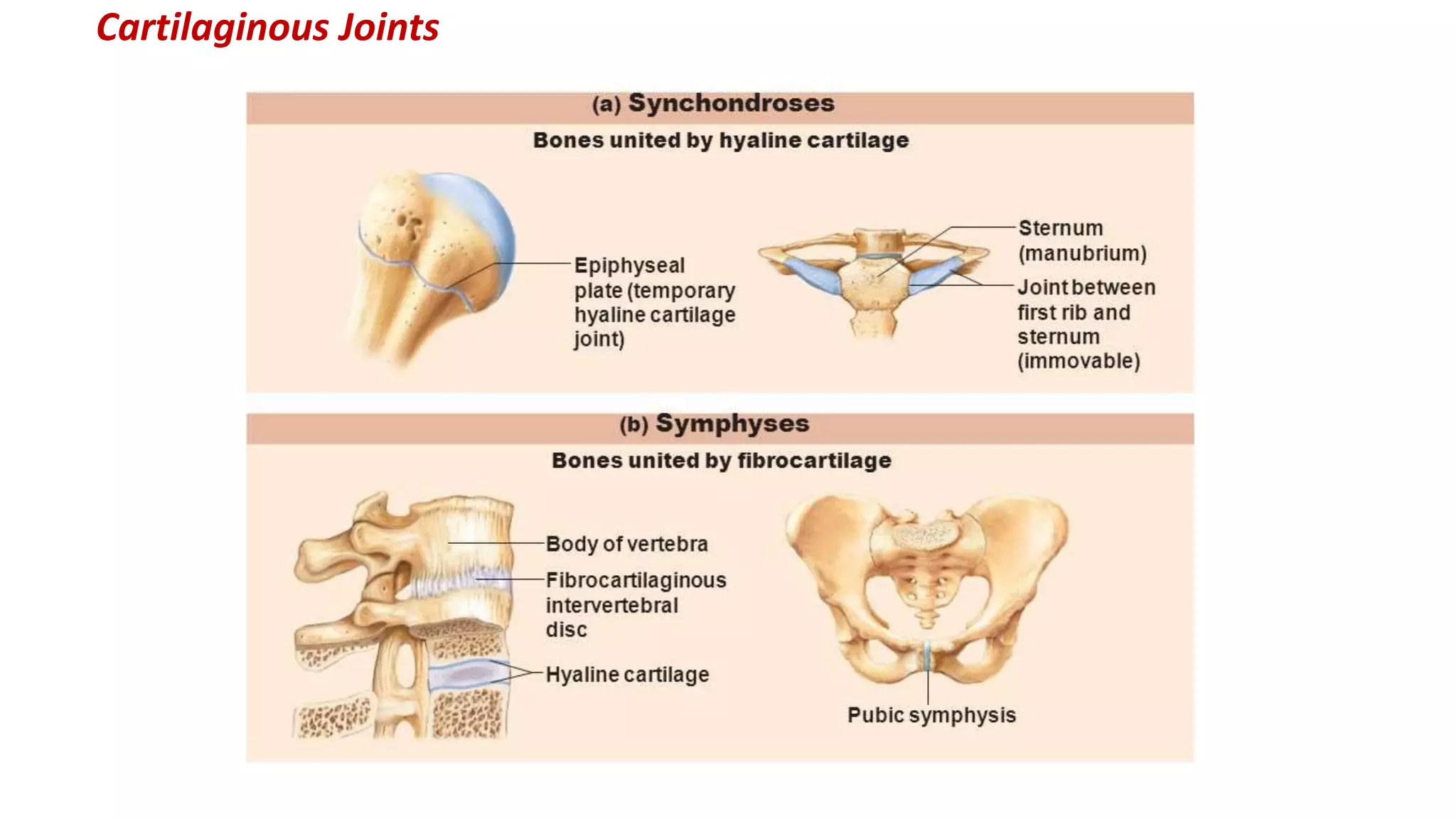
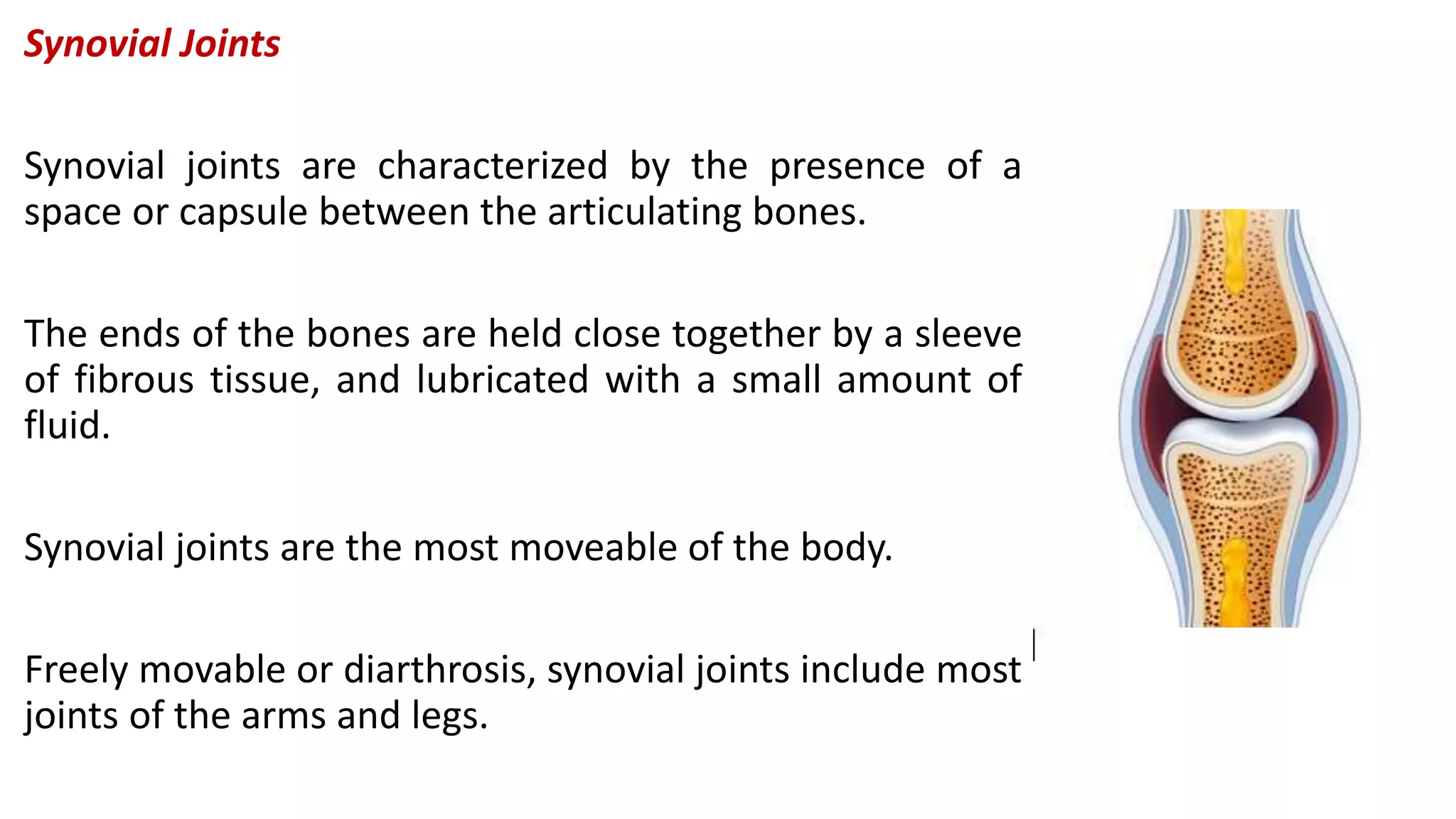
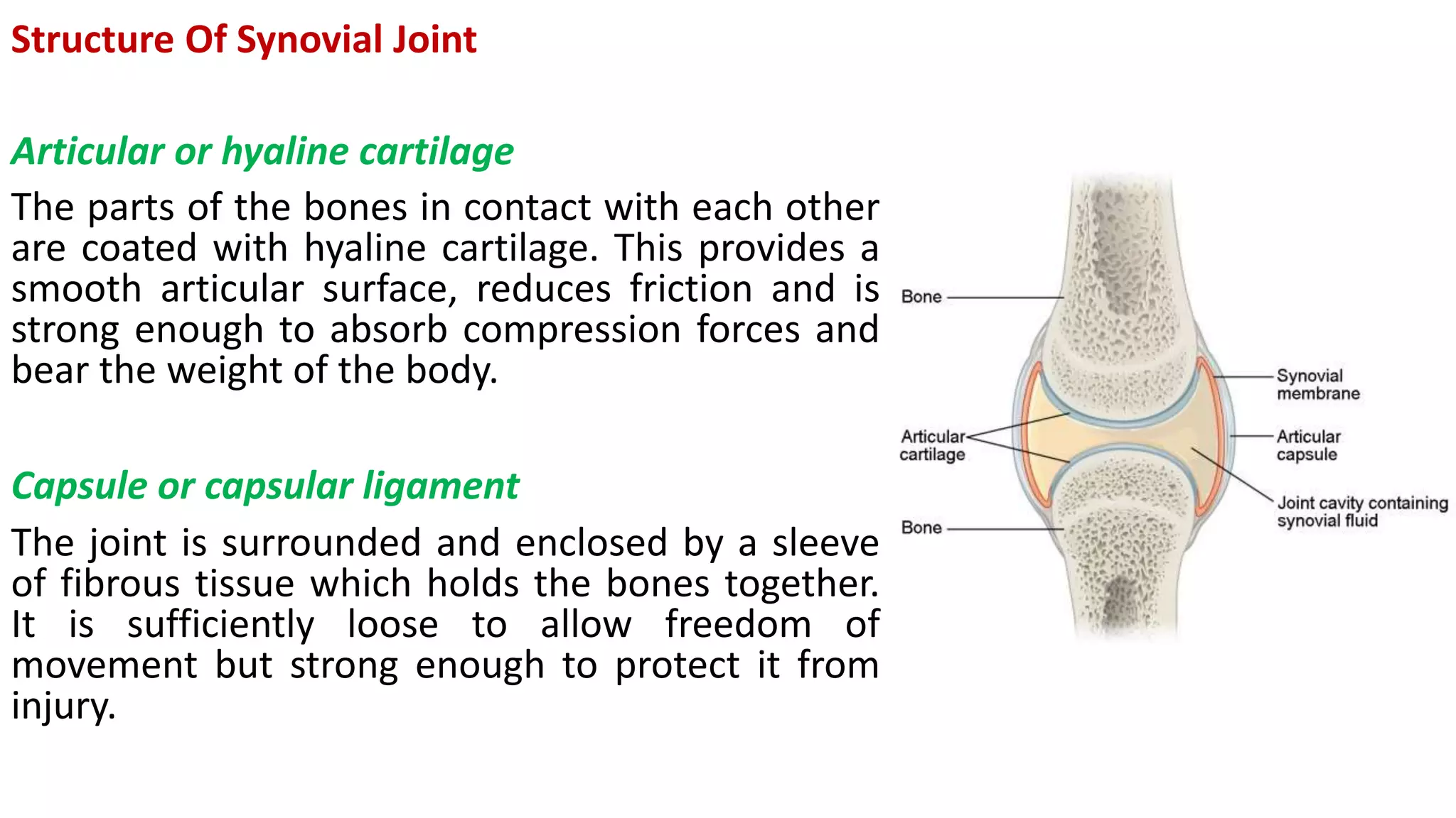
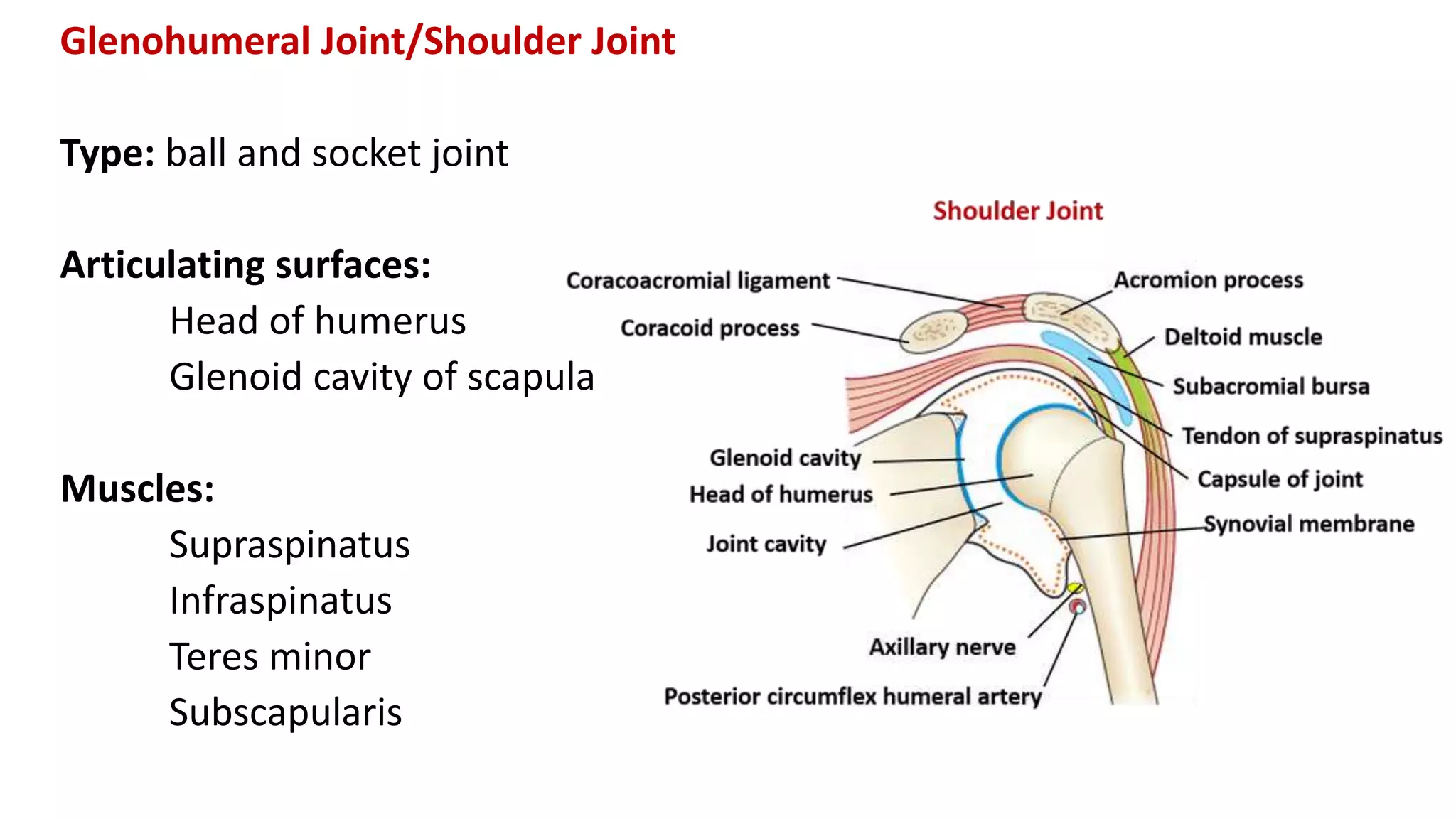
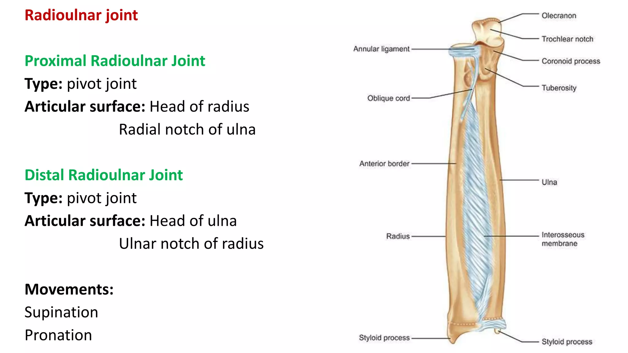
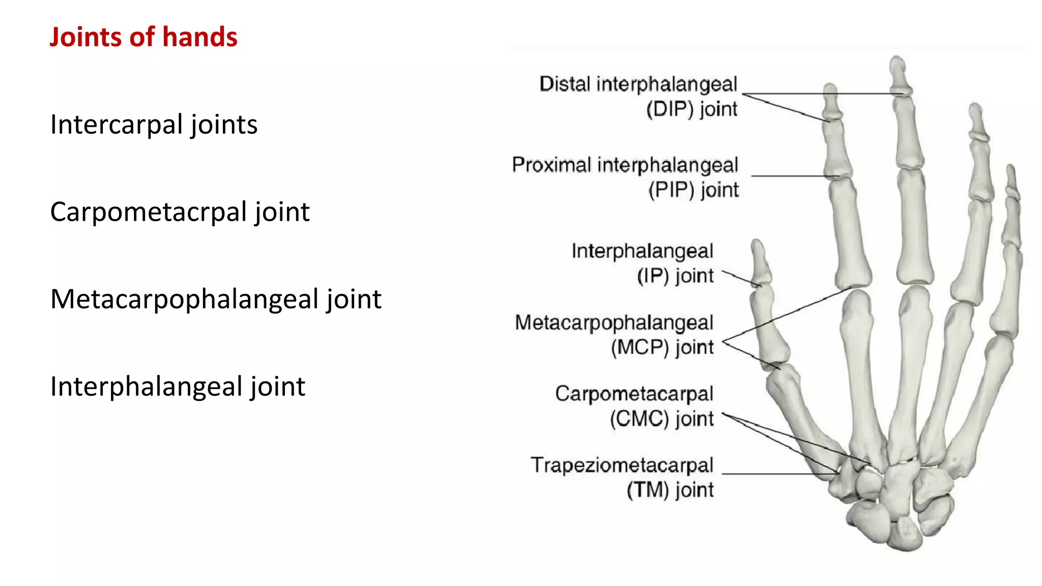
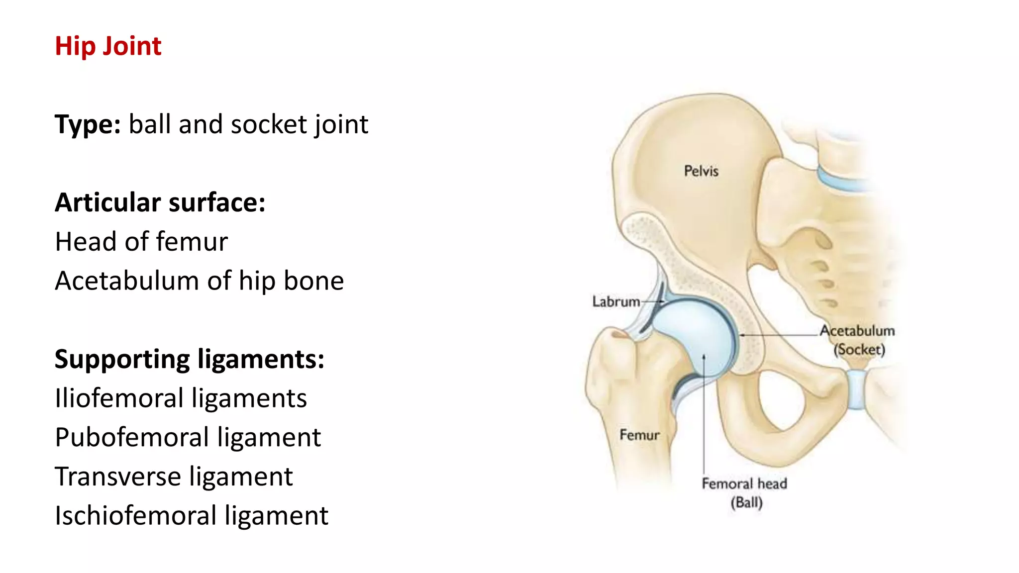
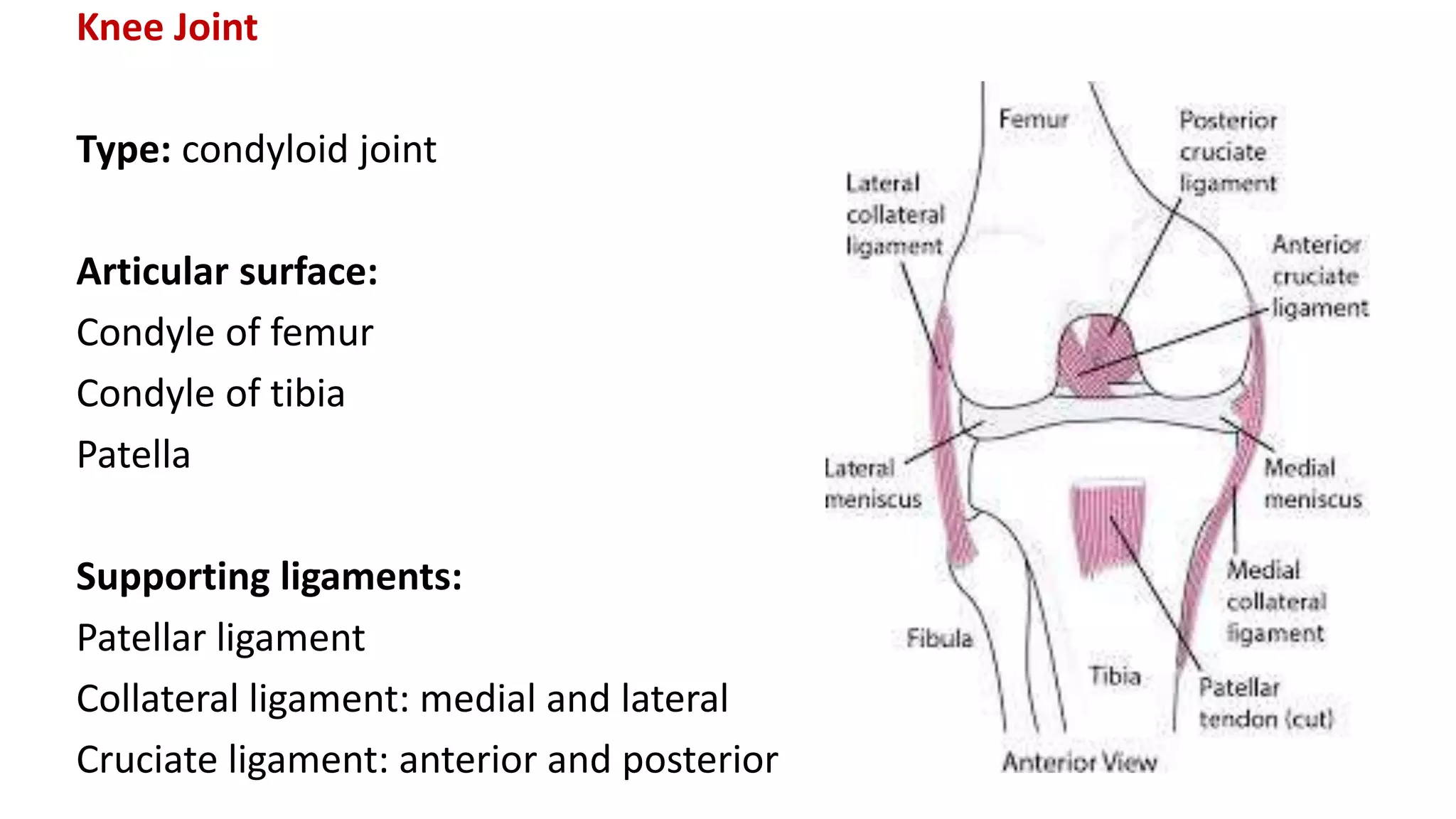
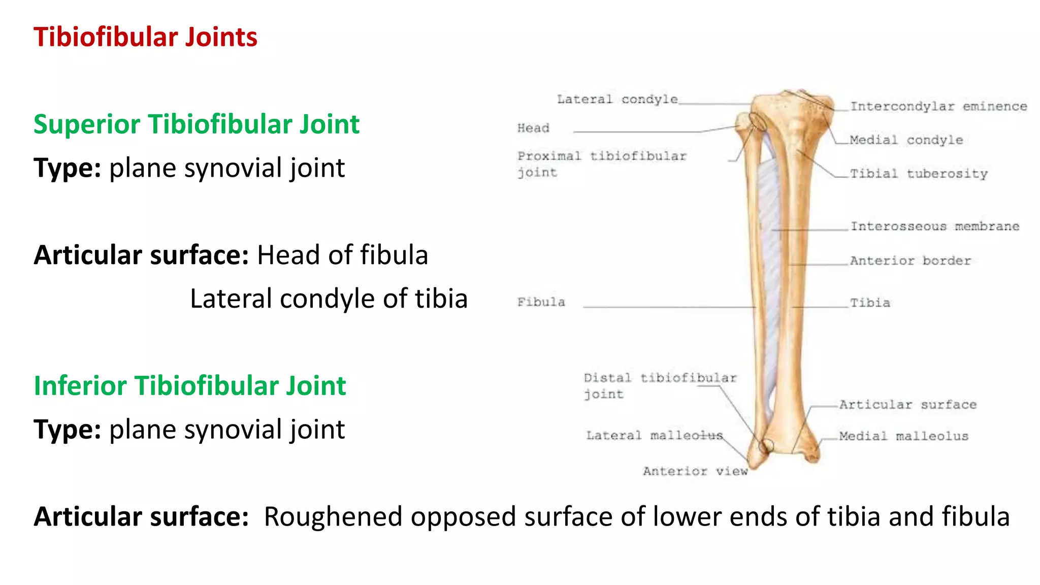
This document discusses the different types of joints in the human body. It begins by defining a joint and classifying joints by function and structure. The three main types by function are synarthrosis (immovable), amphiarthrosis (slightly movable), and diarthrosis (freely movable). The three main types by structure are fibrous, cartilaginous, and synovial joints. It then provides details on the different types of synovial joints and examples of various joints in the body like the shoulder, elbow, hip, knee, ankle joints. It concludes with a brief description of bursae and fontanels.
A joint is an articulation between two bones in the body and are broadly classified by the tissue which connects the bones. The three main types of joints are: synovial, cartilaginous and fibrous.
A joint is the site at which any two or more bones articulate or come together, meaning the ends or edges of the bones are held together by connective tissues.
It may allow flexibility and movement of the skeleton.
With the exception of the Hyoid bone, every bone in the body is connected to or form joints.
There are total 230 joints in the body.
Structural Classification of joints:
FIBROUS JOINT/ FIXED JOINT
CARTILAGENOUS JOINTS
SYNOVIAL JOINTS
The functional classification of joints is determined by the amount of mobility found between the adjacent bones.
Joints are thus functionally classified as a
Synarthrosis or immobile joint,
Amphiarthrosis or slightly moveable joint,
Diarthrosis, which is a freely moveable joint
synovial joint, definition of synovial joint, diarthrodial joints, components of synovial joint, types of synovial joints, hinge joint with examples, pivot joint with examples, condyloid joint with examples, saddle joint with examples, ball and socket joint with examples, gliding joint with examples, features of synovial joint, synovial membrane, synovial fluid, components of synovial membrane, meniscus, true and accessory ligament of synovial joint, bursae, blood supply of synovial joint, innervation of synovial joint
All human joints including axial and appendicular skeleton will clear all the concepts and doubts of learner related to joints and different classification of joints with detail. Also, help student, learner, reader to communicate.
it consist of detailed description about joints, their number and location. we briefly explained about the types of joints with colorful images. easy to understand for students with the help of pictures. language is also simple.it will be helpful for the beginners.
Joints, function and types of joints.pptx10khan113ii
Joints, or articulations, are the structures where two or more bones meet, allowing for movement and providing mechanical support. They are essential for a wide range of motions and functions in the human body. Here's a detailed description of joints:
### Types of Joints Based on Structure:
1. **Fibrous Joints**:
- **Description**: Connected by dense connective tissue rich in collagen.
- **Movement**: Generally immovable (synarthrotic).
- **Examples**: Sutures in the skull, syndesmoses between long bones like the tibia and fibula.
2. **Cartilaginous Joints**:
- **Description**: Bones connected entirely by cartilage (either hyaline or fibrocartilage).
- **Movement**: Allow limited movement (amphiarthrotic).
- **Examples**: Intervertebral discs, pubic symphysis.
3. **Synovial Joints**:
- **Description**: Characterized by a fluid-filled synovial cavity that separates the articulating bones.
- **Movement**: Freely movable (diarthrotic), with various degrees and types of movement depending on the specific joint type.
- **Examples and Subtypes**:
- **Hinge Joints**: Permit flexion and extension (e.g., elbow, knee).
- **Ball-and-Socket Joints**: Allow rotational movement and a wide range of motion (e.g., shoulder, hip).
- **Pivot Joints**: Enable rotational movement around a single axis (e.g., atlantoaxial joint in the neck).
- **Gliding Joints**: Allow sliding or gliding movements (e.g., intercarpal joints in the wrist).
- **Saddle Joints**: Permit movement along two planes (e.g., thumb joint).
- **Condyloid Joints**: Allow movement but no rotation (e.g., wrist joint between the radius and carpal bones).
### Types of Joints Based on Function:
1. **Synarthroses**:
- **Description**: Immovable joints providing strong, stable connections.
- **Examples**: Skull sutures.
2. **Amphiarthroses**:
- **Description**: Slightly movable joints.
- **Examples**: Intervertebral discs, pubic symphysis.
3. **Diarthroses**:
- **Description**: Freely movable joints, synonymous with synovial joints.
- **Examples**: Most limb joints like the knee, hip, shoulder.
### Joint Anatomy:
- **Cartilage**: Covers the ends of bones, providing a smooth surface for movement and cushioning.
- **Synovial Membrane**: Lines the joint capsule and secretes synovial fluid for lubrication.
- **Ligaments**: Strong bands of connective tissue that connect bones and stabilize the joint.
- **Tendons**: Connect muscle to bone, aiding in joint movement.
- **Bursae**: Fluid-filled sacs that reduce friction between moving structures within the joint.
### Functions of Joints:
- **Movement**: Facilitate a range of bodily movements, from gross motor functions like walking and lifting to fine motor skills like writing and typing.
- **Support**: Maintain the body's structure and posture.
- **Protection**: Protect internal structures by providing cushioning and absorbing impact.
This is a PowerPoint about joints in your skeletal system. This biological PowerPoint can be used or accessed by any CXC/CSEC student. This is on the CSEC syllabus. Good luck on your exams!
Powerpoint by - Mr. Lynch
At the completion of this unit, learners will be able to: 1. Define the term joint. 2. List three types of joints I.e. Fibrous Cartilaginous Synovial 3. Describe the common characteristic features of a synovial joint. 4. List the types of synovial joints. 5. Discuss the types of movements possible at synovial joints.
These lecture slides, by Dr Sidra Arshad, offer a quick overview of physiological basis of a normal electrocardiogram.
Learning objectives:
1. Define an electrocardiogram (ECG) and electrocardiography
2. Describe how dipoles generated by the heart produce the waveforms of the ECG
3. Describe the components of a normal electrocardiogram of a typical bipolar leads (limb II)
4. Differentiate between intervals and segments
5. Enlist some common indications for obtaining an ECG
Study Resources:
1. Chapter 11, Guyton and Hall Textbook of Medical Physiology, 14th edition
2. Chapter 9, Human Physiology - From Cells to Systems, Lauralee Sherwood, 9th edition
3. Chapter 29, Ganong’s Review of Medical Physiology, 26th edition
4. Electrocardiogram, StatPearls - https://www.ncbi.nlm.nih.gov/books/NBK549803/
5. ECG in Medical Practice by ABM Abdullah, 4th edition
6. ECG Basics, http://www.nataliescasebook.com/tag/e-c-g-basics
- Video recording of this lecture in English language: https://youtu.be/lK81BzxMqdo
- Video recording of this lecture in Arabic language: https://youtu.be/Ve4P0COk9OI
- Link to download the book free: https://nephrotube.blogspot.com/p/nephrotube-nephrology-books.html
- Link to NephroTube website: www.NephroTube.com
- Link to NephroTube social media accounts: https://nephrotube.blogspot.com/p/join-nephrotube-on-social-media.html
Couples presenting to the infertility clinic- Do they really have infertility...Sujoy Dasgupta
Dr Sujoy Dasgupta presented the study on "Couples presenting to the infertility clinic- Do they really have infertility? – The unexplored stories of non-consummation" in the 13th Congress of the Asia Pacific Initiative on Reproduction (ASPIRE 2024) at Manila on 24 May, 2024.
Ozempic: Preoperative Management of Patients on GLP-1 Receptor Agonists Saeid Safari
Preoperative Management of Patients on GLP-1 Receptor Agonists like Ozempic and Semiglutide
ASA GUIDELINE
NYSORA Guideline
2 Case Reports of Gastric Ultrasound
Lung Cancer: Artificial Intelligence, Synergetics, Complex System Analysis, S...Oleg Kshivets
RESULTS: Overall life span (LS) was 2252.1±1742.5 days and cumulative 5-year survival (5YS) reached 73.2%, 10 years – 64.8%, 20 years – 42.5%. 513 LCP lived more than 5 years (LS=3124.6±1525.6 days), 148 LCP – more than 10 years (LS=5054.4±1504.1 days).199 LCP died because of LC (LS=562.7±374.5 days). 5YS of LCP after bi/lobectomies was significantly superior in comparison with LCP after pneumonectomies (78.1% vs.63.7%, P=0.00001 by log-rank test). AT significantly improved 5YS (66.3% vs. 34.8%) (P=0.00000 by log-rank test) only for LCP with N1-2. Cox modeling displayed that 5YS of LCP significantly depended on: phase transition (PT) early-invasive LC in terms of synergetics, PT N0—N12, cell ratio factors (ratio between cancer cells- CC and blood cells subpopulations), G1-3, histology, glucose, AT, blood cell circuit, prothrombin index, heparin tolerance, recalcification time (P=0.000-0.038). Neural networks, genetic algorithm selection and bootstrap simulation revealed relationships between 5YS and PT early-invasive LC (rank=1), PT N0—N12 (rank=2), thrombocytes/CC (3), erythrocytes/CC (4), eosinophils/CC (5), healthy cells/CC (6), lymphocytes/CC (7), segmented neutrophils/CC (8), stick neutrophils/CC (9), monocytes/CC (10); leucocytes/CC (11). Correct prediction of 5YS was 100% by neural networks computing (area under ROC curve=1.0; error=0.0).
CONCLUSIONS: 5YS of LCP after radical procedures significantly depended on: 1) PT early-invasive cancer; 2) PT N0--N12; 3) cell ratio factors; 4) blood cell circuit; 5) biochemical factors; 6) hemostasis system; 7) AT; 8) LC characteristics; 9) LC cell dynamics; 10) surgery type: lobectomy/pneumonectomy; 11) anthropometric data. Optimal diagnosis and treatment strategies for LC are: 1) screening and early detection of LC; 2) availability of experienced thoracic surgeons because of complexity of radical procedures; 3) aggressive en block surgery and adequate lymph node dissection for completeness; 4) precise prediction; 5) adjuvant chemoimmunoradiotherapy for LCP with unfavorable prognosis.
New Directions in Targeted Therapeutic Approaches for Older Adults With Mantl...i3 Health
i3 Health is pleased to make the speaker slides from this activity available for use as a non-accredited self-study or teaching resource.
This slide deck presented by Dr. Kami Maddocks, Professor-Clinical in the Division of Hematology and
Associate Division Director for Ambulatory Operations
The Ohio State University Comprehensive Cancer Center, will provide insight into new directions in targeted therapeutic approaches for older adults with mantle cell lymphoma.
STATEMENT OF NEED
Mantle cell lymphoma (MCL) is a rare, aggressive B-cell non-Hodgkin lymphoma (NHL) accounting for 5% to 7% of all lymphomas. Its prognosis ranges from indolent disease that does not require treatment for years to very aggressive disease, which is associated with poor survival (Silkenstedt et al, 2021). Typically, MCL is diagnosed at advanced stage and in older patients who cannot tolerate intensive therapy (NCCN, 2022). Although recent advances have slightly increased remission rates, recurrence and relapse remain very common, leading to a median overall survival between 3 and 6 years (LLS, 2021). Though there are several effective options, progress is still needed towards establishing an accepted frontline approach for MCL (Castellino et al, 2022). Treatment selection and management of MCL are complicated by the heterogeneity of prognosis, advanced age and comorbidities of patients, and lack of an established standard approach for treatment, making it vital that clinicians be familiar with the latest research and advances in this area. In this activity chaired by Michael Wang, MD, Professor in the Department of Lymphoma & Myeloma at MD Anderson Cancer Center, expert faculty will discuss prognostic factors informing treatment, the promising results of recent trials in new therapeutic approaches, and the implications of treatment resistance in therapeutic selection for MCL.
Target Audience
Hematology/oncology fellows, attending faculty, and other health care professionals involved in the treatment of patients with mantle cell lymphoma (MCL).
Learning Objectives
1.) Identify clinical and biological prognostic factors that can guide treatment decision making for older adults with MCL
2.) Evaluate emerging data on targeted therapeutic approaches for treatment-naive and relapsed/refractory MCL and their applicability to older adults
3.) Assess mechanisms of resistance to targeted therapies for MCL and their implications for treatment selection
micro teaching on communication m.sc nursing.pdfAnurag Sharma
Microteaching is a unique model of practice teaching. It is a viable instrument for the. desired change in the teaching behavior or the behavior potential which, in specified types of real. classroom situations, tends to facilitate the achievement of specified types of objectives.
Factory Supply Best Quality Pmk Oil CAS 28578–16–7 PMK Powder in Stockrebeccabio
Factory Supply Best Quality Pmk Oil CAS 28578–16–7 PMK Powder in Stock
Telegram: bmksupplier
signal: +85264872720
threema: TUD4A6YC
You can contact me on Telegram or Threema
Communicate promptly and reply
Free of customs clearance, Double Clearance 100% pass delivery to USA, Canada, Spain, Germany, Netherland, Poland, Italy, Sweden, UK, Czech Republic, Australia, Mexico, Russia, Ukraine, Kazakhstan.Door to door service
Hot Selling Organic intermediates
Prix Galien International 2024 Forum ProgramLevi Shapiro
June 20, 2024, Prix Galien International and Jerusalem Ethics Forum in ROME. Detailed agenda including panels:
- ADVANCES IN CARDIOLOGY: A NEW PARADIGM IS COMING
- WOMEN’S HEALTH: FERTILITY PRESERVATION
- WHAT’S NEW IN THE TREATMENT OF INFECTIOUS,
ONCOLOGICAL AND INFLAMMATORY SKIN DISEASES?
- ARTIFICIAL INTELLIGENCE AND ETHICS
- GENE THERAPY
- BEYOND BORDERS: GLOBAL INITIATIVES FOR DEMOCRATIZING LIFE SCIENCE TECHNOLOGIES AND PROMOTING ACCESS TO HEALTHCARE
- ETHICAL CHALLENGES IN LIFE SCIENCES
- Prix Galien International Awards Ceremony
The prostate is an exocrine gland of the male mammalian reproductive system
It is a walnut-sized gland that forms part of the male reproductive system and is located in front of the rectum and just below the urinary bladder
Function is to store and secrete a clear, slightly alkaline fluid that constitutes 10-30% of the volume of the seminal fluid that along with the spermatozoa, constitutes semen
A healthy human prostate measures (4cm-vertical, by 3cm-horizontal, 2cm ant-post ).
It surrounds the urethra just below the urinary bladder. It has anterior, median, posterior and two lateral lobes
It’s work is regulated by androgens which are responsible for male sex characteristics
Generalised disease of the prostate due to hormonal derangement which leads to non malignant enlargement of the gland (increase in the number of epithelial cells and stromal tissue)to cause compression of the urethra leading to symptoms (LUTS
Tom Selleck Health: A Comprehensive Look at the Iconic Actor’s Wellness Journeygreendigital
Tom Selleck, an enduring figure in Hollywood. has captivated audiences for decades with his rugged charm, iconic moustache. and memorable roles in television and film. From his breakout role as Thomas Magnum in Magnum P.I. to his current portrayal of Frank Reagan in Blue Bloods. Selleck's career has spanned over 50 years. But beyond his professional achievements. fans have often been curious about Tom Selleck Health. especially as he has aged in the public eye.
Follow us on: Pinterest
Introduction
Many have been interested in Tom Selleck health. not only because of his enduring presence on screen but also because of the challenges. and lifestyle choices he has faced and made over the years. This article delves into the various aspects of Tom Selleck health. exploring his fitness regimen, diet, mental health. and the challenges he has encountered as he ages. We'll look at how he maintains his well-being. the health issues he has faced, and his approach to ageing .
Early Life and Career
Childhood and Athletic Beginnings
Tom Selleck was born on January 29, 1945, in Detroit, Michigan, and grew up in Sherman Oaks, California. From an early age, he was involved in sports, particularly basketball. which played a significant role in his physical development. His athletic pursuits continued into college. where he attended the University of Southern California (USC) on a basketball scholarship. This early involvement in sports laid a strong foundation for his physical health and disciplined lifestyle.
Transition to Acting
Selleck's transition from an athlete to an actor came with its physical demands. His first significant role in "Magnum P.I." required him to perform various stunts and maintain a fit appearance. This role, which he played from 1980 to 1988. necessitated a rigorous fitness routine to meet the show's demands. setting the stage for his long-term commitment to health and wellness.
Fitness Regimen
Workout Routine
Tom Selleck health and fitness regimen has evolved. adapting to his changing roles and age. During his "Magnum, P.I." days. Selleck's workouts were intense and focused on building and maintaining muscle mass. His routine included weightlifting, cardiovascular exercises. and specific training for the stunts he performed on the show.
Selleck adjusted his fitness routine as he aged to suit his body's needs. Today, his workouts focus on maintaining flexibility, strength, and cardiovascular health. He incorporates low-impact exercises such as swimming, walking, and light weightlifting. This balanced approach helps him stay fit without putting undue strain on his joints and muscles.
Importance of Flexibility and Mobility
In recent years, Selleck has emphasized the importance of flexibility and mobility in his fitness regimen. Understanding the natural decline in muscle mass and joint flexibility with age. he includes stretching and yoga in his routine. These practices help prevent injuries, improve posture, and maintain mobilit
Title: Sense of Taste
Presenter: Dr. Faiza, Assistant Professor of Physiology
Qualifications:
MBBS (Best Graduate, AIMC Lahore)
FCPS Physiology
ICMT, CHPE, DHPE (STMU)
MPH (GC University, Faisalabad)
MBA (Virtual University of Pakistan)
Learning Objectives:
Describe the structure and function of taste buds.
Describe the relationship between the taste threshold and taste index of common substances.
Explain the chemical basis and signal transduction of taste perception for each type of primary taste sensation.
Recognize different abnormalities of taste perception and their causes.
Key Topics:
Significance of Taste Sensation:
Differentiation between pleasant and harmful food
Influence on behavior
Selection of food based on metabolic needs
Receptors of Taste:
Taste buds on the tongue
Influence of sense of smell, texture of food, and pain stimulation (e.g., by pepper)
Primary and Secondary Taste Sensations:
Primary taste sensations: Sweet, Sour, Salty, Bitter, Umami
Chemical basis and signal transduction mechanisms for each taste
Taste Threshold and Index:
Taste threshold values for Sweet (sucrose), Salty (NaCl), Sour (HCl), and Bitter (Quinine)
Taste index relationship: Inversely proportional to taste threshold
Taste Blindness:
Inability to taste certain substances, particularly thiourea compounds
Example: Phenylthiocarbamide
Structure and Function of Taste Buds:
Composition: Epithelial cells, Sustentacular/Supporting cells, Taste cells, Basal cells
Features: Taste pores, Taste hairs/microvilli, and Taste nerve fibers
Location of Taste Buds:
Found in papillae of the tongue (Fungiform, Circumvallate, Foliate)
Also present on the palate, tonsillar pillars, epiglottis, and proximal esophagus
Mechanism of Taste Stimulation:
Interaction of taste substances with receptors on microvilli
Signal transduction pathways for Umami, Sweet, Bitter, Sour, and Salty tastes
Taste Sensitivity and Adaptation:
Decrease in sensitivity with age
Rapid adaptation of taste sensation
Role of Saliva in Taste:
Dissolution of tastants to reach receptors
Washing away the stimulus
Taste Preferences and Aversions:
Mechanisms behind taste preference and aversion
Influence of receptors and neural pathways
Impact of Sensory Nerve Damage:
Degeneration of taste buds if the sensory nerve fiber is cut
Abnormalities of Taste Detection:
Conditions: Ageusia, Hypogeusia, Dysgeusia (parageusia)
Causes: Nerve damage, neurological disorders, infections, poor oral hygiene, adverse drug effects, deficiencies, aging, tobacco use, altered neurotransmitter levels
Neurotransmitters and Taste Threshold:
Effects of serotonin (5-HT) and norepinephrine (NE) on taste sensitivity
Supertasters:
25% of the population with heightened sensitivity to taste, especially bitterness
Increased number of fungiform papillae
2. Joints
A joint is the site at which any two or more bones articulate or come
together.
Joints allow flexibility and movement of the skeleton and allow
attachment between bones.
Joints can be classified by function (extent of movement) or by structure
(what they’re made of).
The body has three major types of joints classified by function and three
major types classified by structure.
3. Functional Classification
By function, a joint may be classified as:
1. Synarthrosis (immovable)
2. Amphiarthrosis (slightly movable)
3. Diarthrosis (freely movable).
Structural Classification
By structure, a joint may be classified as:
1. Fibrous Joint
2. Cartilaginous Joint
3. Synovial Joint.
4. Fibrous joints
With fibrous joints, the articular surfaces of the two bones are bound closely
by fibrous connective tissue, and little movement is possible.
Fibrous joints include
Sutures (such as in skull)
syndesmosis (such as the radioulnar joints)
gomphosis (such as the dental alveolar joint).
5. Cartilaginous Joints
These joints are formed by a pad of tough fibrocartilage that acts as a shock
absorber. Cartilaginous joints allow slight movement.
They occur as:
Primary cartilaginous joint or synchondroses, which are typically temporary
joints in which the intervening hyaline cartilage converts to bone by
adulthood— for example, the epiphyseal plates of long bones
Secondary cartilaginous joint or symphyses, which are joints with an
intervening pad of fibrocartilage— for example, the symphysis pubis.
7. Synovial Joints
Synovial joints are characterized by the presence of a
space or capsule between the articulating bones.
The ends of the bones are held close together by a sleeve
of fibrous tissue, and lubricated with a small amount of
fluid.
Synovial joints are the most moveable of the body.
Freely movable or diarthrosis, synovial joints include most
joints of the arms and legs.
8. Structure Of Synovial Joint
Articular or hyaline cartilage
The parts of the bones in contact with each other
are coated with hyaline cartilage. This provides a
smooth articular surface, reduces friction and is
strong enough to absorb compression forces and
bear the weight of the body.
Capsule or capsular ligament
The joint is surrounded and enclosed by a sleeve
of fibrous tissue which holds the bones together.
It is sufficiently loose to allow freedom of
movement but strong enough to protect it from
injury.
9. Structure Of Synovial Joint
Synovial membrane
This epithelial layer lines the capsule and
covers all non weight- bearing surfaces
inside the joint. It secretes synovial fluid.
Other intracapsular structures
Some joints have structures within the
capsule to pad and stabilize the joint, e.g.
fat pads and menisci in the knee joint.
10. Structure Of Synovial Joint
Extracapsular structures
Ligaments that blend with the capsule
stabilize the joint.
Muscles or their tendons also provide
stability and stretch across the joints they
move. When the muscle contracts it shortens,
pulling one bone towards the other.
joint cavity: a potential space that separates
the articulating surfaces of the two bones
11. Synovial Fluid
This is a thick sticky fluid, of egg-white consistency, which fills the synovial
cavity.
Functions Of Synovial Fluid:
nourishes the structures within the joint cavity
contains phagocytes, which remove microbes and cellular debris
acts as a lubricant
maintains joint stability
prevents the ends of the bones from being separated, as does a little
water between two glass surfaces.
12. Movements At Synovial Joints
Movement at any given joint depends on various factors, such as the
tightness of the ligaments holding the joint together, how well the bones
fit and the presence or absence of intracapsular structures.
Generally, the more stable the joint, the less mobile it is.
13.
14.
15.
16. Types Of Synovial Joints
Gliding Joint/Plane Joint
Hinge Joint
Pivot Joint
Condylar Joint/Ellipsoid Joint
Saddle Joint and
Ball and socket Joint
17. Gliding Joints
Gliding joints have flat or slightly curved
articular surfaces and allow gliding
movements.
However, because they’re bound by
ligaments, they may not allow movement in
all directions.
Examples: intertarsal and intercarpal joints
of the feet and hands.
18. Hinge Joints
With hinge joints, a convex portion of one bone fits into a concave
portion of another.
The articulating ends of the bones fit together like a hinge on a door,
and movement is therefore restricted to flexion and extension.
Examples: elbow joint and knee joint.
19. Pivot Joints
A rounded portion of one bone in a pivot joint fits into a groove in another
bone. One bone fits into a hoop-shaped ligament that holds it close to
another bone and allows it to rotate in the ring thus formed.
These joints allow a bone or a limb to rotate. Pivot joints allow only uniaxial
rotation of the first bone around the second.
Example: proximal radioulnar joint
20. Condyloid Joints
A condyle is a smooth, rounded projection on a
bone and in a condyloid joint it sits within a cup-
shaped depression on the other bone.
Condylar joints allow flexion, extension,
abduction, adduction, and circumduction.
Examples: radiocarpal and metacarpophalangeal
joints of the hand.
21. Saddle Joints
The articulating bones fit together like a man sitting on a saddle.
Saddle joints resemble condylar joints but allow greater freedom of
movement.
Example: carpometacarpal joints of the thumb.
22. Ball And Socket Joints
The head of one bone is ball-shaped and articulates with a cup-shaped
socket of another.
The joint allows for a wide range of movement, including flexion,
extension, adduction, abduction, rotation and circumduction.
Examples: shoulder joint and hip joint.
23. Bursae
Bursae are small synovial fluid sacs that are
located at friction points around joints between
tendons, ligaments, and bones.
Bursae act as cushions to decrease stress on
adjacent structures.
Examples of bursae include the subacromial bursa
(located in the shoulder) and the prepatellar
bursa (located in the knee).
24. Which statement about cartilage is true?
A. It receives a generous blood supply.
B. It protects body structures.
C. It’s completely flexible.
D. It cushions and absorbs shock.
The type of joint that permits free movement is classified as:
A. synarthrosis.
B. cartilaginous.
C. diarthrosis.
D. fibrous.
The carpometacarpal joints of the thumb are classified as:
A. pivot joints.
B. saddle joints.
C. hinge joints.
D. gliding joints.
25. Temporomandibular Joint
Type: condylar joint
Articular surface:
Upper part: articular tubercle and mandibular fossa of temporal bone
Lower part: head of mandible
Movements:
Depression
Elevation
Protrusion
Retraction
26. Glenohumeral Joint/Shoulder Joint
Type: ball and socket joint
Articulating surfaces:
Head of humerus
Glenoid cavity of scapula
Muscles:
Supraspinatus
Infraspinatus
Teres minor
Subscapularis
28. Elbow Joint
Type: hinge joint
Articular surface:
capitulum and trochlea of humerus
trochlear notch of ulna and head of radius
Movements:
flexion
extension
29. Radioulnar joint
Proximal Radioulnar Joint
Type: pivot joint
Articular surface: Head of radius
Radial notch of ulna
Distal Radioulnar Joint
Type: pivot joint
Articular surface: Head of ulna
Ulnar notch of radius
Movements:
Supination
Pronation
30. Wrist Joint
Type: ellipsoid joint
Articular surface:
Upper: inferior surface of lower end of radius articular disc of inferior
radioulnar joint
Lower: scaphoid lunate triquetral bones
Movements:
Flexion
Extension
Abduction
Adduction
32. Hip Joint
Type: ball and socket joint
Articular surface:
Head of femur
Acetabulum of hip bone
Supporting ligaments:
Iliofemoral ligaments
Pubofemoral ligament
Transverse ligament
Ischiofemoral ligament
36. Tibiofibular Joints
Superior Tibiofibular Joint
Type: plane synovial joint
Articular surface: Head of fibula
Lateral condyle of tibia
Inferior Tibiofibular Joint
Type: plane synovial joint
Articular surface: Roughened opposed surface of lower ends of tibia and fibula
37. Ankle Joint
Type: hinge joint
Articular surface:
Lower end of tibia with its medial malleolus
Lateral malleolus of fibula
Talus
Movements:
Dorsiflexion
Plantarflexion
39. Fontanels
A fontanelle is an anatomical feature of the infant human skull
comprising any of the soft membranous gaps (sutures) between the
cranial bones that make up the calvaria of a fetus or an infant.