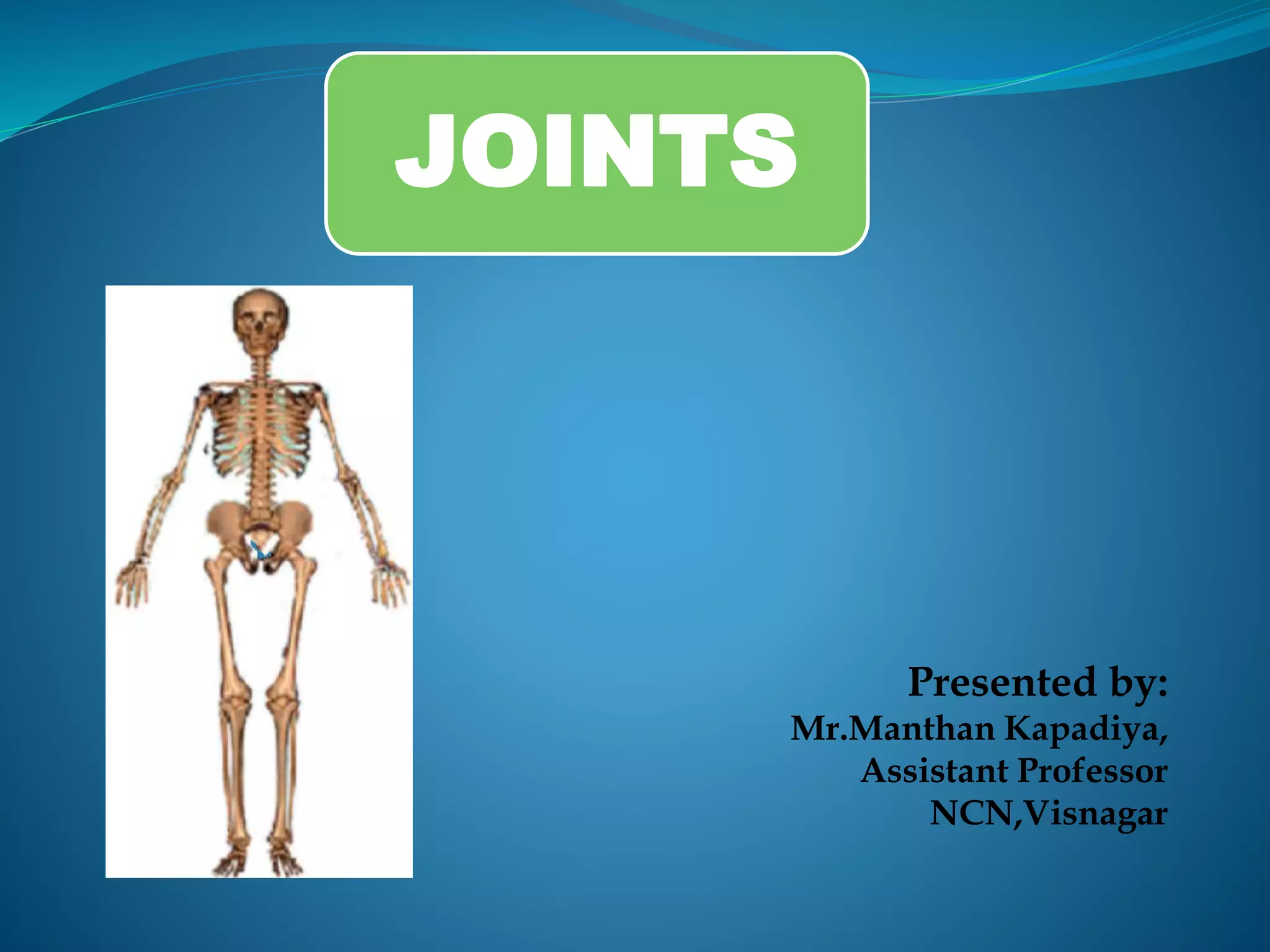
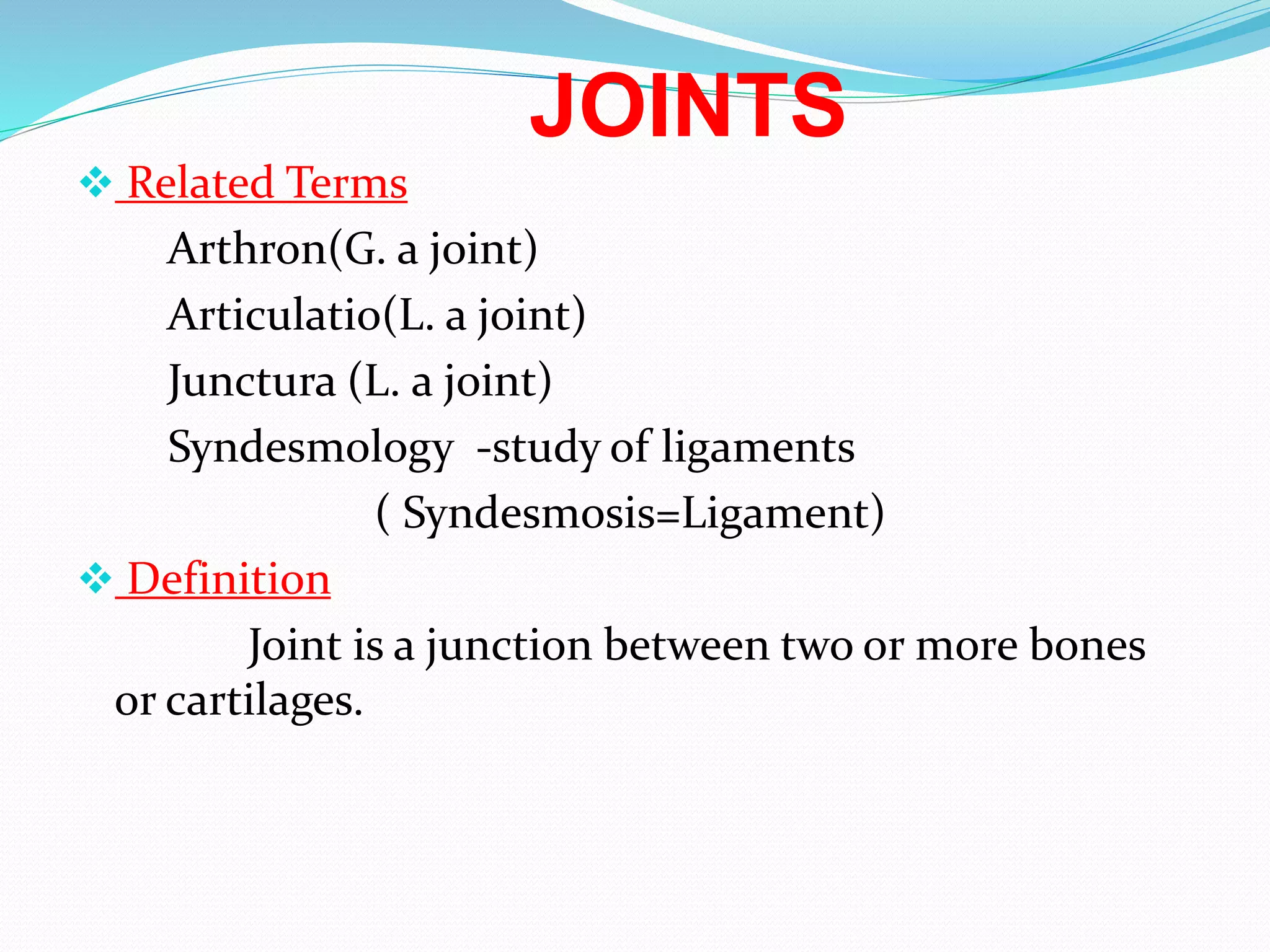
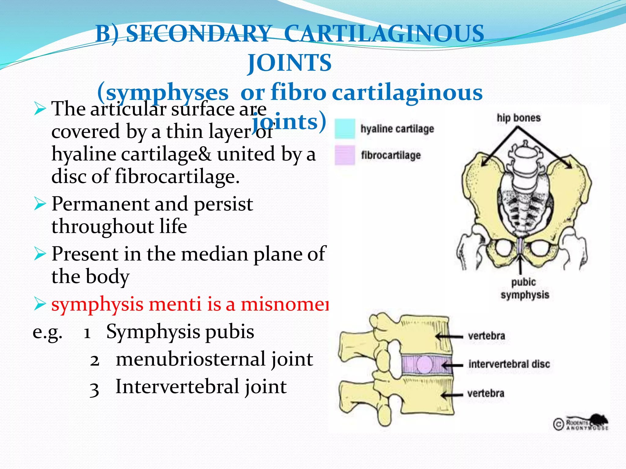
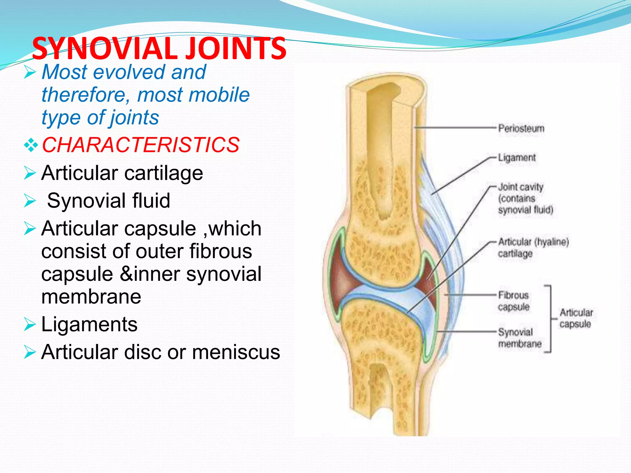
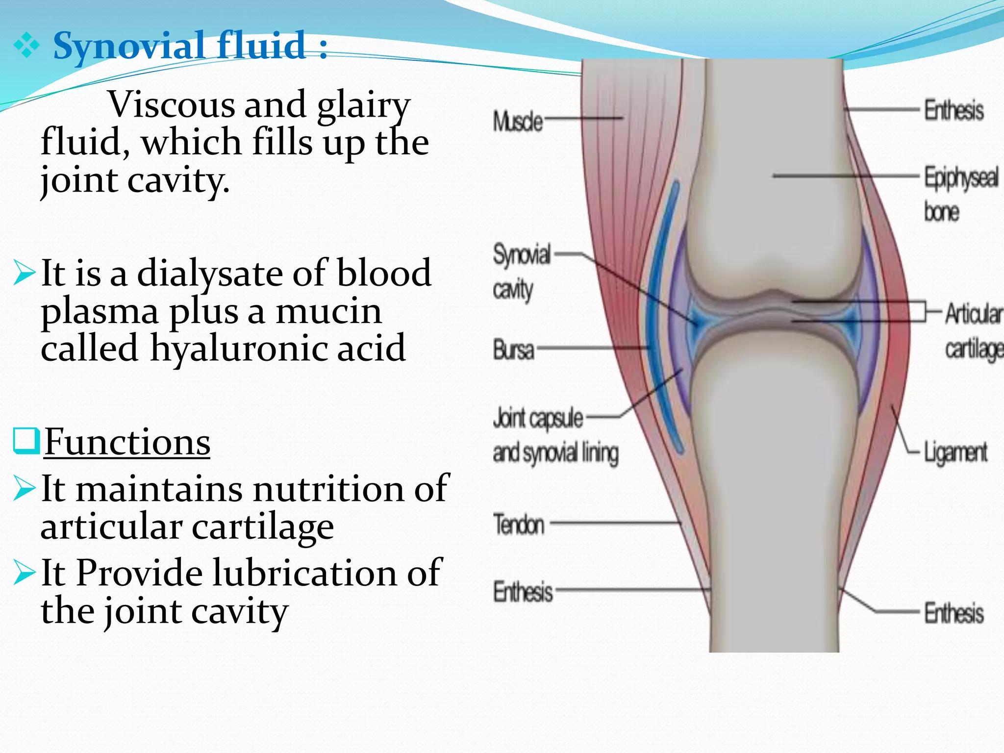
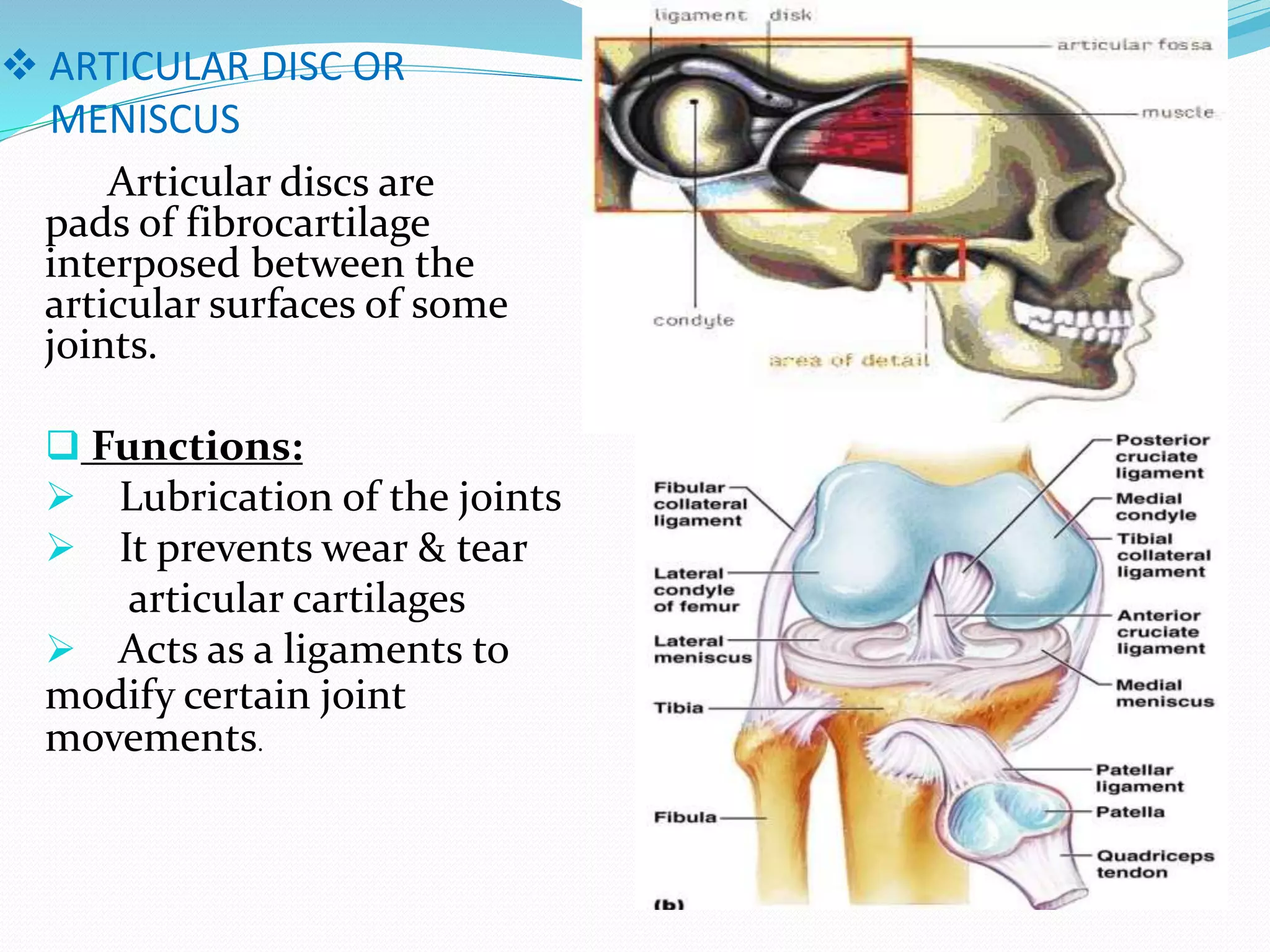
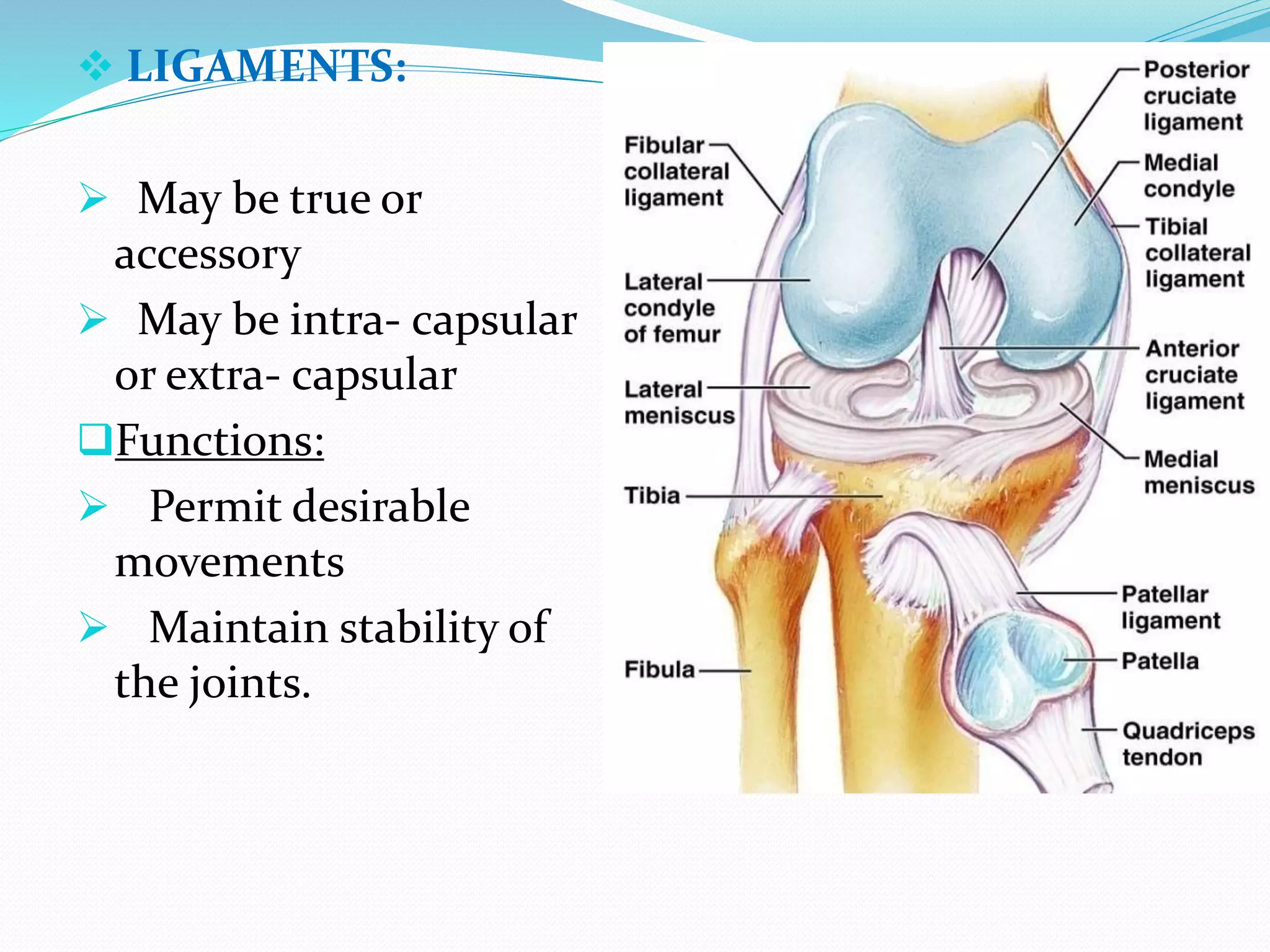
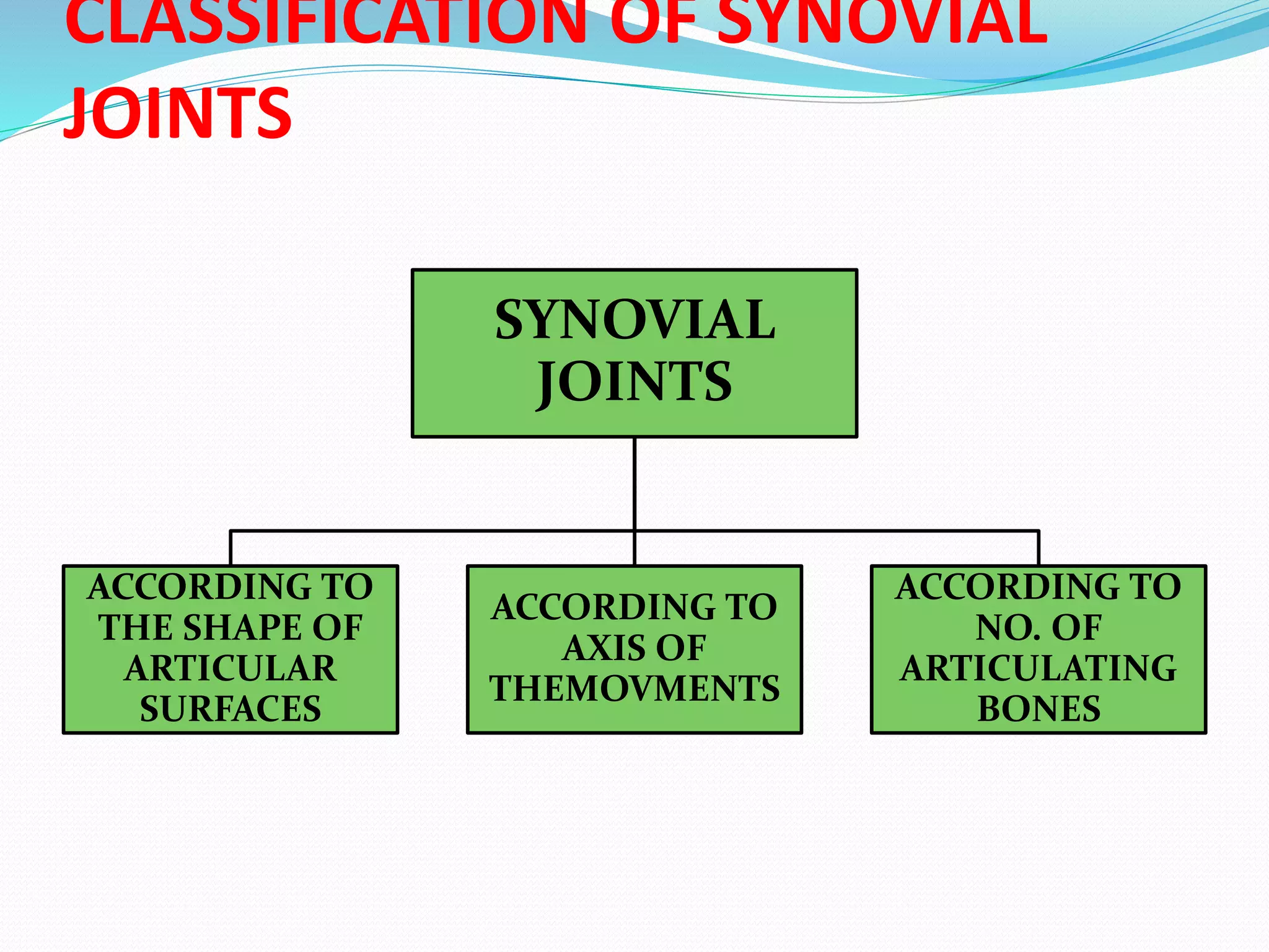
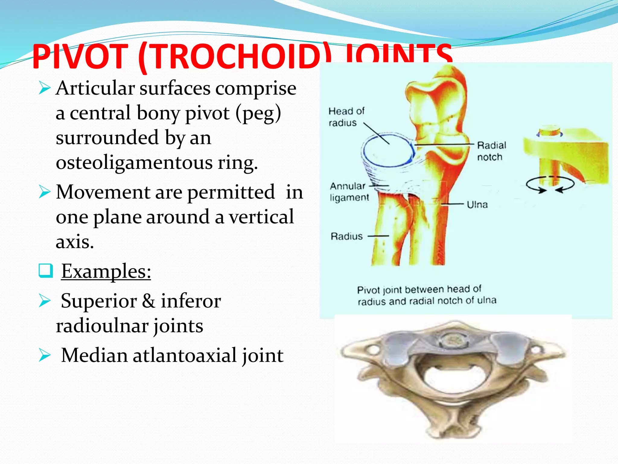
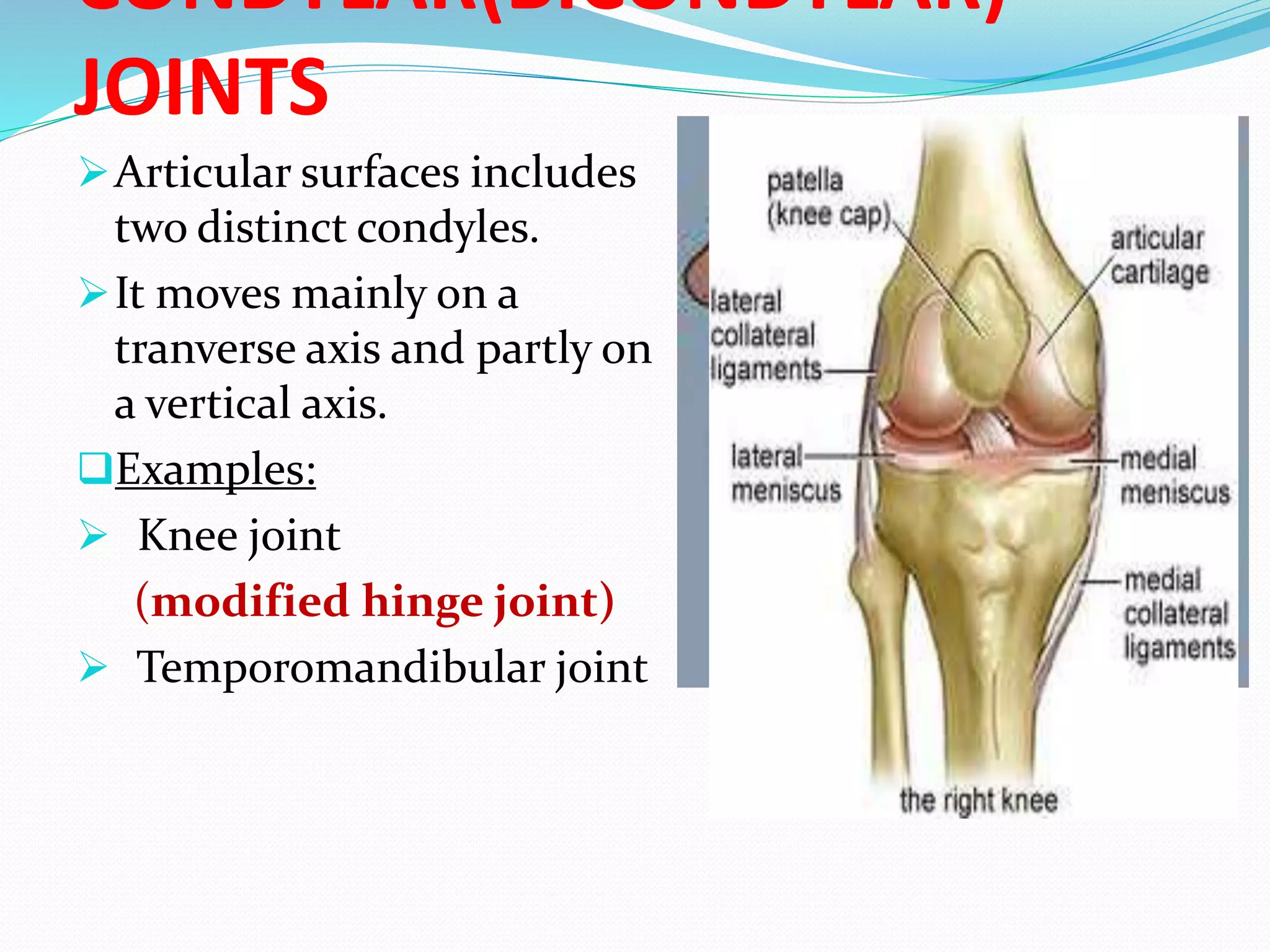
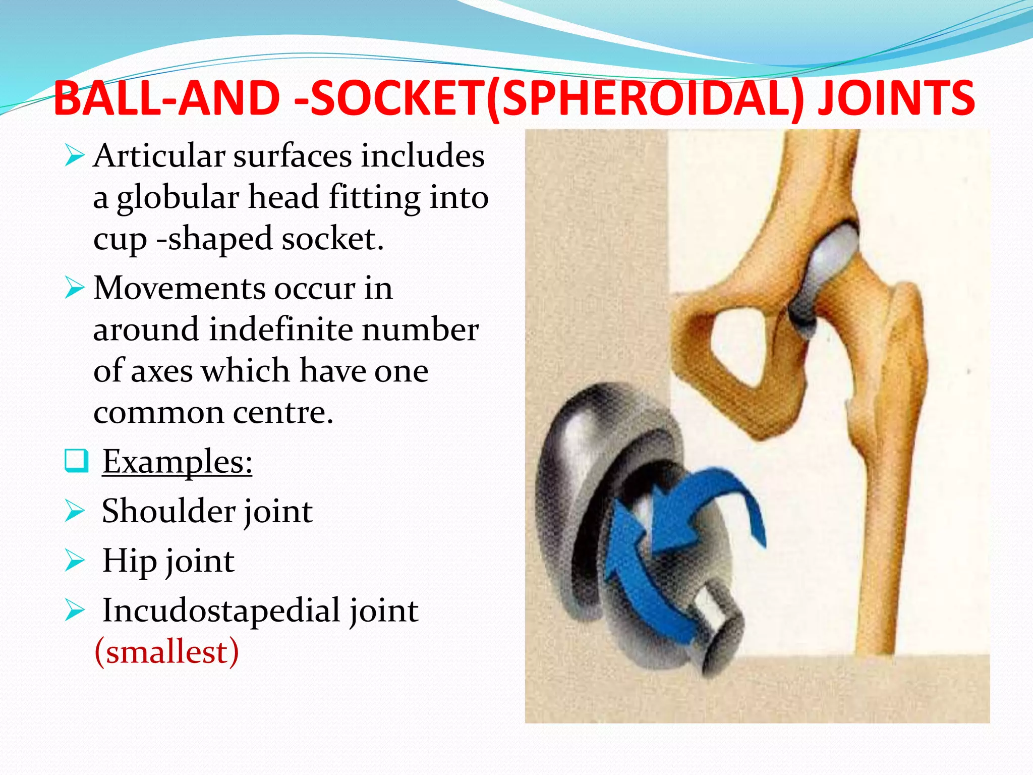
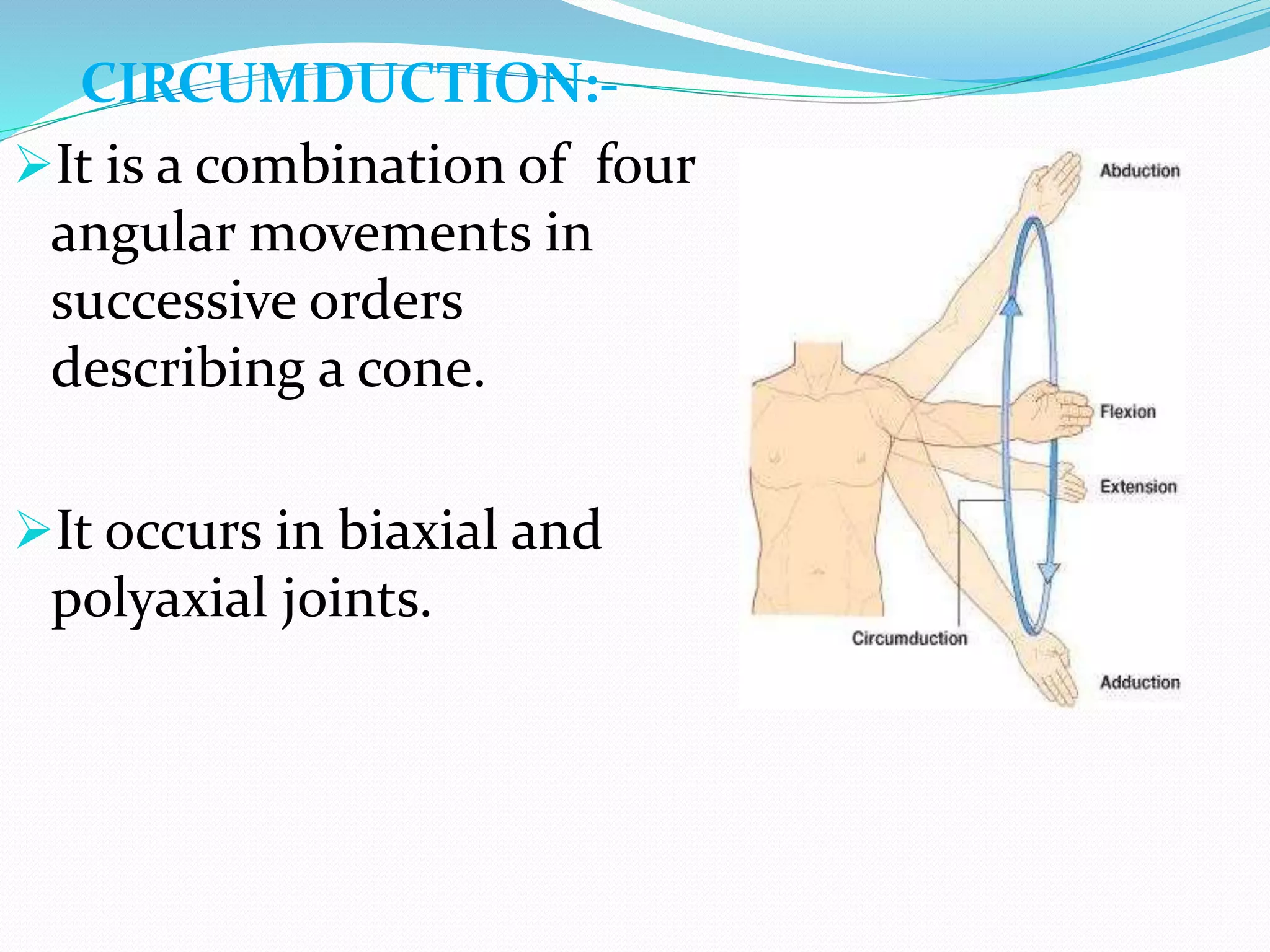
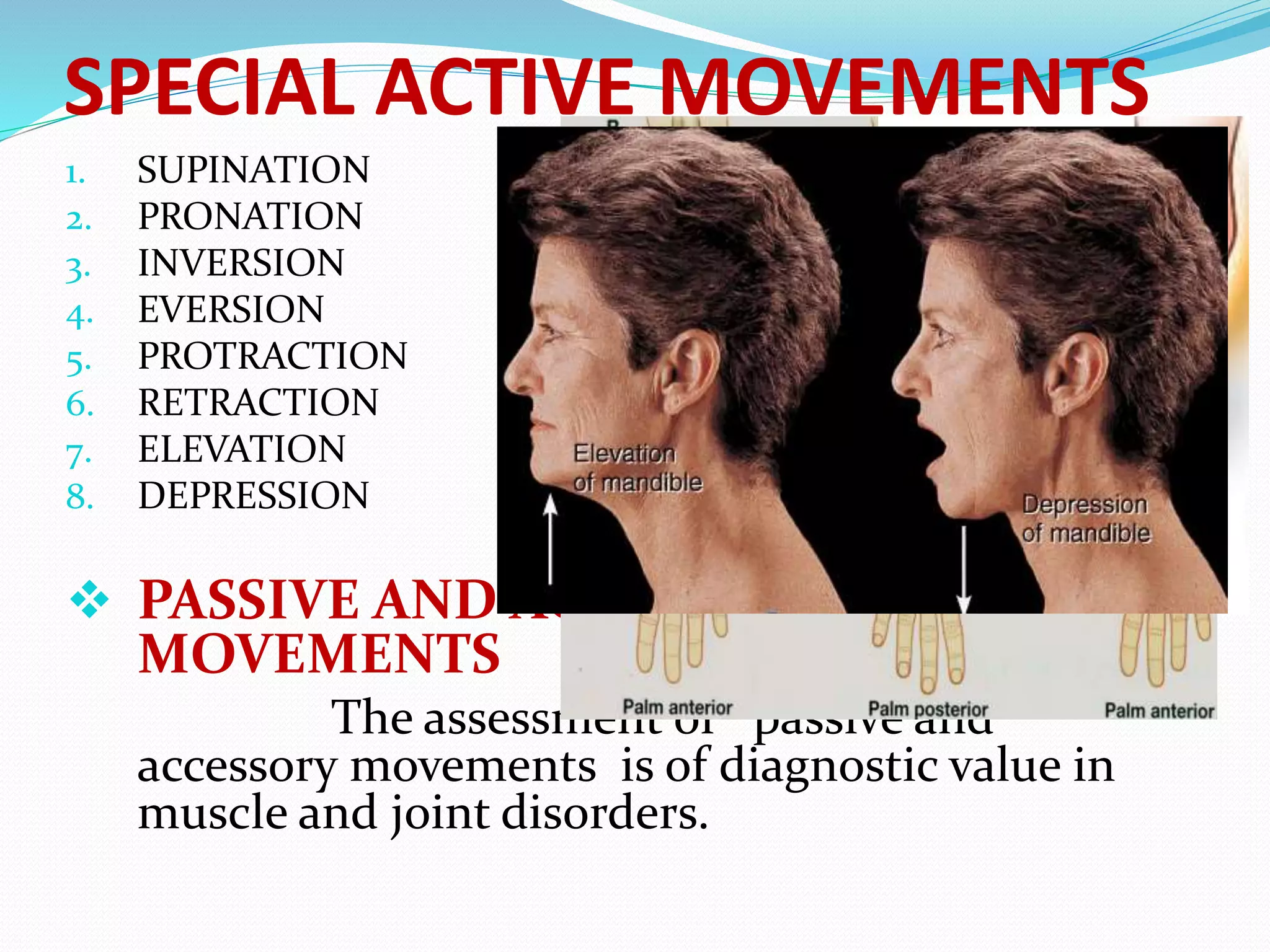
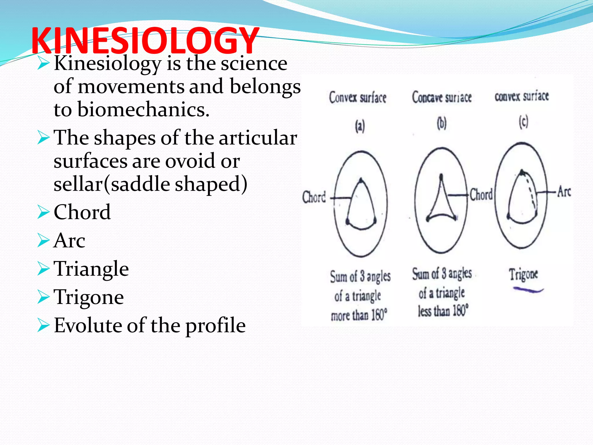
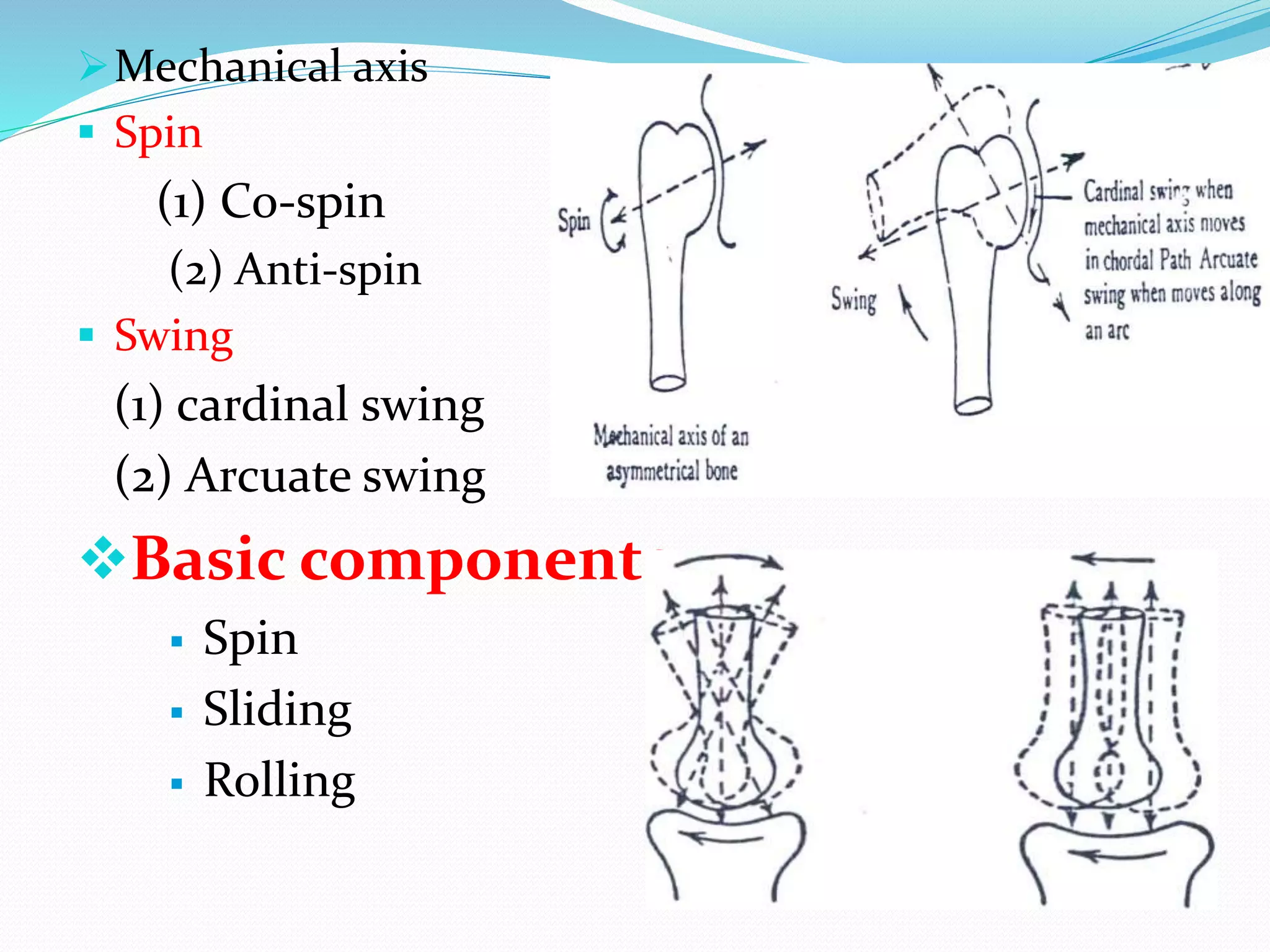
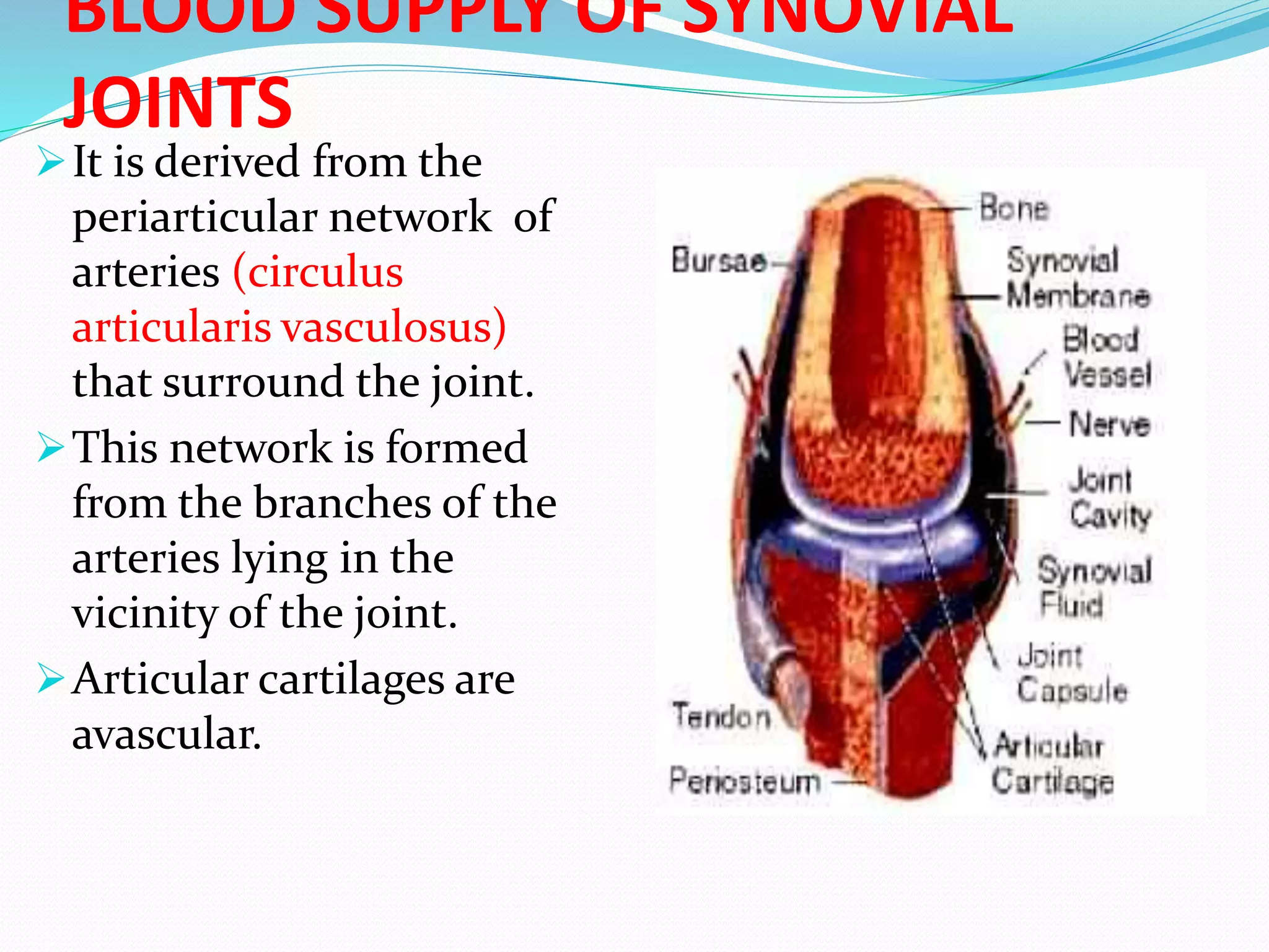
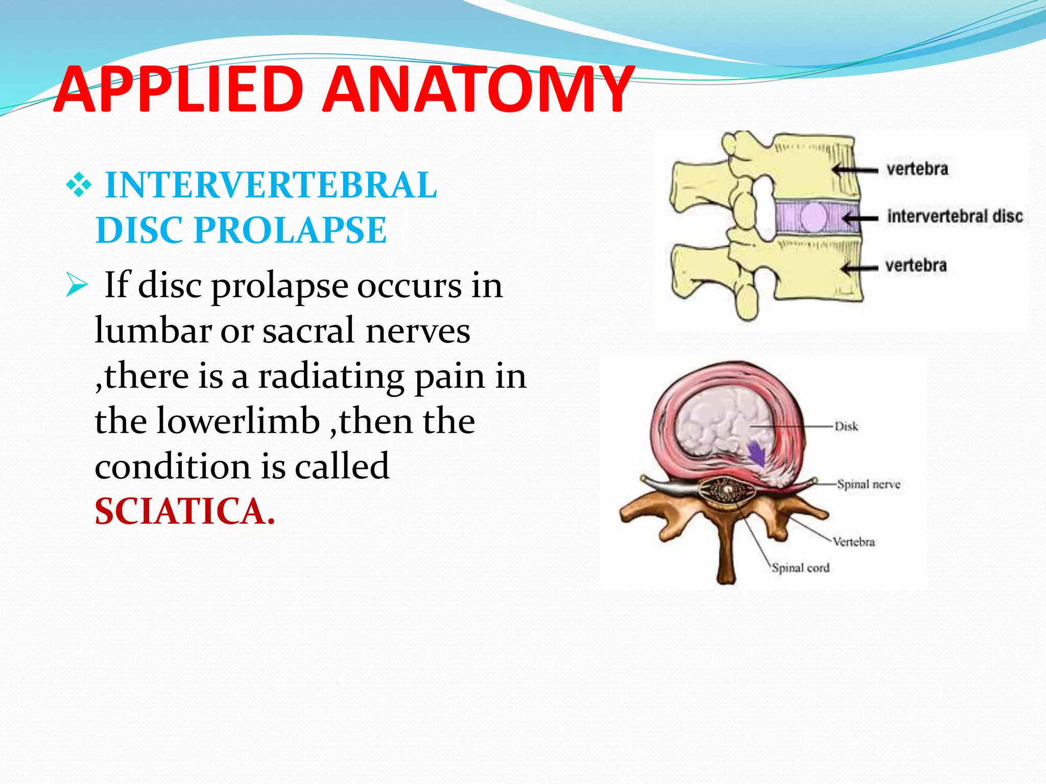
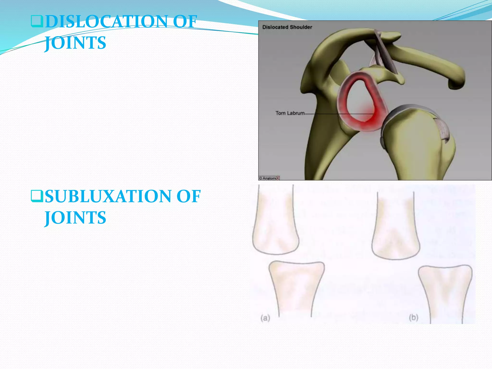
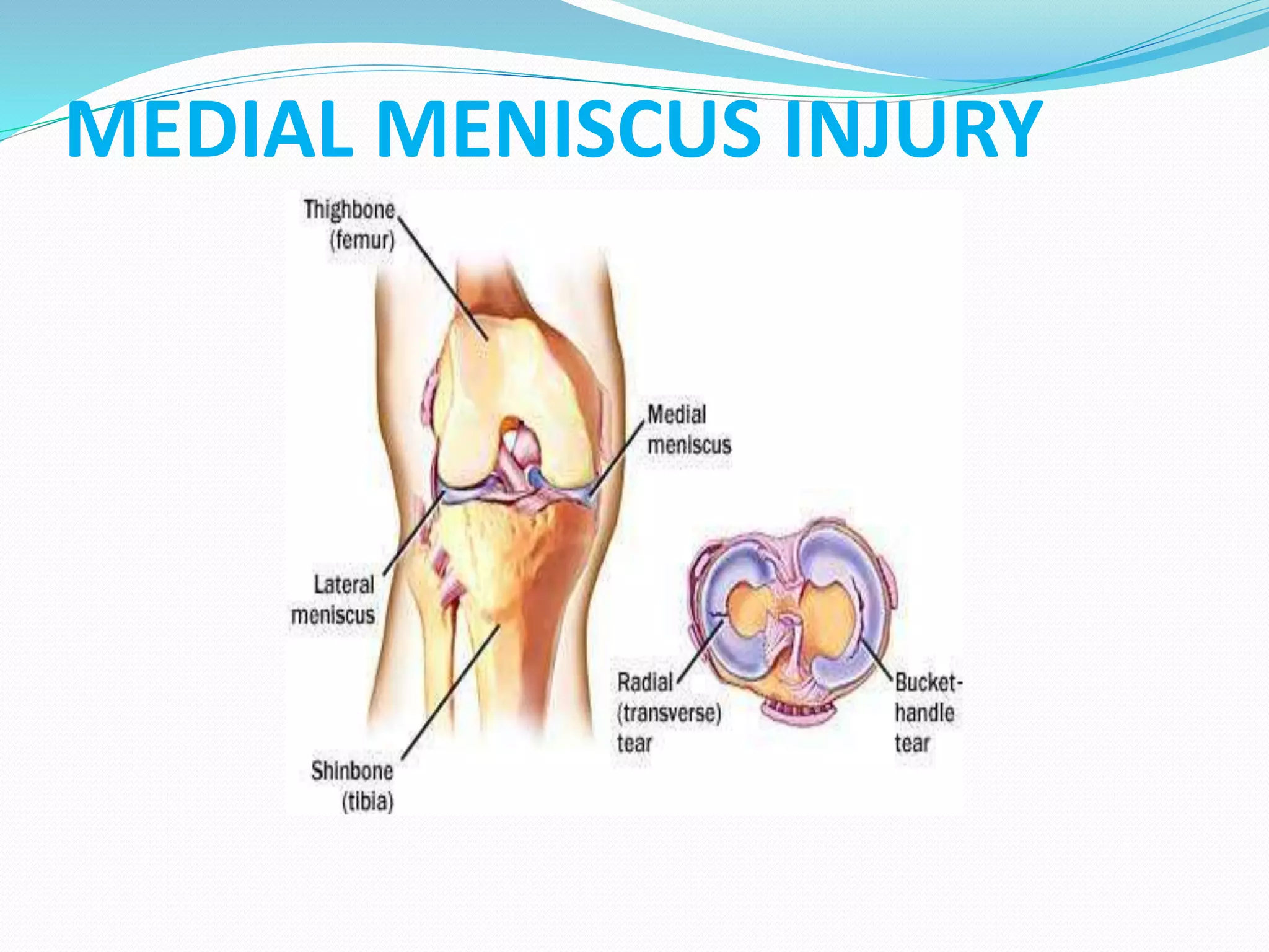
JOINTS
Presented by:
Mr.Manthan Kapadiya,
Assistant Professor
NCN,Visnagar
JOINTS
 Related Terms
Arthron(G. a joint)
Articulatio(L. a joint)
Junctura (L. a joint)
Syndesmology -study of ligaments
( Syndesmosis=Ligament)
 Definition
Joint is a junction between two or more bones
or cartilages.
CLASSIFICATION OF JOINTS
 Structural classification
 Functional classification
 Regional classification
Structural
classification
Fibrous joints Cartilaginous
joints
Synovial joints
Sutures
Syndesmosis
Gomphosis
Primary cartilagenous joints
(synchondrosis)
Secondary cartilagenous
joints (symphysis)
Plane joints
Hinge joints
Pivot joints
Condylar joints
Ellipsoid joints
Saddle joints
Ball and socket joint
Functional
classification
Synarthrosis
(immovable)
like fibrous
joints
Amphiarthrosis
(slightly
movable) like
cartilaginous
joints
Diarthrosis
(freely movable)
like synovial
joints
Regional
classification
Skull type
(immovable)
Vertebral type
(slightly
movable)
Limb type
(Freely
movable)
Fibrous joints
The bones are joined by fibrous tissue
Immovable ,lacks joint cavity
Synarthrosis
present in joints of skull
In children they permit slight
Movement
 Sutural ligament
 Appear between those bones which
ossify in membranes
SUTURES
TYPES OF SUTURES
FONTANELLES
At six specific points on
the sutures in new born
skull,there are membrane
filled gaps called
“FONTANELLES”.
 They are temporary in
nature.
All these fontanelles
become bone by 18
months.
SYNDESMOSIS
 Surfaces of bones are
united by interosseous
ligaments
 Such ligaments persist
throughout life
 Slight amount of
movement is possible
e.g. Introsseous membranes
of the forearm
Inferior tibiofibular
joint
Gomphosis(peg and socket joint)
The roots of the teeth fit
in the socket of the jaw
and are united by fibrous
tissue
 Periodontal ligaments
CARTILAGINOUS JOINTS
A) PRIMARY
CARTILAGINOUS JOINTS
(synchondrosis or hyaline
cartilage joints)
 The bones are united by a
plate of hyaline cartilage
 Joint is immovable and strong
 Temporary in nature
 (Synostosis)
 e.g. Sphenoccipital joint
1st chondrosternal joint
Costochondral joints
 The articular surface are
covered by a thin layer of
hyaline cartilage& united by a
disc of fibrocartilage.
 Permanent and persist
throughout life
 Present in the median plane of
the body
 symphysis menti is a misnomer
e.g. 1 Symphysis pubis
2 menubriosternal joint
3 Intervertebral joint
B) SECONDARY CARTILAGINOUS
JOINTS
(symphyses or fibro cartilaginous
joints)
SYNOVIAL JOINTS
 Most evolved and
therefore, most mobile
type of joints
CHARACTERISTICS
 Articular cartilage
 Synovial fluid
 Articular capsule ,which
consist of outer fibrous
capsule &inner synovial
membrane
 Ligaments
 Articular disc or meniscus
COMPONENTS OF SYNOVIAL
JOINTSArticular capsule: it
consists of outer fibrous
capsule & inner synovial
membrane
Fibrous capsule:
formed by bundle of
collagen fibers
 Functions
 It binds the
articulating bones
together
 It supports the
synovial membrane
 Watch dog action
 Synovial membrane
It is highly vascular
and cellular connective
tissue membrane.
 Functions
Secretes synovial
fluids
Liberates hyaluronic
acid
Phagocytic activity
(Articular cartilages are
not covered by synovial
membrane)
 Synovial fluid :
Viscous and glairy
fluid, which fills up the
joint cavity.
It is a dialysate of blood
plasma plus a mucin
called hyaluronic acid
Functions
It maintains nutrition of
articular cartilage
It Provide lubrication of
the joint cavity
ARTICULAR CARTILAGE
Articular cartilage
of most joints is
hyaline except in those
bones which are
ossified in membrane
where it is fibro-
cartilage.
 Hyaline articular
cartilage is avascular,
non nervous and
elastic.
 Cartilage consists of
proteoglycan-
hyaluronic acid
aggregates.
Functions
Provide smooth gliding surface and
reduces friction between two bones
during movement.
Regulate the growth of epiphysis
 CHANGES IN ARTICULAR CARTILAGE WITH
AGE:-
 Degenerative changes
 Proliferative changes
Nutrition :
 Synovial fluid;
 Diffusion from capillaries;
 Diffusion from the adjacent epiphyseal
blood vessels
 ARTICULAR DISC OR
MENISCUS
Articular discs are
pads of fibrocartilage
interposed between the
articular surfaces of some
joints.
 Functions:
 Lubrication of the joints
 It prevents wear & tear
articular cartilages
 Acts as a ligaments to
modify certain joint
movements.
 LIGAMENTS:
 May be true or
accessory
 May be intra- capsular
or extra- capsular
Functions:
 Permit desirable
movements
 Maintain stability of
the joints.
BURSAE:
These are pouch
like sacs of connective
tissue filled with
synovial fluid
 Locations
 Functions
 Reduces friction.
 Acts as a cushion.
(Synovial tendon
sheath)
 FAT PADS (Haversian glands):
These are pads of fat placed
between synovial membrane and
fibrous capsule or between
synovial membrane and bone.
 It acts as a vacuum filler
e.g. Acetabular fat of hip joint
Infrapatellar fat-pad of
knee joint.
CLASSIFICATION OF SYNOVIAL
JOINTS
SYNOVIAL
JOINTS
ACCORDING TO
THE SHAPE OF
ARTICULAR
SURFACES
ACCORDING TO
AXIS OF
THEMOVMENTS
ACCORDING TO
NO. OF
ARTICULATING
BONES
ACCORDING TO THE SHAPE OF
ARTICULAR SURFACES
Plane
joints
Hinge
joints
(Ginglymi
)
Pivot
joints
(Trochoid )
Condylar
joints
Ellipsoidal
joints
Saddle
joints
Ball-
and-
socket
joints
ACCORDING TO
AXIS OF THE MOVMENTS
Uniaxial
joints
e.g.
Hinge joints
Pivot joints
Biaxial joints
e.g.
Condylar joints
Ellipsoid joints
Multi axial joints
e.g.
saddle joints
Ball and socket
joints
ACCORDING TO NO. OF
ARTICULATING BONES
Simple joints
(only two bones)
e.g.
Interphalangeal
joints
Compound joints
(more than two
bones)
e.g.
Radio carpal joint,
Ankle joint
Complex
joints
Ex.temporom
andibular
joint,
sernoclavicul
ar joint
PLANE SYNOVIAL JOINTS
Articular surfaces are nearly
flat
They permit gliding
movements in various
directions
Examples
 Intercarpal joints
 Intertarsal joints
 Carpometacarpal
(except first) etc…
HINGE JOINTS(GINGLYMI)
Articular surfaces are
pulley shaped.
Movement are
permitted in one plane
around a transverse
axis.
 Examples:
 Elbow joint
 Ankle joint
 Interphalangeal
joints
PIVOT (TROCHOID) JOINTS
 Articular surfaces comprise
a central bony pivot (peg)
surrounded by an
osteoligamentous ring.
 Movement are permitted in
one plane around a vertical
axis.
 Examples:
 Superior & inferor
radioulnar joints
 Median atlantoaxial joint
CONDYLAR(BICONDYLAR)
JOINTS
Articular surfaces includes
two distinct condyles.
It moves mainly on a
tranverse axis and partly on
a vertical axis.
Examples:
 Knee joint
(modified hinge joint)
 Temporomandibular joint
ELLIPSOID JOINTS
 The elliptical convex surface
of one bone articulates with
the elliptical concave surface
of another bone.
 Movements are permitted in
two directions
 Combination of movements
produces circumduction.
 Examples:
 Wrist joint
 Metacarpophalangeal joints
 Atlanto-occipital joints
SADDLE(SELLAR) JOINTS
Articular surfaces are
reciprocally concavoconvex.
Movements are similar to
ellipsoid joint.
Conjunct rotation is present
 Examples:
 First carpometacarpal joint
 Sternoclavicular joint ,etc..
BALL-AND -SOCKET(SPHEROIDAL) JOINTS
 Articular surfaces includes
a globular head fitting into
cup -shaped socket.
 Movements occur in
around indefinite number
of axes which have one
common centre.
 Examples:
 Shoulder joint
 Hip joint
 Incudostapedial joint
(smallest)
 TERMINOLOGY AND DEFINITION
 HUMAN KINESIOLOGY
 MALE SURFACE
 FEMALE SURFACE
 SIMPLE JOINTS
 COMPOUND JOINTS
 DEGREES OF FREEDOM
 UNIAXIAL
 BIAXIAL
 MULTI-AXIAL
 TRANSLATION
 SHAPE OF ARTICULAR SURFACE
 OVOID
 SELLAR / SADDLE- SHAPED
MOVEMENTS OF SYNOVIAL
JOINTS
Active movements
GLIDING
(TRANSLATION)
Sliding movement of one
articulating surface over
the other.
This movements occur in
plane joints.
 ANGULAR MOVEMENT
 Movements leading to
diminution or increase in
angle between two adjoining
bones.
(1) FLEXION AND
EXTENSION
Flexion means bending ,and
extension signifies
straightening.
Movements occur around
transverse axis
(2)ABDUCTION AND
ADDUCTION
In adduction part moves
towards the median
plane.
In abduction it deviates
away from the middle
line.
Take place around an
antero-posterior axis
Except…Carpo-metacarpal
joint of thumb
(3)ROTARY (CIRCULAR)
MOVEMENTS
ROTATION:-
Bone moves around a lonitudinal
axis.
 ADJUNCT ROTATION
Independent rotation
e.g. Rotations at hip, shoulder
and atlantoaxial joints
 CONJUNCT ROTATION
Rotation which accompany
other movements.
e.g. Locking and unlocking of
knee joint
Carpo-metacarpal joint of
thumb
CIRCUMDUCTION:-
It is a combination of four
angular movements in
successive orders
describing a cone.
It occurs in biaxial and
polyaxial joints.
SPECIAL ACTIVE MOVEMENTS
1. SUPINATION
2. PRONATION
3. INVERSION
4. EVERSION
5. PROTRACTION
6. RETRACTION
7. ELEVATION
8. DEPRESSION
 PASSIVE AND ACCESSORY
MOVEMENTS
The assessment of passive and
accessory movements is of diagnostic value in
muscle and joint disorders.
 FACTOR LIMITING THE RANGE OF
MOVEMENT
Shape of articulating bones
Reflex contraction of antagonistic muscles
Due to stimulation of mechanoreceptors in articular
tissue
Tension of ligaments
The approximation of soft parts
 FACTORS MAINTAINING STABILITY OF JOINTS
 Bony configuration
 Ligaments
 Muscles
 Atmospheric pressure and force of cohesion
 MECHANISM OF LUBRICATION OF SYNOVIAL JOINT
 Synovial fluid
 Hyaline cartilage
 Intra-articular fibrocartilage
 Haversian fatty pads
 Bursa
KINESIOLOGYKinesiology is the science
of movements and belongs
to biomechanics.
The shapes of the articular
surfaces are ovoid or
sellar(saddle shaped)
Chord
Arc
Triangle
Trigone
Evolute of the profile
Mechanical axis
 Spin
(1) Co-spin
(2) Anti-spin
 Swing
(1) cardinal swing
(2) Arcuate swing
Basic components
 Spin
 Sliding
 Rolling
JOINT POSITION
CLOSED PACKED POSITION
It is the position of joints,in which the
articular surfaces are fully congruent and have
maximum area of contact.
e.g. Temporomandibular joint –Clenched teeth
LOOSE PACKED POSITION
The position in which articular surfaces are
incongruent.
e.g. Shoulder –semiabduction
BLOOD SUPPLY OF SYNOVIAL
JOINTS
It is derived from the
periarticular network of
arteries (circulus
articularis vasculosus)
that surround the joint.
This network is formed
from the branches of the
arteries lying in the
vicinity of the joint.
Articular cartilages are
avascular.
Fibrous capsule and ligament have poor blood supply.
The synovial membrane is richly supplied by blood
 The rich vascular plexus of synovial membrane also
helps to supply nutrition to the periphery of the
articular cartilages.
 After epiphysial fusion ,communications between
circulus vasculosus and the end arteries of metaphysis
are established ,thus minimizing the chances of
osteomyelitis in the metaphysis.
 HILTON’S LAW
 e.g. Axillary nerve
 GARDNER’S OBSERVATION
 e.g. Inferomedial part of capsule of
hip joint
LYMPHATIC DRAINAGE OF
SYNOVIAL JOINTS
Lymphatics form a plexus in the
subintima of the synovial
membrane.
APPLIED ANATOMY INTERVERTEBRAL DISC PROLAPSE
 DISLOCATION OF JOINTS
 SUBLUXATION OF JOINTS
 SPRAIN
 RHEUMATIC FEVER
 ARTHRITIS
 RHEUMATOID ARTHRITIS
 OSTEOARTHRITIS
 SEPTIC ARTHRITIS
 SPONDYLITIS
 INJURY TO A JOINT AND ITS STRUCTURES
 STIFNESS OF JOINTS
 NEUROPATHIC JOINTS
 ARTHROSCOPY
 ARTHROGRAPHY
 ARTHROPLASTY
APPLIED ANATOMY
 INTERVERTEBRAL
DISC PROLAPSE
 If disc prolapse occurs in
lumbar or sacral nerves
,there is a radiating pain in
the lowerlimb ,then the
condition is called
SCIATICA.
DISLOCATION OF
JOINTS
SUBLUXATION OF
JOINTS
SPRAIN
ARTHRITIS
SEPTIC ARTHRITIS
INTRACAPSULAR
METAPHYSIS
(BLACK AREAS)
SPONDYLITIS
INJURY TO A JOINT
MEDIAL MENISCUS INJURY
ARTHROSCOPY
ARTHROPLASTY
Joints anatomy and physiology

Joints anatomy and physiology

  • 1.
  • 2.
    JOINTS  Related Terms Arthron(G.a joint) Articulatio(L. a joint) Junctura (L. a joint) Syndesmology -study of ligaments ( Syndesmosis=Ligament)  Definition Joint is a junction between two or more bones or cartilages.
  • 3.
    CLASSIFICATION OF JOINTS Structural classification  Functional classification  Regional classification
  • 4.
    Structural classification Fibrous joints Cartilaginous joints Synovialjoints Sutures Syndesmosis Gomphosis Primary cartilagenous joints (synchondrosis) Secondary cartilagenous joints (symphysis) Plane joints Hinge joints Pivot joints Condylar joints Ellipsoid joints Saddle joints Ball and socket joint
  • 5.
  • 6.
  • 7.
    Fibrous joints The bonesare joined by fibrous tissue Immovable ,lacks joint cavity Synarthrosis present in joints of skull In children they permit slight Movement  Sutural ligament  Appear between those bones which ossify in membranes SUTURES
  • 8.
  • 9.
    FONTANELLES At six specificpoints on the sutures in new born skull,there are membrane filled gaps called “FONTANELLES”.  They are temporary in nature. All these fontanelles become bone by 18 months.
  • 10.
    SYNDESMOSIS  Surfaces ofbones are united by interosseous ligaments  Such ligaments persist throughout life  Slight amount of movement is possible e.g. Introsseous membranes of the forearm Inferior tibiofibular joint
  • 11.
    Gomphosis(peg and socketjoint) The roots of the teeth fit in the socket of the jaw and are united by fibrous tissue  Periodontal ligaments
  • 12.
    CARTILAGINOUS JOINTS A) PRIMARY CARTILAGINOUSJOINTS (synchondrosis or hyaline cartilage joints)  The bones are united by a plate of hyaline cartilage  Joint is immovable and strong  Temporary in nature  (Synostosis)  e.g. Sphenoccipital joint 1st chondrosternal joint Costochondral joints
  • 13.
     The articularsurface are covered by a thin layer of hyaline cartilage& united by a disc of fibrocartilage.  Permanent and persist throughout life  Present in the median plane of the body  symphysis menti is a misnomer e.g. 1 Symphysis pubis 2 menubriosternal joint 3 Intervertebral joint B) SECONDARY CARTILAGINOUS JOINTS (symphyses or fibro cartilaginous joints)
  • 14.
    SYNOVIAL JOINTS  Mostevolved and therefore, most mobile type of joints CHARACTERISTICS  Articular cartilage  Synovial fluid  Articular capsule ,which consist of outer fibrous capsule &inner synovial membrane  Ligaments  Articular disc or meniscus
  • 15.
    COMPONENTS OF SYNOVIAL JOINTSArticularcapsule: it consists of outer fibrous capsule & inner synovial membrane Fibrous capsule: formed by bundle of collagen fibers  Functions  It binds the articulating bones together  It supports the synovial membrane  Watch dog action
  • 16.
     Synovial membrane Itis highly vascular and cellular connective tissue membrane.  Functions Secretes synovial fluids Liberates hyaluronic acid Phagocytic activity (Articular cartilages are not covered by synovial membrane)
  • 17.
     Synovial fluid: Viscous and glairy fluid, which fills up the joint cavity. It is a dialysate of blood plasma plus a mucin called hyaluronic acid Functions It maintains nutrition of articular cartilage It Provide lubrication of the joint cavity
  • 18.
    ARTICULAR CARTILAGE Articular cartilage ofmost joints is hyaline except in those bones which are ossified in membrane where it is fibro- cartilage.  Hyaline articular cartilage is avascular, non nervous and elastic.  Cartilage consists of proteoglycan- hyaluronic acid aggregates.
  • 19.
    Functions Provide smooth glidingsurface and reduces friction between two bones during movement. Regulate the growth of epiphysis  CHANGES IN ARTICULAR CARTILAGE WITH AGE:-  Degenerative changes  Proliferative changes Nutrition :  Synovial fluid;  Diffusion from capillaries;  Diffusion from the adjacent epiphyseal blood vessels
  • 20.
     ARTICULAR DISCOR MENISCUS Articular discs are pads of fibrocartilage interposed between the articular surfaces of some joints.  Functions:  Lubrication of the joints  It prevents wear & tear articular cartilages  Acts as a ligaments to modify certain joint movements.
  • 21.
     LIGAMENTS:  Maybe true or accessory  May be intra- capsular or extra- capsular Functions:  Permit desirable movements  Maintain stability of the joints.
  • 22.
    BURSAE: These are pouch likesacs of connective tissue filled with synovial fluid  Locations  Functions  Reduces friction.  Acts as a cushion. (Synovial tendon sheath)
  • 23.
     FAT PADS(Haversian glands): These are pads of fat placed between synovial membrane and fibrous capsule or between synovial membrane and bone.  It acts as a vacuum filler e.g. Acetabular fat of hip joint Infrapatellar fat-pad of knee joint.
  • 24.
    CLASSIFICATION OF SYNOVIAL JOINTS SYNOVIAL JOINTS ACCORDINGTO THE SHAPE OF ARTICULAR SURFACES ACCORDING TO AXIS OF THEMOVMENTS ACCORDING TO NO. OF ARTICULATING BONES
  • 25.
    ACCORDING TO THESHAPE OF ARTICULAR SURFACES Plane joints Hinge joints (Ginglymi ) Pivot joints (Trochoid ) Condylar joints Ellipsoidal joints Saddle joints Ball- and- socket joints
  • 26.
    ACCORDING TO AXIS OFTHE MOVMENTS Uniaxial joints e.g. Hinge joints Pivot joints Biaxial joints e.g. Condylar joints Ellipsoid joints Multi axial joints e.g. saddle joints Ball and socket joints
  • 27.
    ACCORDING TO NO.OF ARTICULATING BONES Simple joints (only two bones) e.g. Interphalangeal joints Compound joints (more than two bones) e.g. Radio carpal joint, Ankle joint Complex joints Ex.temporom andibular joint, sernoclavicul ar joint
  • 28.
    PLANE SYNOVIAL JOINTS Articularsurfaces are nearly flat They permit gliding movements in various directions Examples  Intercarpal joints  Intertarsal joints  Carpometacarpal (except first) etc…
  • 29.
    HINGE JOINTS(GINGLYMI) Articular surfacesare pulley shaped. Movement are permitted in one plane around a transverse axis.  Examples:  Elbow joint  Ankle joint  Interphalangeal joints
  • 30.
    PIVOT (TROCHOID) JOINTS Articular surfaces comprise a central bony pivot (peg) surrounded by an osteoligamentous ring.  Movement are permitted in one plane around a vertical axis.  Examples:  Superior & inferor radioulnar joints  Median atlantoaxial joint
  • 31.
    CONDYLAR(BICONDYLAR) JOINTS Articular surfaces includes twodistinct condyles. It moves mainly on a tranverse axis and partly on a vertical axis. Examples:  Knee joint (modified hinge joint)  Temporomandibular joint
  • 32.
    ELLIPSOID JOINTS  Theelliptical convex surface of one bone articulates with the elliptical concave surface of another bone.  Movements are permitted in two directions  Combination of movements produces circumduction.  Examples:  Wrist joint  Metacarpophalangeal joints  Atlanto-occipital joints
  • 33.
    SADDLE(SELLAR) JOINTS Articular surfacesare reciprocally concavoconvex. Movements are similar to ellipsoid joint. Conjunct rotation is present  Examples:  First carpometacarpal joint  Sternoclavicular joint ,etc..
  • 34.
    BALL-AND -SOCKET(SPHEROIDAL) JOINTS Articular surfaces includes a globular head fitting into cup -shaped socket.  Movements occur in around indefinite number of axes which have one common centre.  Examples:  Shoulder joint  Hip joint  Incudostapedial joint (smallest)
  • 35.
     TERMINOLOGY ANDDEFINITION  HUMAN KINESIOLOGY  MALE SURFACE  FEMALE SURFACE  SIMPLE JOINTS  COMPOUND JOINTS  DEGREES OF FREEDOM  UNIAXIAL  BIAXIAL  MULTI-AXIAL  TRANSLATION  SHAPE OF ARTICULAR SURFACE  OVOID  SELLAR / SADDLE- SHAPED
  • 36.
    MOVEMENTS OF SYNOVIAL JOINTS Activemovements GLIDING (TRANSLATION) Sliding movement of one articulating surface over the other. This movements occur in plane joints.
  • 37.
     ANGULAR MOVEMENT Movements leading to diminution or increase in angle between two adjoining bones. (1) FLEXION AND EXTENSION Flexion means bending ,and extension signifies straightening. Movements occur around transverse axis
  • 38.
    (2)ABDUCTION AND ADDUCTION In adductionpart moves towards the median plane. In abduction it deviates away from the middle line. Take place around an antero-posterior axis Except…Carpo-metacarpal joint of thumb
  • 39.
    (3)ROTARY (CIRCULAR) MOVEMENTS ROTATION:- Bone movesaround a lonitudinal axis.  ADJUNCT ROTATION Independent rotation e.g. Rotations at hip, shoulder and atlantoaxial joints  CONJUNCT ROTATION Rotation which accompany other movements. e.g. Locking and unlocking of knee joint Carpo-metacarpal joint of thumb
  • 40.
    CIRCUMDUCTION:- It is acombination of four angular movements in successive orders describing a cone. It occurs in biaxial and polyaxial joints.
  • 41.
    SPECIAL ACTIVE MOVEMENTS 1.SUPINATION 2. PRONATION 3. INVERSION 4. EVERSION 5. PROTRACTION 6. RETRACTION 7. ELEVATION 8. DEPRESSION  PASSIVE AND ACCESSORY MOVEMENTS The assessment of passive and accessory movements is of diagnostic value in muscle and joint disorders.
  • 42.
     FACTOR LIMITINGTHE RANGE OF MOVEMENT Shape of articulating bones Reflex contraction of antagonistic muscles Due to stimulation of mechanoreceptors in articular tissue Tension of ligaments The approximation of soft parts
  • 43.
     FACTORS MAINTAININGSTABILITY OF JOINTS  Bony configuration  Ligaments  Muscles  Atmospheric pressure and force of cohesion  MECHANISM OF LUBRICATION OF SYNOVIAL JOINT  Synovial fluid  Hyaline cartilage  Intra-articular fibrocartilage  Haversian fatty pads  Bursa
  • 44.
    KINESIOLOGYKinesiology is thescience of movements and belongs to biomechanics. The shapes of the articular surfaces are ovoid or sellar(saddle shaped) Chord Arc Triangle Trigone Evolute of the profile
  • 45.
    Mechanical axis  Spin (1)Co-spin (2) Anti-spin  Swing (1) cardinal swing (2) Arcuate swing Basic components  Spin  Sliding  Rolling
  • 46.
    JOINT POSITION CLOSED PACKEDPOSITION It is the position of joints,in which the articular surfaces are fully congruent and have maximum area of contact. e.g. Temporomandibular joint –Clenched teeth LOOSE PACKED POSITION The position in which articular surfaces are incongruent. e.g. Shoulder –semiabduction
  • 47.
    BLOOD SUPPLY OFSYNOVIAL JOINTS It is derived from the periarticular network of arteries (circulus articularis vasculosus) that surround the joint. This network is formed from the branches of the arteries lying in the vicinity of the joint. Articular cartilages are avascular.
  • 48.
    Fibrous capsule andligament have poor blood supply. The synovial membrane is richly supplied by blood  The rich vascular plexus of synovial membrane also helps to supply nutrition to the periphery of the articular cartilages.  After epiphysial fusion ,communications between circulus vasculosus and the end arteries of metaphysis are established ,thus minimizing the chances of osteomyelitis in the metaphysis.
  • 49.
     HILTON’S LAW e.g. Axillary nerve  GARDNER’S OBSERVATION  e.g. Inferomedial part of capsule of hip joint LYMPHATIC DRAINAGE OF SYNOVIAL JOINTS Lymphatics form a plexus in the subintima of the synovial membrane.
  • 50.
    APPLIED ANATOMY INTERVERTEBRALDISC PROLAPSE  DISLOCATION OF JOINTS  SUBLUXATION OF JOINTS  SPRAIN  RHEUMATIC FEVER  ARTHRITIS  RHEUMATOID ARTHRITIS  OSTEOARTHRITIS  SEPTIC ARTHRITIS  SPONDYLITIS  INJURY TO A JOINT AND ITS STRUCTURES  STIFNESS OF JOINTS  NEUROPATHIC JOINTS  ARTHROSCOPY  ARTHROGRAPHY  ARTHROPLASTY
  • 51.
    APPLIED ANATOMY  INTERVERTEBRAL DISCPROLAPSE  If disc prolapse occurs in lumbar or sacral nerves ,there is a radiating pain in the lowerlimb ,then the condition is called SCIATICA.
  • 52.
  • 53.
  • 54.
  • 55.
  • 56.
  • 57.
  • 58.
  • 59.
  • 60.