
Download as PDF, PPTX

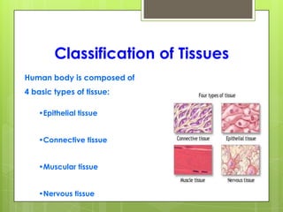


The document provides an overview of the four basic types of tissues in the human body: epithelial, connective, muscular, and nervous tissue. It describes their general characteristics, classifications, and locations. Epithelial tissue forms the protective outer layer of organs and lines body cavities. Connective tissue connects and supports other tissues. Muscular tissue enables movement. Nervous tissue receives and transmits signals throughout the body. The document focuses in detail on epithelial tissues, their functions in protection, secretion, and absorption, and classifications based on cell shape and layering.


