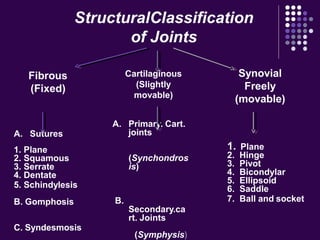
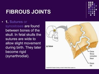
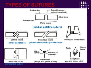
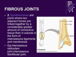
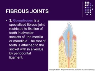
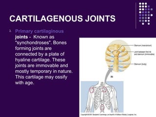
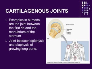
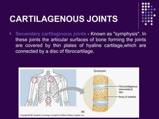
Joints
Prepared by
Mr. Abhay Rajpoot
HOD (Dep. of Medical Surgical)
abhayrajpoot5591@gmail.com
Introduction of Joints
Joint (or articulation)-
the junction
between two or more bones.
With the exception of the
hyoid bone, every bone in the
body is connected to or forms a joint.
There are 230 joints in the body
DEFINITION
 A joint is the location at which two or more bones
make contact. They are constructed to allow
movement and provide mechanical support, and
are classified structurally and functionally.
 Structural classification is determined by how the
bones connect to each other, while functional
classification is determined by the degree of
movement between the articulating bones
Joint Functions
1. Hold the skeletal bones together
2. Allow the skeleton some flexibility so gross
movement can occur
3. Make bone growth possible
CLASSIFICATION OF
JOINTS
A. Sutures
1. Plane
2. Squamous
3. Serrate
4. Dentate
5. Schindylesis
B. Gomphosis
C. Syndesmosis
A. Primary. Cart.
joints
(Synchondros
is)
B.
Secondary.ca
rt. Joints
(Symphysis)
1. Plane
2. Hinge
3. Pivot
4. Bicondylar
5. Ellipsoid
6. Saddle
7. Ball and socket
Synovial
Freely
(movable)
Cartilaginous
(Slightly
movable)
Fibrous
(Fixed)
StructuralClassification
of Joints
STRUCTURAL CLASSIFICATION
 Fibrous/Immovable
Fibrous/Immovable bones are connected
by dense connective tissue, consisting
mainly of collagen. The fibrous joints are
further divided into three types
 Cartilaginous
Cartilaginous are connected entirely by
cartilage. Cartilaginous joints allow more
movement between bones than a fibrous joint
but less than the highly mobile synovial joint.
 Synovial
Synovial joints have a space between the
articulating bones for synovial fluid. This
classification contains joints that are the most
mobile of the three, and includes the knee and
shoulder.
FIBROUS JOINTS
 1. Sutures or
synostoses are found
between bones of the
skull. In fetal skulls the
sutures are wide to
allow slight movement
during birth. They later
become rigid
(synarthrodial).
TYPES OF SUTURES
(median palatine suture)
(inter parietal s.)
(lambdoid suture)
(between temporal & parietal bone)
FIBROUS JOINTS

 2. Syndesmoses are
joints where two
adjacent bones are
linked together by a
considerably greater
amount of connective
tissue than in sutures in
the form of
interosseous ligaments
and membranes
Eg-interosseous
radioulanr
joint,interosseous
tibiofibular joint.
FIBROUS JOINTS
 3. Gomphoses is a
specialized fibrous joint
restricted to fixation of
teeth in alveolar
sockets of the maxilla
or mandible. The root of
tooth is attached to the
socket with in alveolus
by periodontal
ligament.
CARTILAGENOUS JOINTS
 Cartilaginous joints are those joints in which
the bone forming joints are united by means
of either hyaline cartilage or fibro cartilage
 The cartilaginous joints also lack the joint
cavity.
 There are two types of cartilagenous joints:
 Primary
 Secondary
CARTILAGENOUS JOINTS
 Primary cartilaginous
joints - Known as
"synchondroses". Bones
forming joints are
connected by a plate of
hyaline cartilage. These
joints are immovable and
mostly temporary in nature.
This cartilage may ossify
with age.
CARTILAGENOUS JOINTS
 Examples in humans
are the joint between
the first rib and the
manubrium of the
sternum
 Joint between epiphysis
and diaphysis of
growing long bone.
CARTILAGENOUS JOINTS
 Secondary cartilaginous joints - Known as "symphysis". In
these joints the articular surfaces of bone forming the joints
are covered by thin plates of hyaline cartilage,which are
connected by a disc of fibrocartilage.
 Example:-symphysis pubis
 Intervertebral disc
 Manubriosternal joint
 Symphysis menti.
SYNOVIAL JOINTS
These joints possess a cavity and the
articular ends of bones forming the joint
are enclosed in a fibrous capsule.As a
result they are seprated by a narrow
cavity,the articular cavity,which is filled
with a fluid called synovial fluid. The
synovial fluid is like an egg albumin,hence
name synovial joint.
Characteristic features
 The articular surfaces are
covered by a thin plate of
hyaline cartilage
 The joint cavity is
enveloped by an articular
capsule which consists of
outer fibrous capsule and
inner synovial membrane.
 .
 The cavity of joint is lined everywhere by
synovial membrane except over articular
cartilages.
 The cavity is filled with synovial fluid
secreted by synovial membrane which
provides nutrition to articular cartilage and
lubrication of articular surfaces.
 Some joint cavity completely or
incompletely divided by articular disc/
menisci
There are seven types of
synovial joints…
 Plane Joint
 Hinge Joint
 Pivot Joint
 Condylar Joint
 Ellipsoid joint
 Saddle Joint
 Ball-and-Socket Joint
PLANE JOINT
 Articular surfaces are more or less flat.They
permit gliding movements in various
directions.
 Examples; intercarpal joints,intertarsal
joints,jts between articular process of
adjacent vertebrae.
PLANE JOINT
Hinge Joint

s

Hinge Joint: the articular surface
are pulley shaped. This type of
joint permits movement in one
plane around transverse axis. Thi
movement consists of flexion and
extension.
These joints have stong collateral
ligaments to prevent other
movements.
Two examples are the elbow joint,
knee joint, interphalangeal joint
,ankle joint.
.
HINGE JOINT
Pivot Joint


The articular surface of one
bone is rounded and fits into
concavity of another
bone.further,rounded part is
surrounded by a ligamentous
ring. Movement is limited to
the rotation around a central
axis.
Examples of this type of joint
are the joints between the
proximal ends of the radius
and ulna .atlanto axial joint.
PIVOT JOINT
Condlyar Joint
 The round articular surface of
one bone fits into socket type
articular surfae of another
bone.the end of bone bearing
round articular surface is
called condyle .these joint
permit movements in 2
direction
Examples – right and left
temporomandibular joints.
knee joint
ELLIPSOID JOINT
 Elliptical convex surface of one bone
articulates with elliptical concave surface of
another bone.
 The movements are permitted in two
directions.
 Eg; wrist joint , atlanto occipital
joint,metacarpo phalangeal joints,metatarso
phalangeal joint.
3. ELLIPSOIDAL JOINTS
Biaxial
Elliptical convex surface of one
bone
articulates with elliptical
concave surface of other bone
Eg. Radio-Carpal Joint
Atlanto Occipital Joint
Meta-tarso phalangeal
Joint
Meta-carpophalangeal
Joint
Saddle Joint
 Saddle Joint: The articular
surfaces are reciprocally
saddle shaped i.e .concavo
-convex.this unique artiulation
is modified condyloid joint that
allows a wide range of
movement.
An example would be the joint
between the trapezium and the
metacarpal bones of the
thumb,sternoclavicular joint.
SADDLE JOINT
Ball-and-Socket Joint
 Ball-and-Socket Joint: consists of
a bone with a ball-shaped head
that attaches with the cup-shaped
cavity of another bone. This type
of joint allows for a wider range
of motion than any other kind. It
permits movement in all planes,
and a rotational movement around
a central axis. Two examples of
this type of joint would be the hip,
shoulder joints and
incudostapedial joint.
BALL & SOCKET JOINT
FUNCTIONAL CLASSIFICATION
 Synarthrosis
 Amphiarthrosis
 Diarthrosis
FUNCTIONAL CLASSIFICATION
 1. Synarthrosis
Synarthroses permit little or no mobility. Most
synarthrosis joints are fibrous joints.Eg-
cranial sutures in adult.
FUNCTIONAL CLASSIFICATION
 2. Amphiarthrosis
Amphiarthroses permit slight mobility. The
two bone surfaces at the joint are both
covered in hyaline cartilage and joined by
strands of fibrocartilage. eg: secondary
cartilaginous joints.
FUNCTIONAL
CLASSIFICATION
 3. Diarthroses
Permit a variety of movements. Only synovial
joints are diarthrodial.
Classification According to
number of articulating bones
 Simple Joint: 2 articulation surfaces (eg.
shoulder joint, hip joint)
 Compound Joint: 3 or more articulation
surfaces (eg. radiocarpal joint)
 Complex Joint: 3 or more articulation
surfaces and an articular disc or meniscus
(eg . knee joint)
TERMS RELATING TO JOINT
MOVEMENTS
 Extension Increasing the angle. In the
anatomical position everything is extended.
 Hyperextension To increase the angle beyond
the anatomical position.
 Flexion Decreasing the angle.
 Plantar Flexion To point the toe.
 Dorsiflexion Pulls the toes up.
 Abduction Moving a limb away from the trunk of
the body.
TERMS RELATING TO JOINT
MOVEMENTS
 Adduction Moving a limb toward the trunk of the
body.
 Rotation Movement of a bone around an axis.
 Circumduction Circular movement of the distal
end of a limb.
 Supination The palm in the anatomical position
(palms forward).
 Pronation Palms backward.
 Inversion Turning the sole of the foot inward.
 Eversion Turning the sole of the foot outward.
PARTS OF A JOINT
 A). fibrous Capsule
B). Reinforcing Ligaments
 C). Synovial membrane
 D). Articular Cartilage
 E). Articular Discs
 F). Fatty Pads or
 G). Bursae Flattened sacs that contain synovial fluid.
Function to reduce friction
PARTS OF A JOINT
PARTS OF A JOINT
FACTORS INFLUENCING JOINT
STABILITY
 A). The bone.
 B). Ligaments
 C). Muscles
The Joints

The Joints

  • 1.
    Joints Prepared by Mr. AbhayRajpoot HOD (Dep. of Medical Surgical) abhayrajpoot5591@gmail.com
  • 2.
    Introduction of Joints Joint(or articulation)- the junction between two or more bones. With the exception of the hyoid bone, every bone in the body is connected to or forms a joint. There are 230 joints in the body
  • 3.
    DEFINITION  A jointis the location at which two or more bones make contact. They are constructed to allow movement and provide mechanical support, and are classified structurally and functionally.  Structural classification is determined by how the bones connect to each other, while functional classification is determined by the degree of movement between the articulating bones
  • 4.
    Joint Functions 1. Holdthe skeletal bones together 2. Allow the skeleton some flexibility so gross movement can occur 3. Make bone growth possible
  • 5.
  • 6.
    A. Sutures 1. Plane 2.Squamous 3. Serrate 4. Dentate 5. Schindylesis B. Gomphosis C. Syndesmosis A. Primary. Cart. joints (Synchondros is) B. Secondary.ca rt. Joints (Symphysis) 1. Plane 2. Hinge 3. Pivot 4. Bicondylar 5. Ellipsoid 6. Saddle 7. Ball and socket Synovial Freely (movable) Cartilaginous (Slightly movable) Fibrous (Fixed) StructuralClassification of Joints
  • 7.
    STRUCTURAL CLASSIFICATION  Fibrous/Immovable Fibrous/Immovablebones are connected by dense connective tissue, consisting mainly of collagen. The fibrous joints are further divided into three types
  • 8.
     Cartilaginous Cartilaginous areconnected entirely by cartilage. Cartilaginous joints allow more movement between bones than a fibrous joint but less than the highly mobile synovial joint.
  • 9.
     Synovial Synovial jointshave a space between the articulating bones for synovial fluid. This classification contains joints that are the most mobile of the three, and includes the knee and shoulder.
  • 10.
    FIBROUS JOINTS  1.Sutures or synostoses are found between bones of the skull. In fetal skulls the sutures are wide to allow slight movement during birth. They later become rigid (synarthrodial).
  • 11.
    TYPES OF SUTURES (medianpalatine suture) (inter parietal s.) (lambdoid suture) (between temporal & parietal bone)
  • 12.
    FIBROUS JOINTS   2.Syndesmoses are joints where two adjacent bones are linked together by a considerably greater amount of connective tissue than in sutures in the form of interosseous ligaments and membranes Eg-interosseous radioulanr joint,interosseous tibiofibular joint.
  • 13.
    FIBROUS JOINTS  3.Gomphoses is a specialized fibrous joint restricted to fixation of teeth in alveolar sockets of the maxilla or mandible. The root of tooth is attached to the socket with in alveolus by periodontal ligament.
  • 14.
    CARTILAGENOUS JOINTS  Cartilaginousjoints are those joints in which the bone forming joints are united by means of either hyaline cartilage or fibro cartilage  The cartilaginous joints also lack the joint cavity.
  • 15.
     There aretwo types of cartilagenous joints:  Primary  Secondary
  • 16.
    CARTILAGENOUS JOINTS  Primarycartilaginous joints - Known as "synchondroses". Bones forming joints are connected by a plate of hyaline cartilage. These joints are immovable and mostly temporary in nature. This cartilage may ossify with age.
  • 17.
    CARTILAGENOUS JOINTS  Examplesin humans are the joint between the first rib and the manubrium of the sternum  Joint between epiphysis and diaphysis of growing long bone.
  • 18.
    CARTILAGENOUS JOINTS  Secondarycartilaginous joints - Known as "symphysis". In these joints the articular surfaces of bone forming the joints are covered by thin plates of hyaline cartilage,which are connected by a disc of fibrocartilage.
  • 19.
     Example:-symphysis pubis Intervertebral disc  Manubriosternal joint  Symphysis menti.
  • 20.
    SYNOVIAL JOINTS These jointspossess a cavity and the articular ends of bones forming the joint are enclosed in a fibrous capsule.As a result they are seprated by a narrow cavity,the articular cavity,which is filled with a fluid called synovial fluid. The synovial fluid is like an egg albumin,hence name synovial joint.
  • 21.
    Characteristic features  Thearticular surfaces are covered by a thin plate of hyaline cartilage  The joint cavity is enveloped by an articular capsule which consists of outer fibrous capsule and inner synovial membrane.  .
  • 22.
     The cavityof joint is lined everywhere by synovial membrane except over articular cartilages.  The cavity is filled with synovial fluid secreted by synovial membrane which provides nutrition to articular cartilage and lubrication of articular surfaces.  Some joint cavity completely or incompletely divided by articular disc/ menisci
  • 23.
    There are seventypes of synovial joints…  Plane Joint  Hinge Joint  Pivot Joint  Condylar Joint  Ellipsoid joint  Saddle Joint  Ball-and-Socket Joint
  • 24.
    PLANE JOINT  Articularsurfaces are more or less flat.They permit gliding movements in various directions.  Examples; intercarpal joints,intertarsal joints,jts between articular process of adjacent vertebrae.
  • 25.
  • 26.
    Hinge Joint  s  Hinge Joint:the articular surface are pulley shaped. This type of joint permits movement in one plane around transverse axis. Thi movement consists of flexion and extension. These joints have stong collateral ligaments to prevent other movements. Two examples are the elbow joint, knee joint, interphalangeal joint ,ankle joint. .
  • 27.
  • 28.
    Pivot Joint   The articularsurface of one bone is rounded and fits into concavity of another bone.further,rounded part is surrounded by a ligamentous ring. Movement is limited to the rotation around a central axis. Examples of this type of joint are the joints between the proximal ends of the radius and ulna .atlanto axial joint.
  • 29.
  • 30.
    Condlyar Joint  Theround articular surface of one bone fits into socket type articular surfae of another bone.the end of bone bearing round articular surface is called condyle .these joint permit movements in 2 direction Examples – right and left temporomandibular joints. knee joint
  • 31.
    ELLIPSOID JOINT  Ellipticalconvex surface of one bone articulates with elliptical concave surface of another bone.  The movements are permitted in two directions.  Eg; wrist joint , atlanto occipital joint,metacarpo phalangeal joints,metatarso phalangeal joint.
  • 32.
    3. ELLIPSOIDAL JOINTS Biaxial Ellipticalconvex surface of one bone articulates with elliptical concave surface of other bone Eg. Radio-Carpal Joint Atlanto Occipital Joint Meta-tarso phalangeal Joint Meta-carpophalangeal Joint
  • 33.
    Saddle Joint  SaddleJoint: The articular surfaces are reciprocally saddle shaped i.e .concavo -convex.this unique artiulation is modified condyloid joint that allows a wide range of movement. An example would be the joint between the trapezium and the metacarpal bones of the thumb,sternoclavicular joint.
  • 34.
  • 35.
    Ball-and-Socket Joint  Ball-and-SocketJoint: consists of a bone with a ball-shaped head that attaches with the cup-shaped cavity of another bone. This type of joint allows for a wider range of motion than any other kind. It permits movement in all planes, and a rotational movement around a central axis. Two examples of this type of joint would be the hip, shoulder joints and incudostapedial joint.
  • 36.
  • 38.
    FUNCTIONAL CLASSIFICATION  Synarthrosis Amphiarthrosis  Diarthrosis
  • 39.
    FUNCTIONAL CLASSIFICATION  1.Synarthrosis Synarthroses permit little or no mobility. Most synarthrosis joints are fibrous joints.Eg- cranial sutures in adult.
  • 40.
    FUNCTIONAL CLASSIFICATION  2.Amphiarthrosis Amphiarthroses permit slight mobility. The two bone surfaces at the joint are both covered in hyaline cartilage and joined by strands of fibrocartilage. eg: secondary cartilaginous joints.
  • 41.
    FUNCTIONAL CLASSIFICATION  3. Diarthroses Permita variety of movements. Only synovial joints are diarthrodial.
  • 42.
    Classification According to numberof articulating bones  Simple Joint: 2 articulation surfaces (eg. shoulder joint, hip joint)  Compound Joint: 3 or more articulation surfaces (eg. radiocarpal joint)  Complex Joint: 3 or more articulation surfaces and an articular disc or meniscus (eg . knee joint)
  • 43.
    TERMS RELATING TOJOINT MOVEMENTS  Extension Increasing the angle. In the anatomical position everything is extended.  Hyperextension To increase the angle beyond the anatomical position.  Flexion Decreasing the angle.  Plantar Flexion To point the toe.  Dorsiflexion Pulls the toes up.  Abduction Moving a limb away from the trunk of the body.
  • 44.
    TERMS RELATING TOJOINT MOVEMENTS  Adduction Moving a limb toward the trunk of the body.  Rotation Movement of a bone around an axis.  Circumduction Circular movement of the distal end of a limb.  Supination The palm in the anatomical position (palms forward).  Pronation Palms backward.  Inversion Turning the sole of the foot inward.  Eversion Turning the sole of the foot outward.
  • 45.
    PARTS OF AJOINT  A). fibrous Capsule B). Reinforcing Ligaments  C). Synovial membrane  D). Articular Cartilage  E). Articular Discs  F). Fatty Pads or  G). Bursae Flattened sacs that contain synovial fluid. Function to reduce friction
  • 46.
  • 47.
  • 48.
    FACTORS INFLUENCING JOINT STABILITY A). The bone.  B). Ligaments  C). Muscles