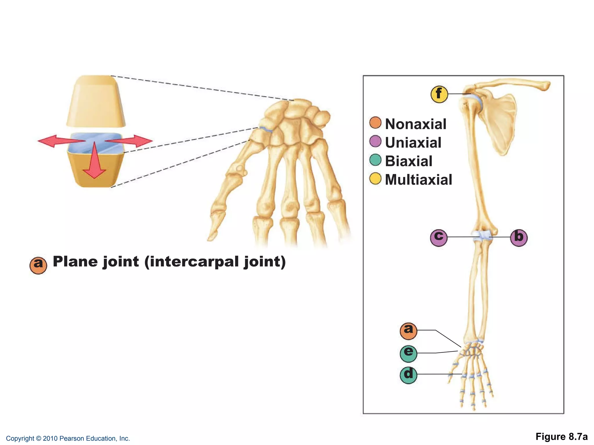
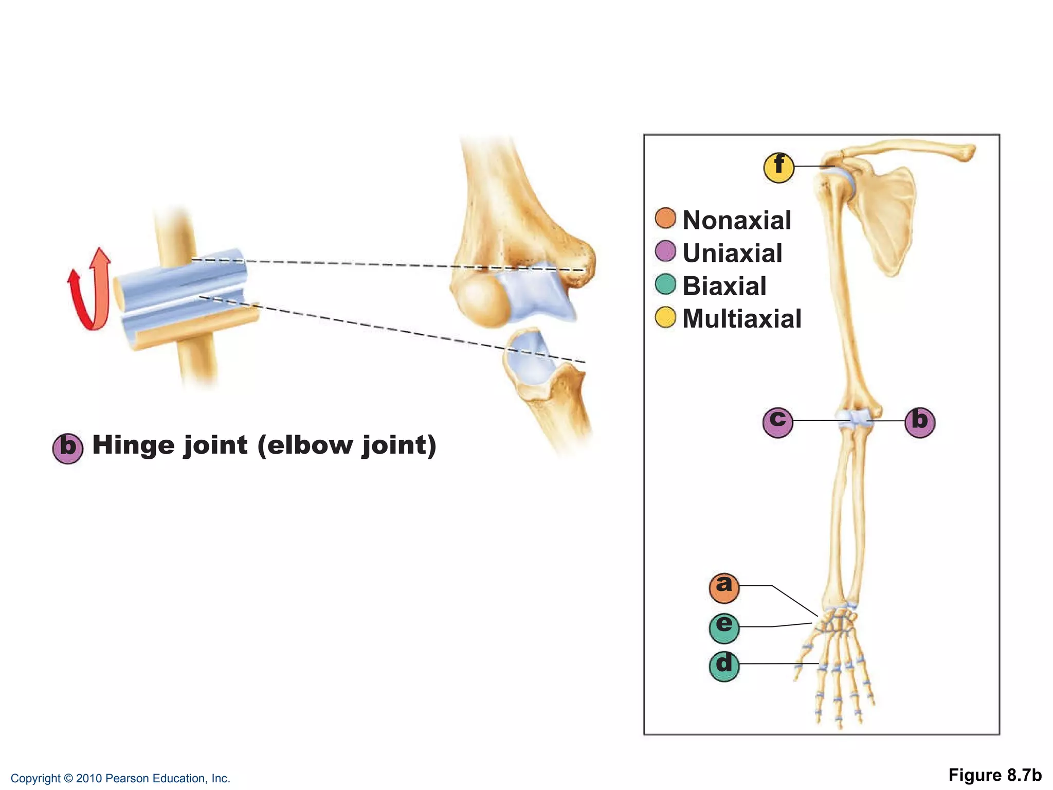
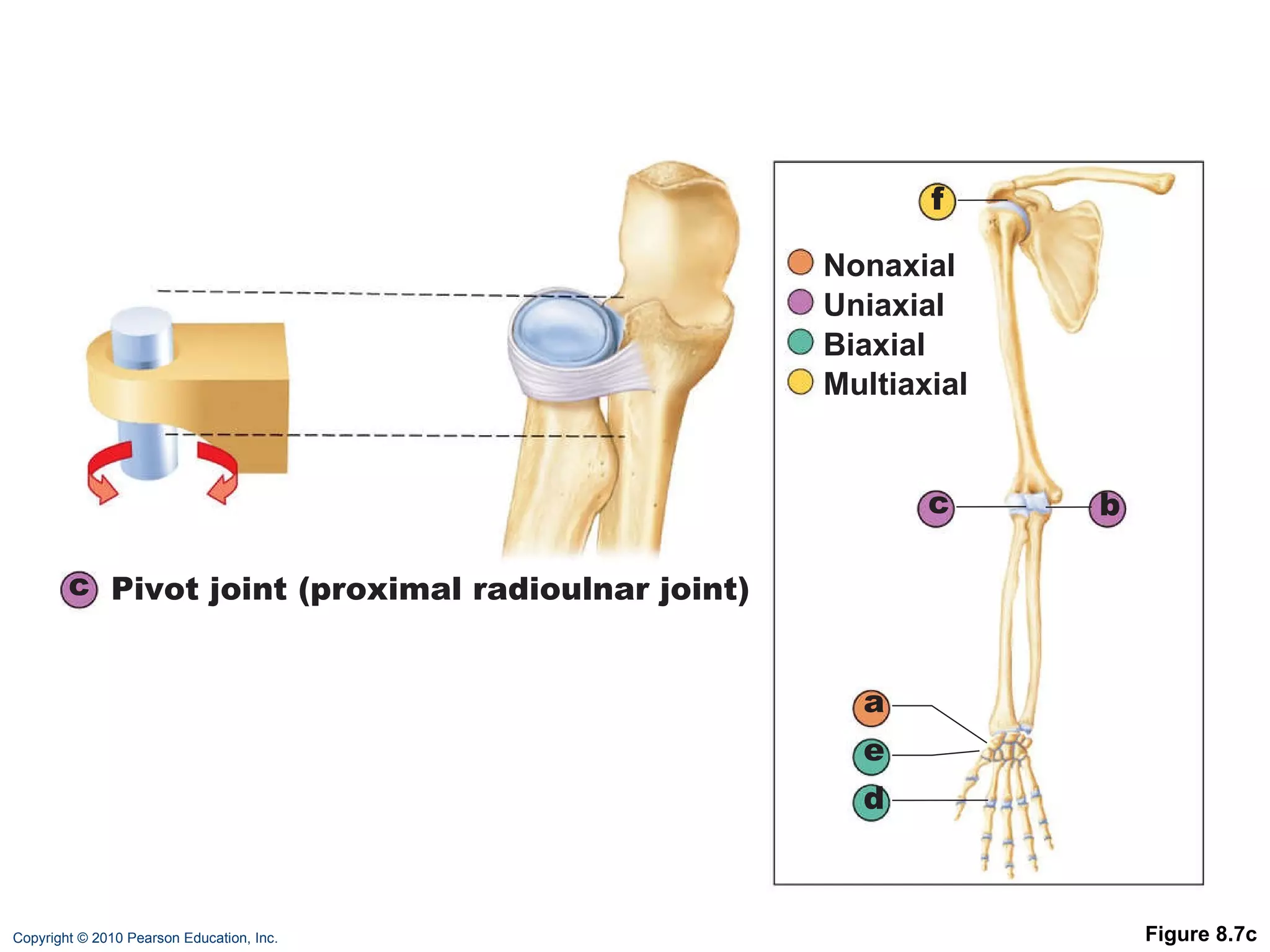
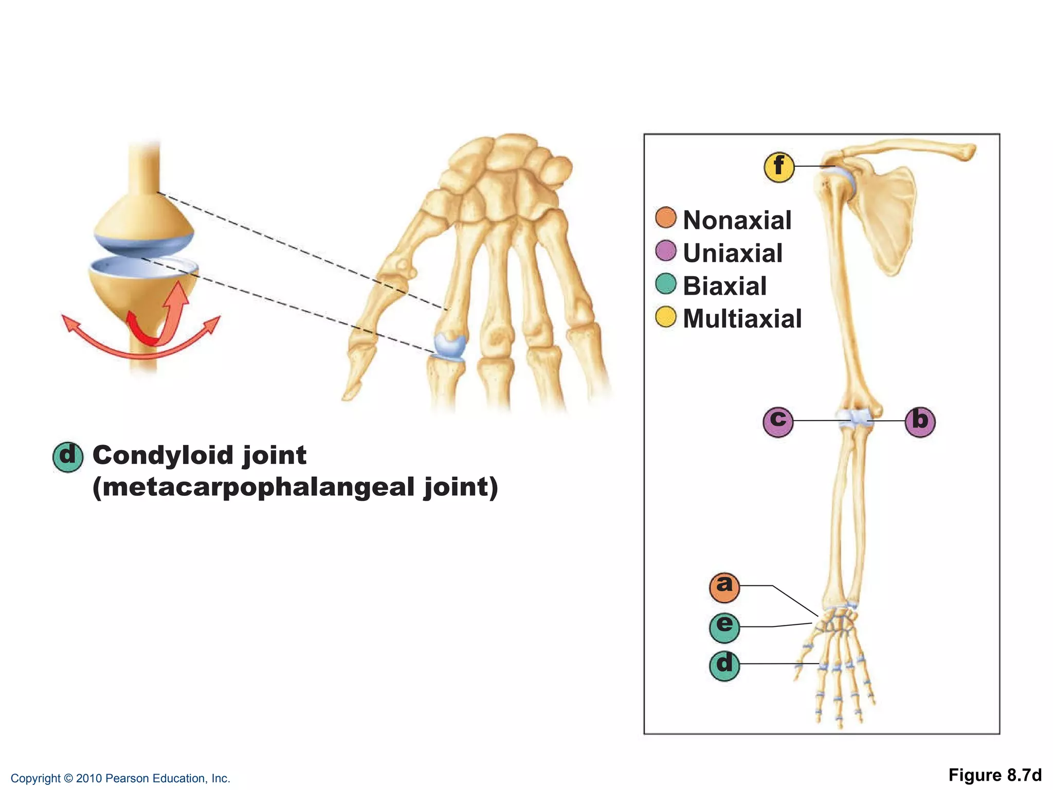
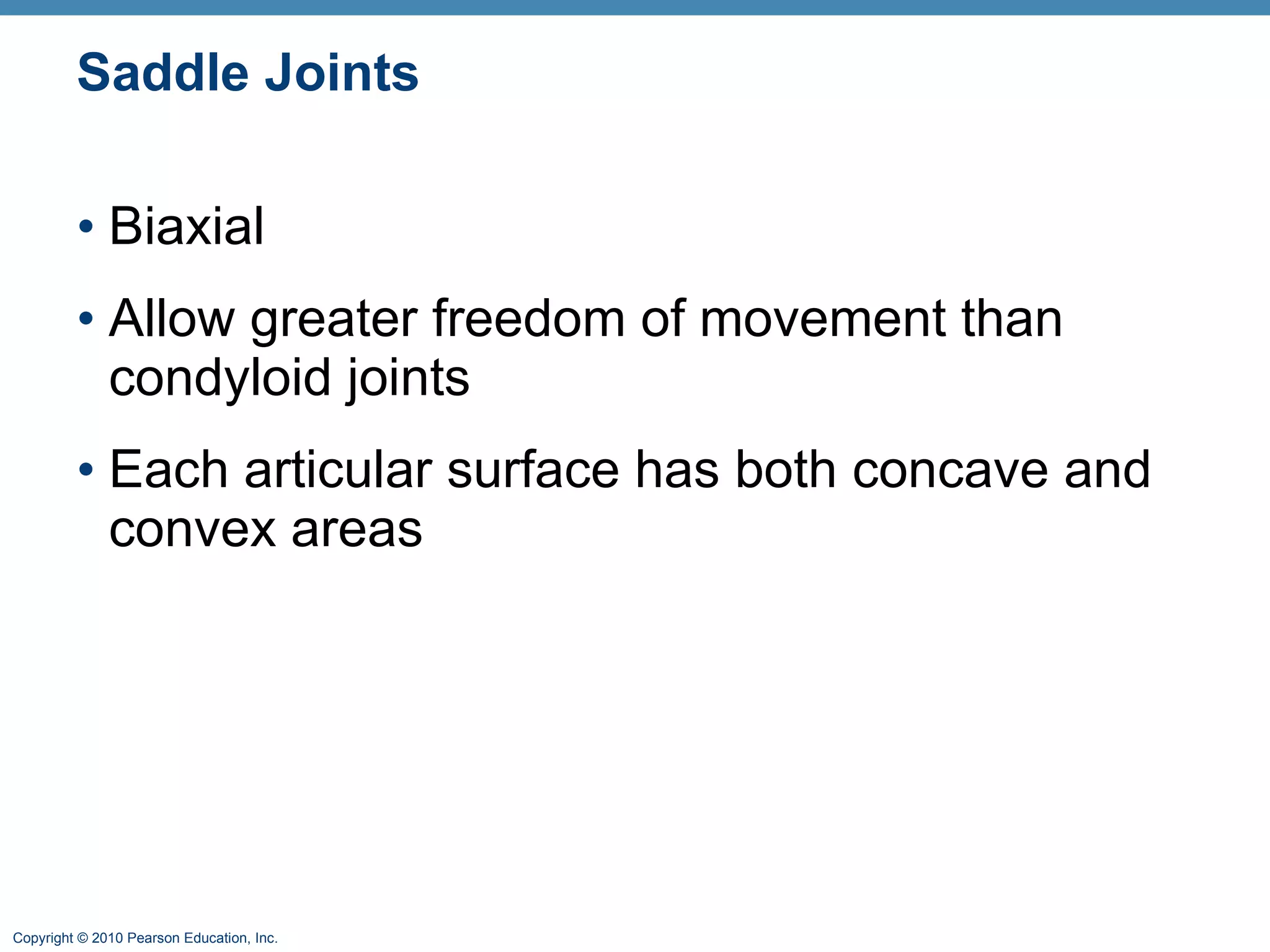
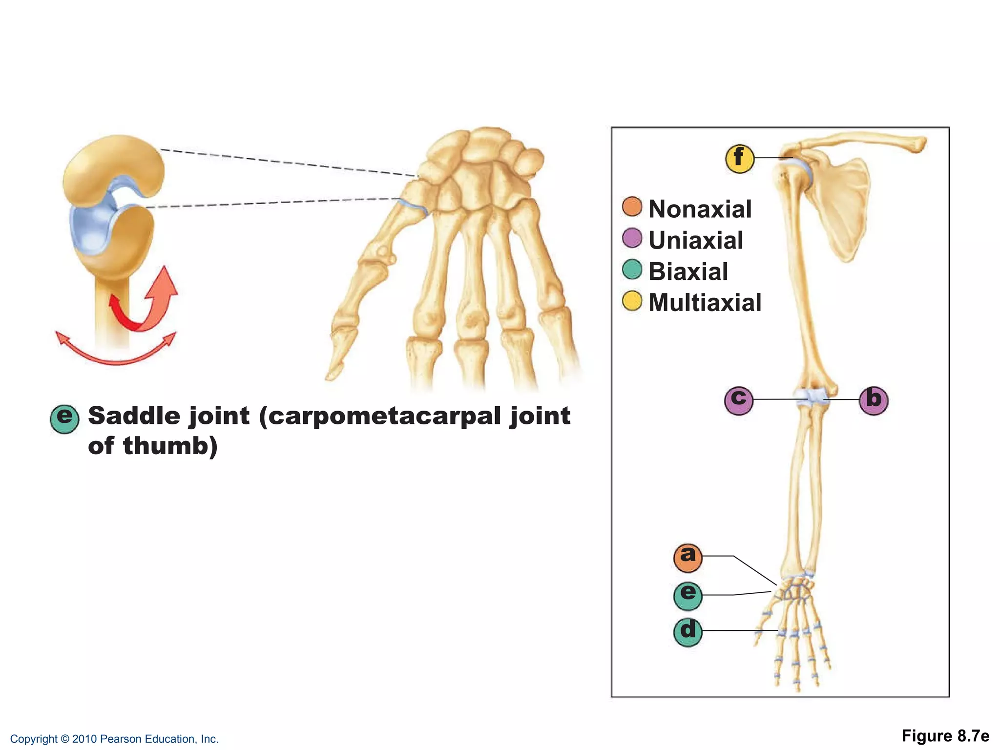
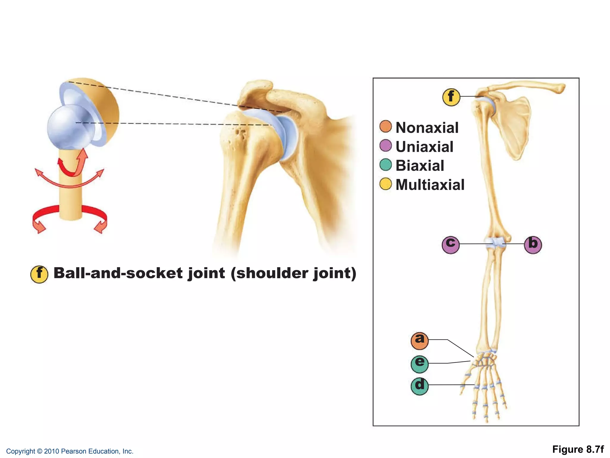
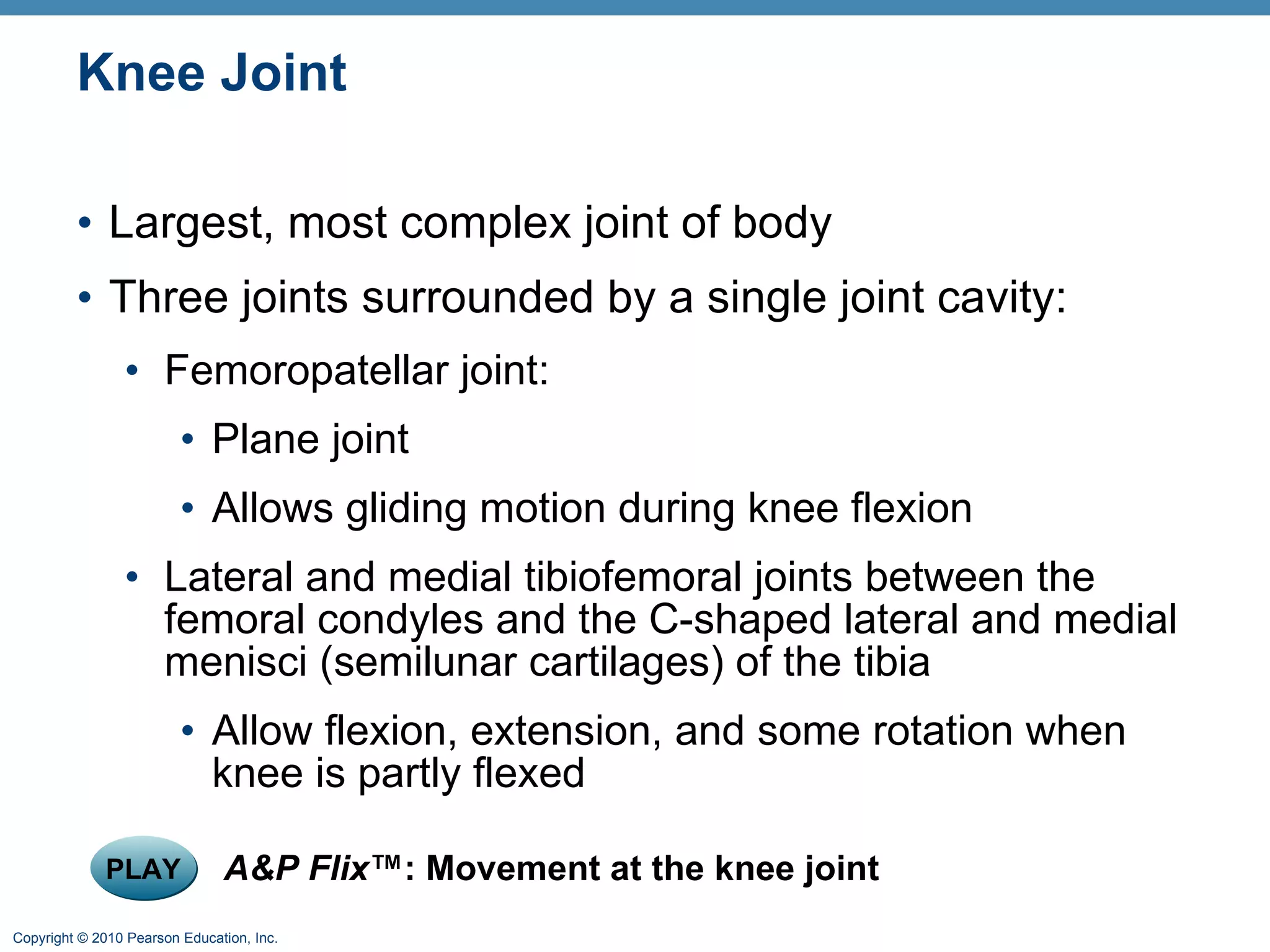
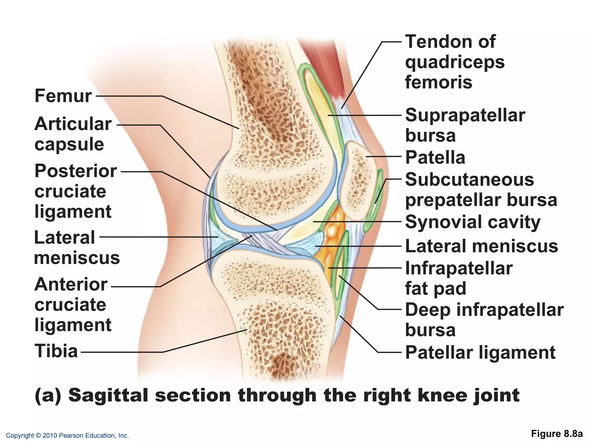
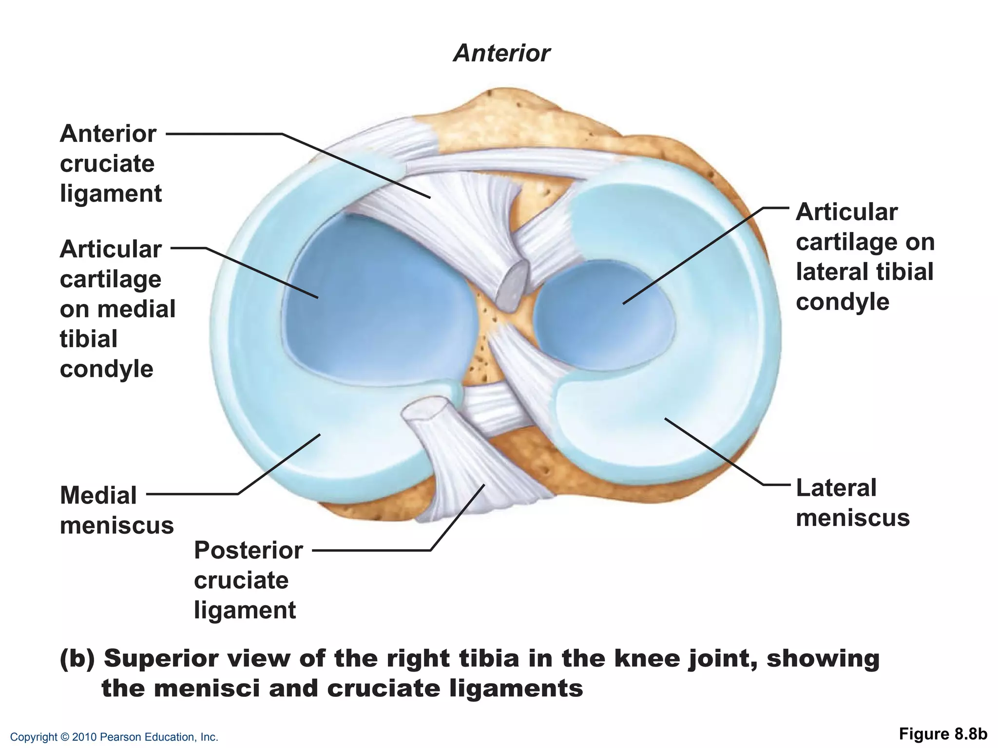
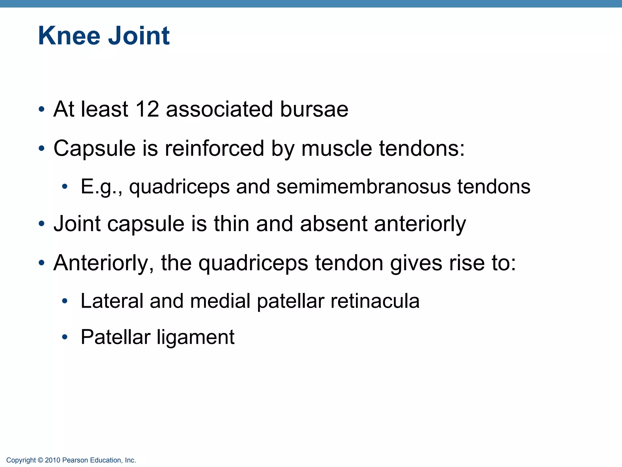
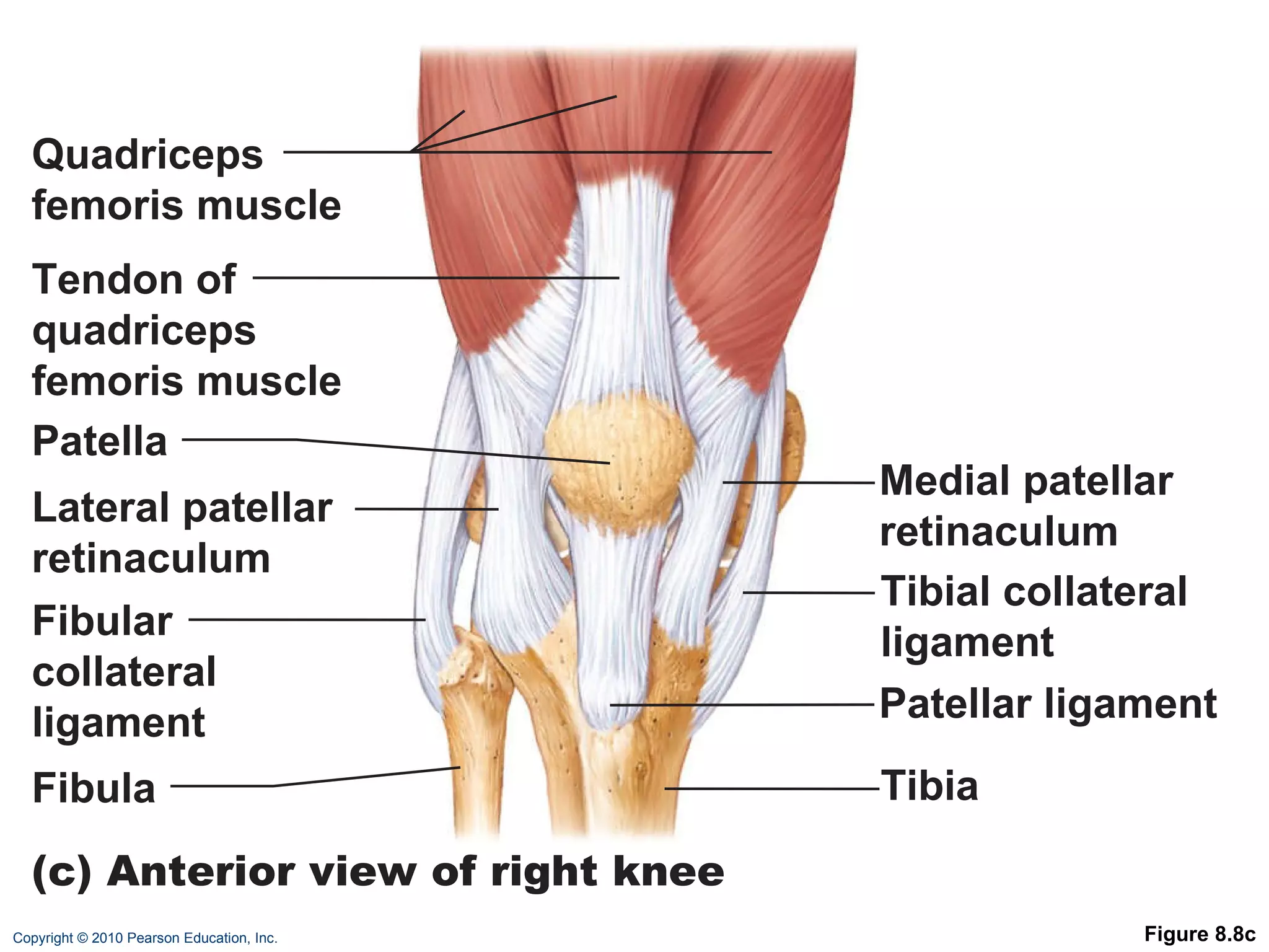
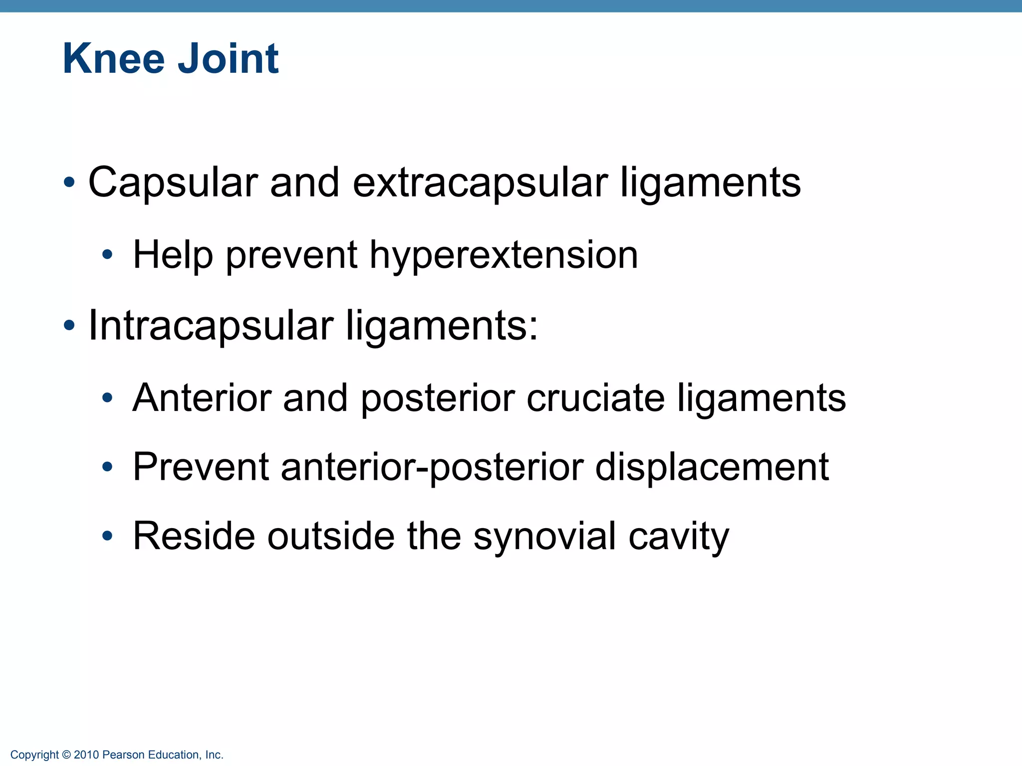
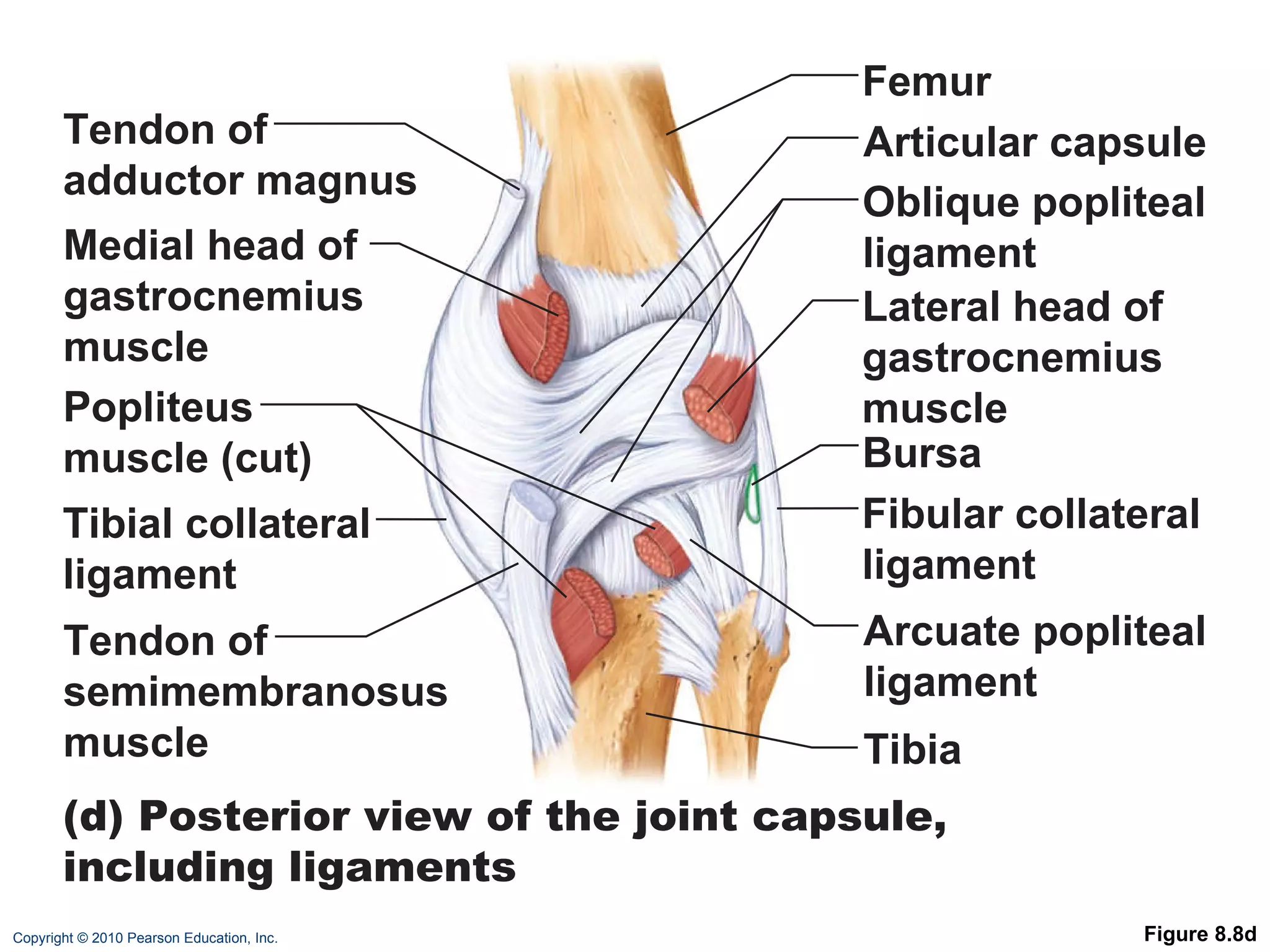
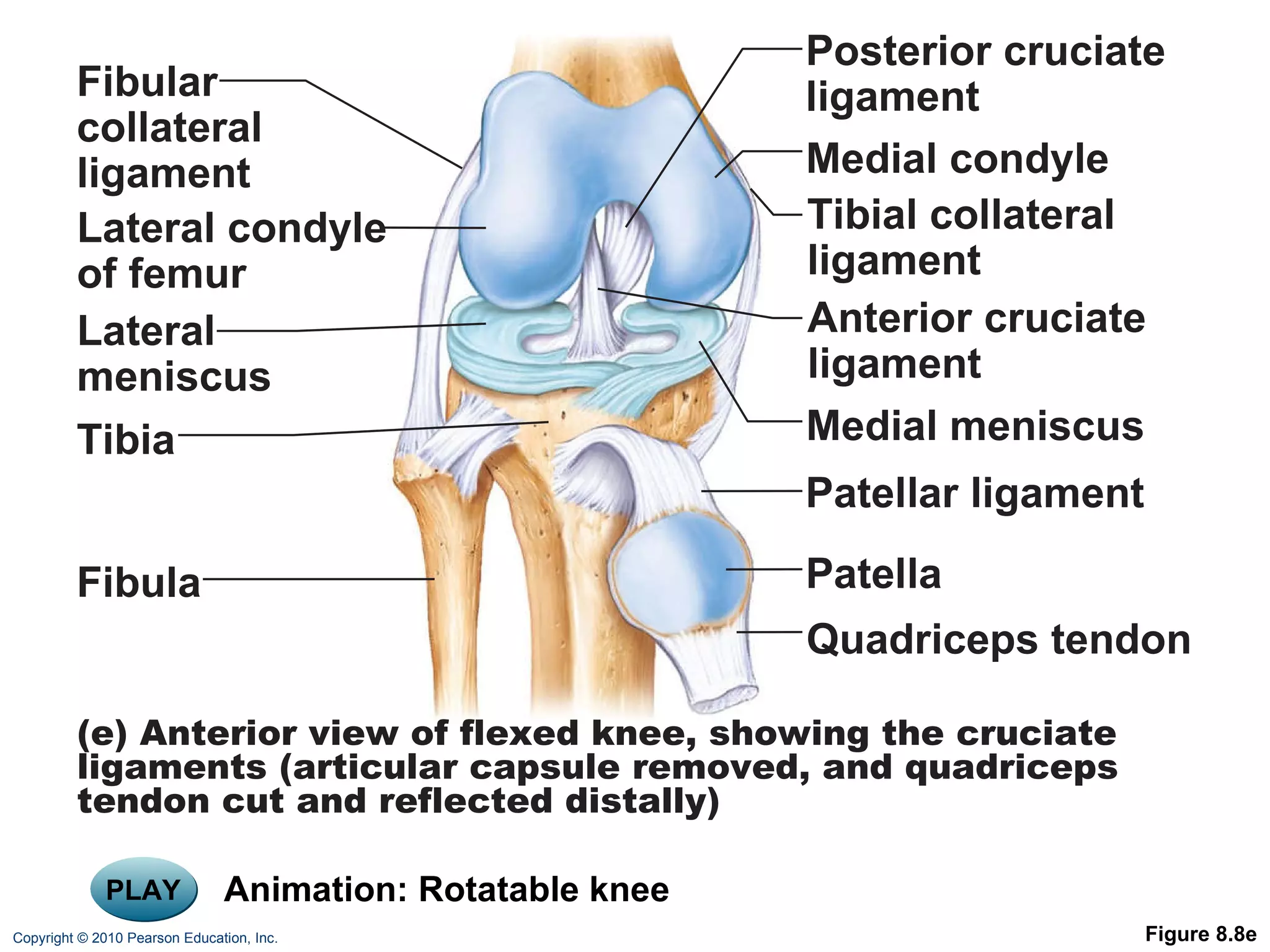
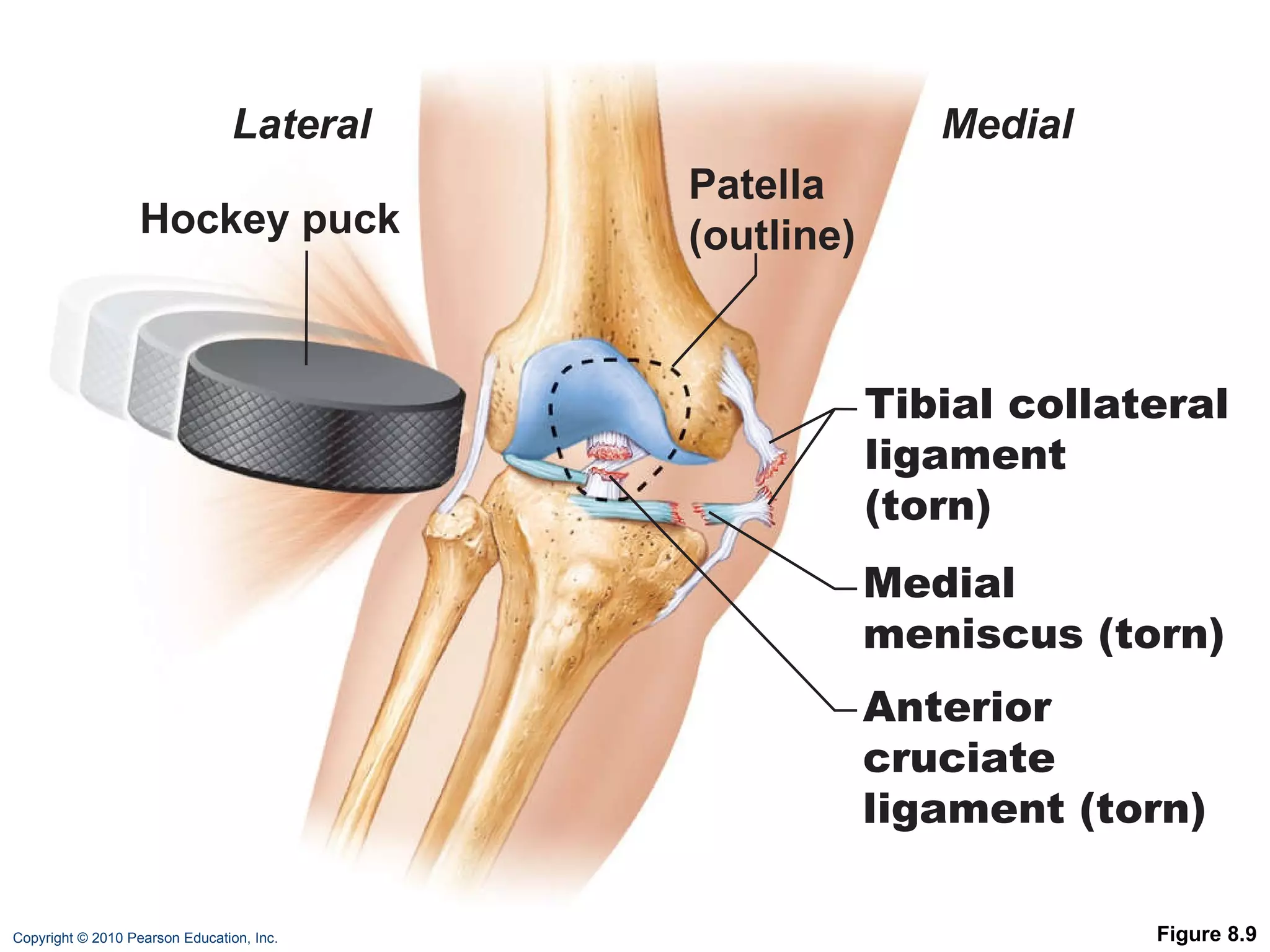
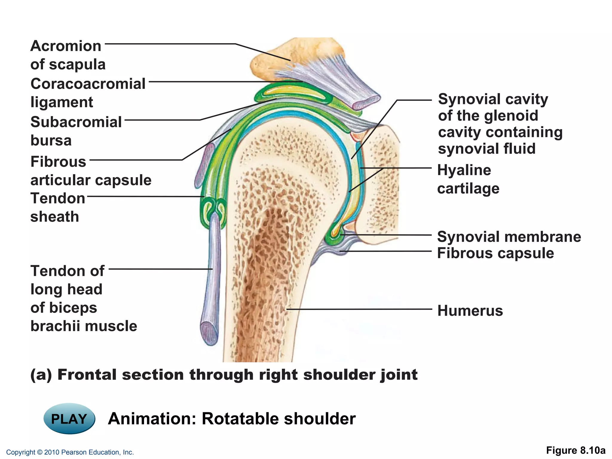
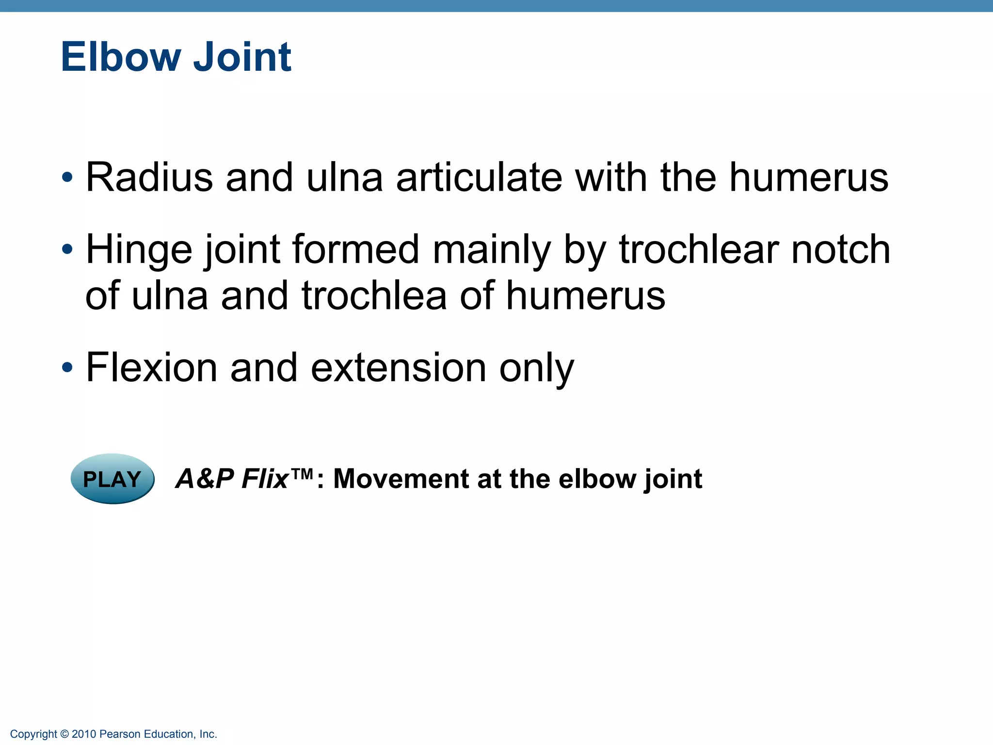
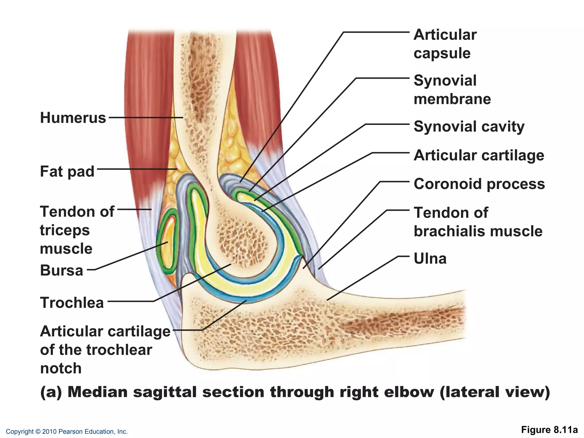
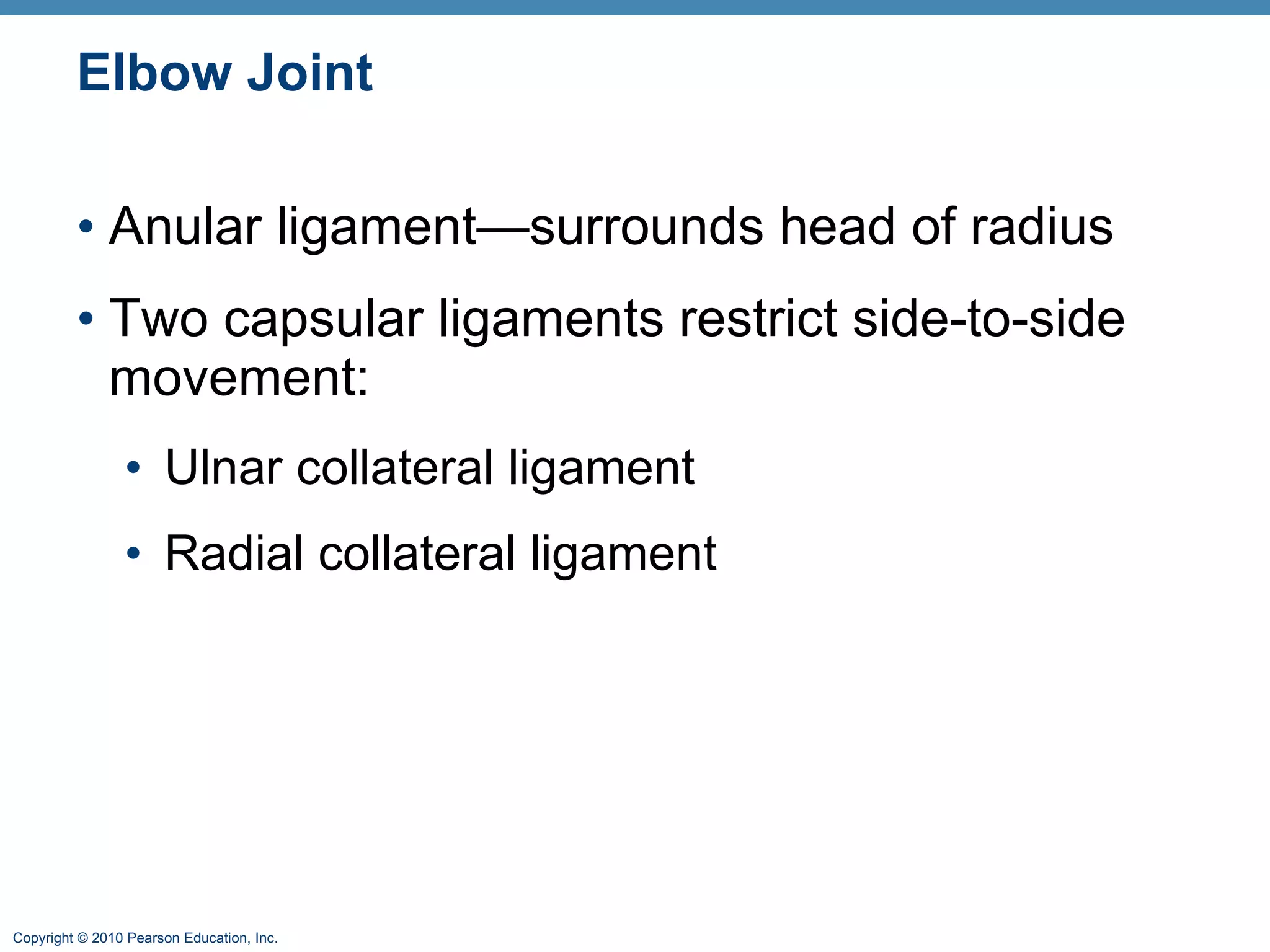
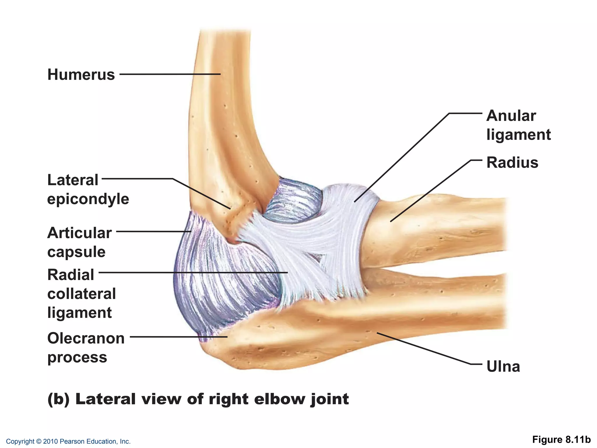
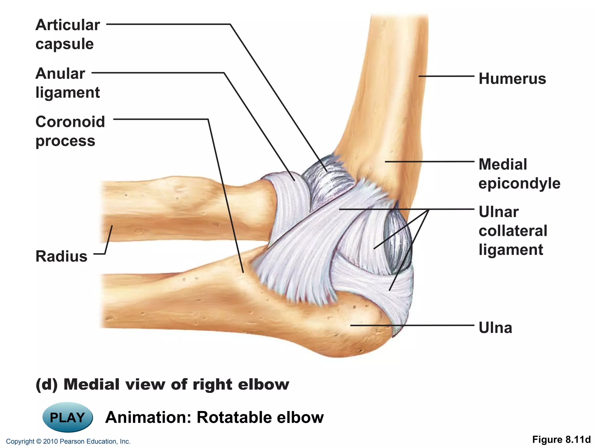
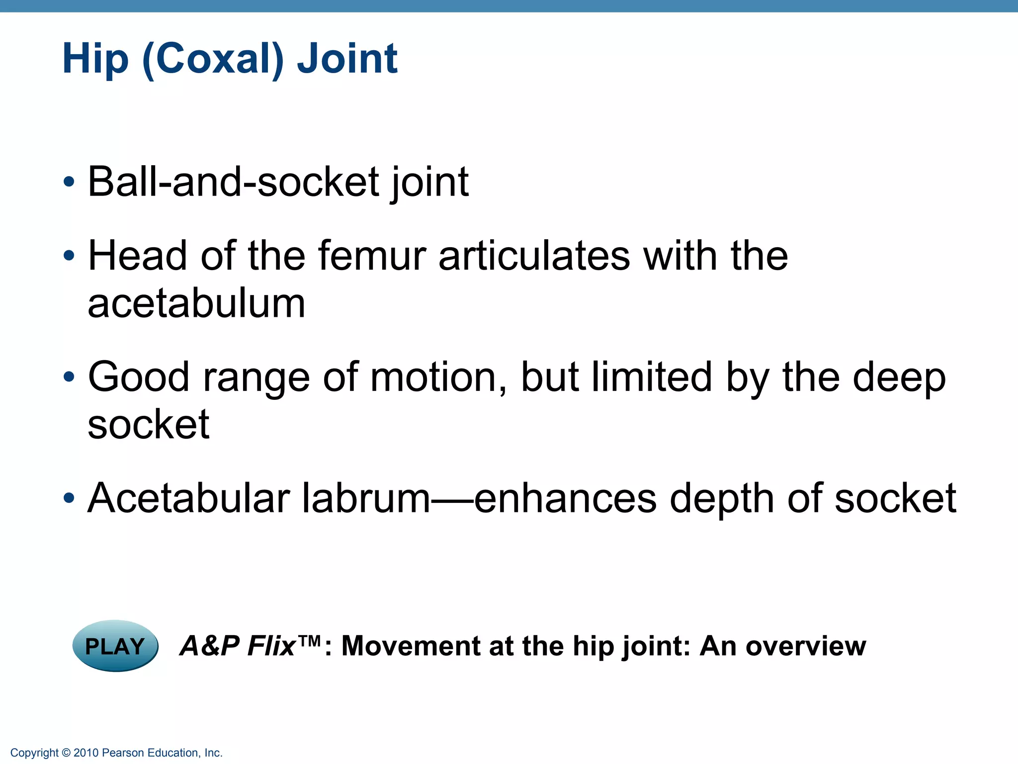
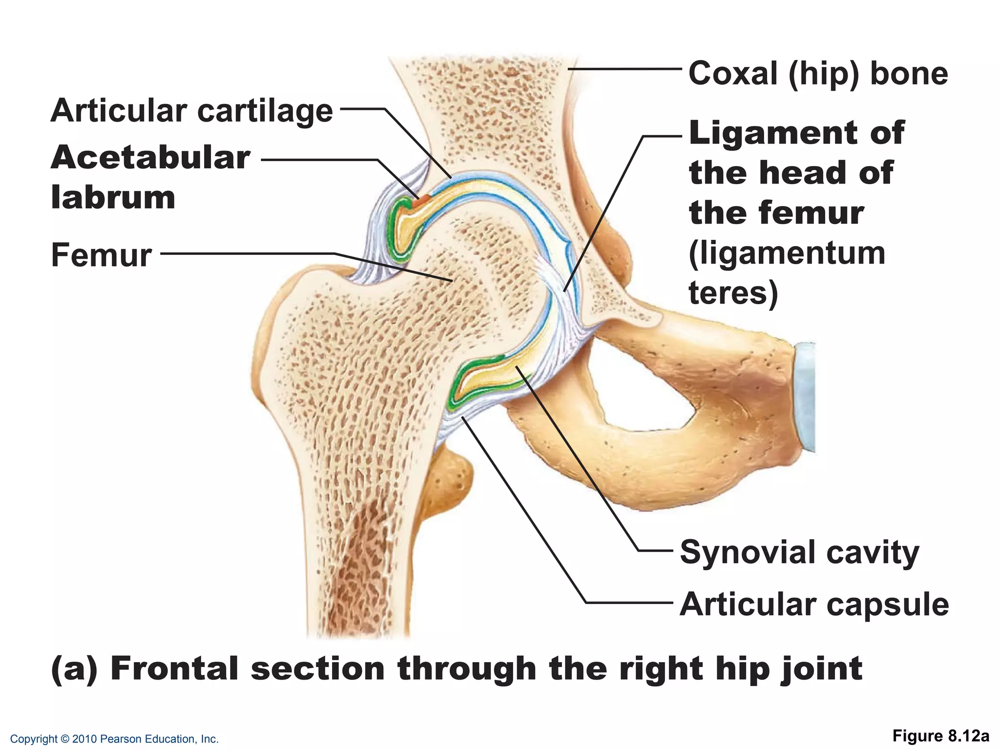
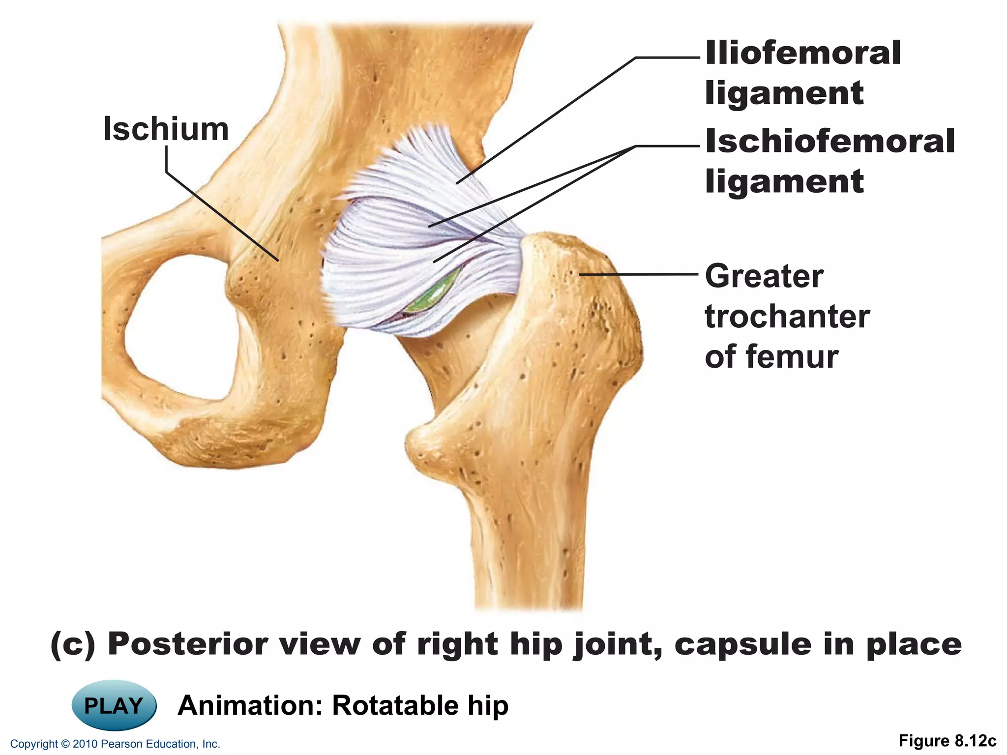
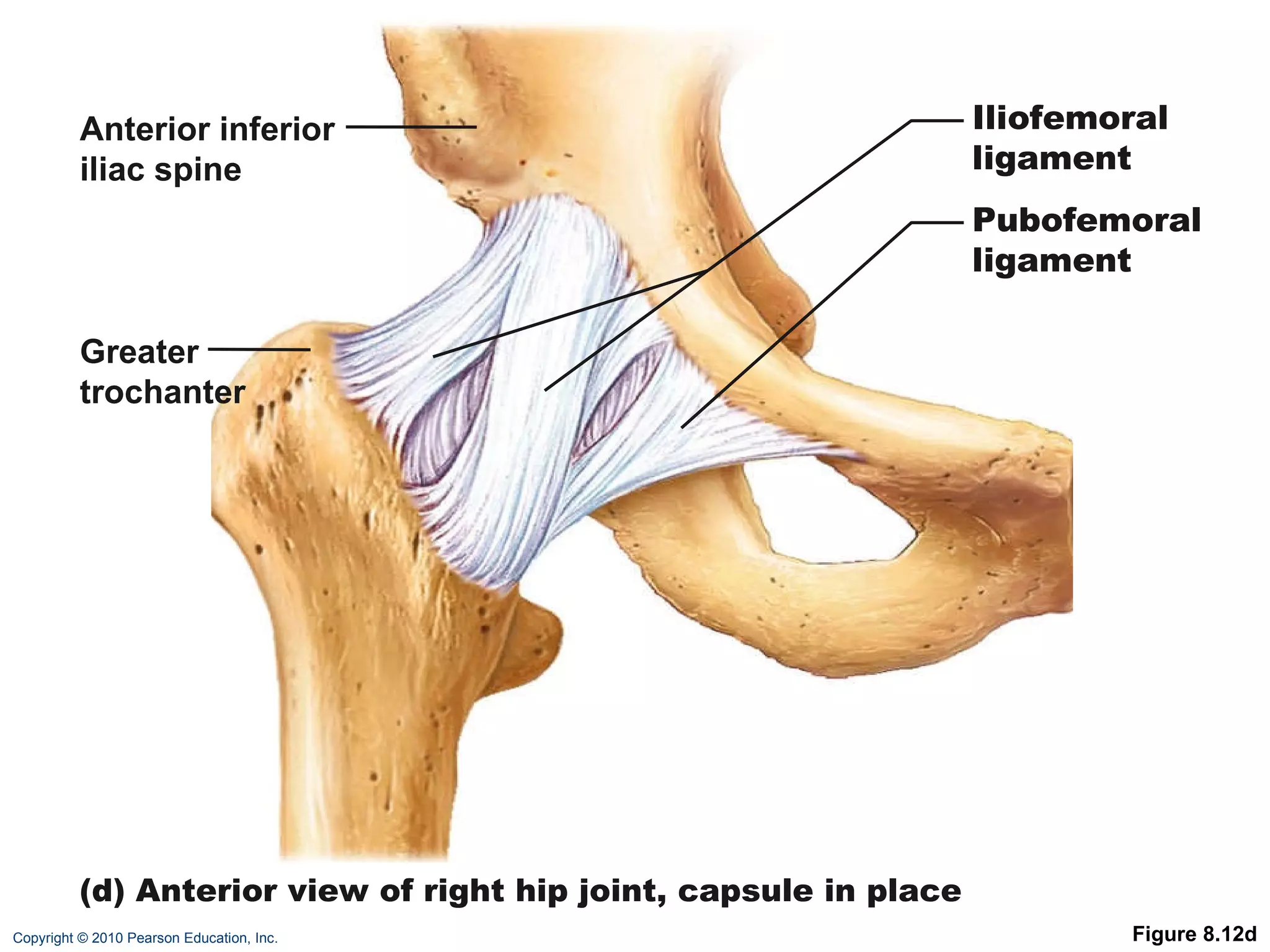
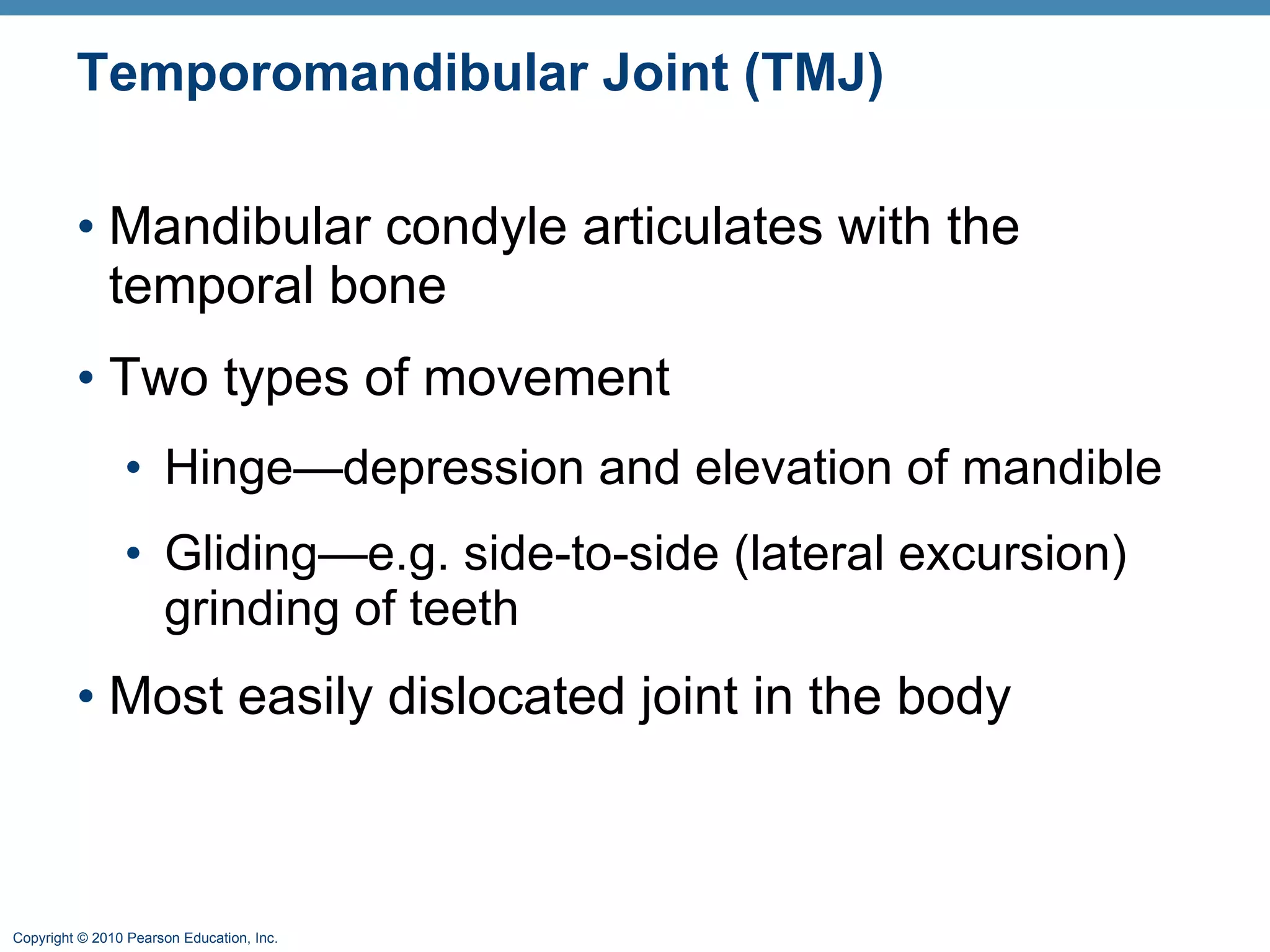
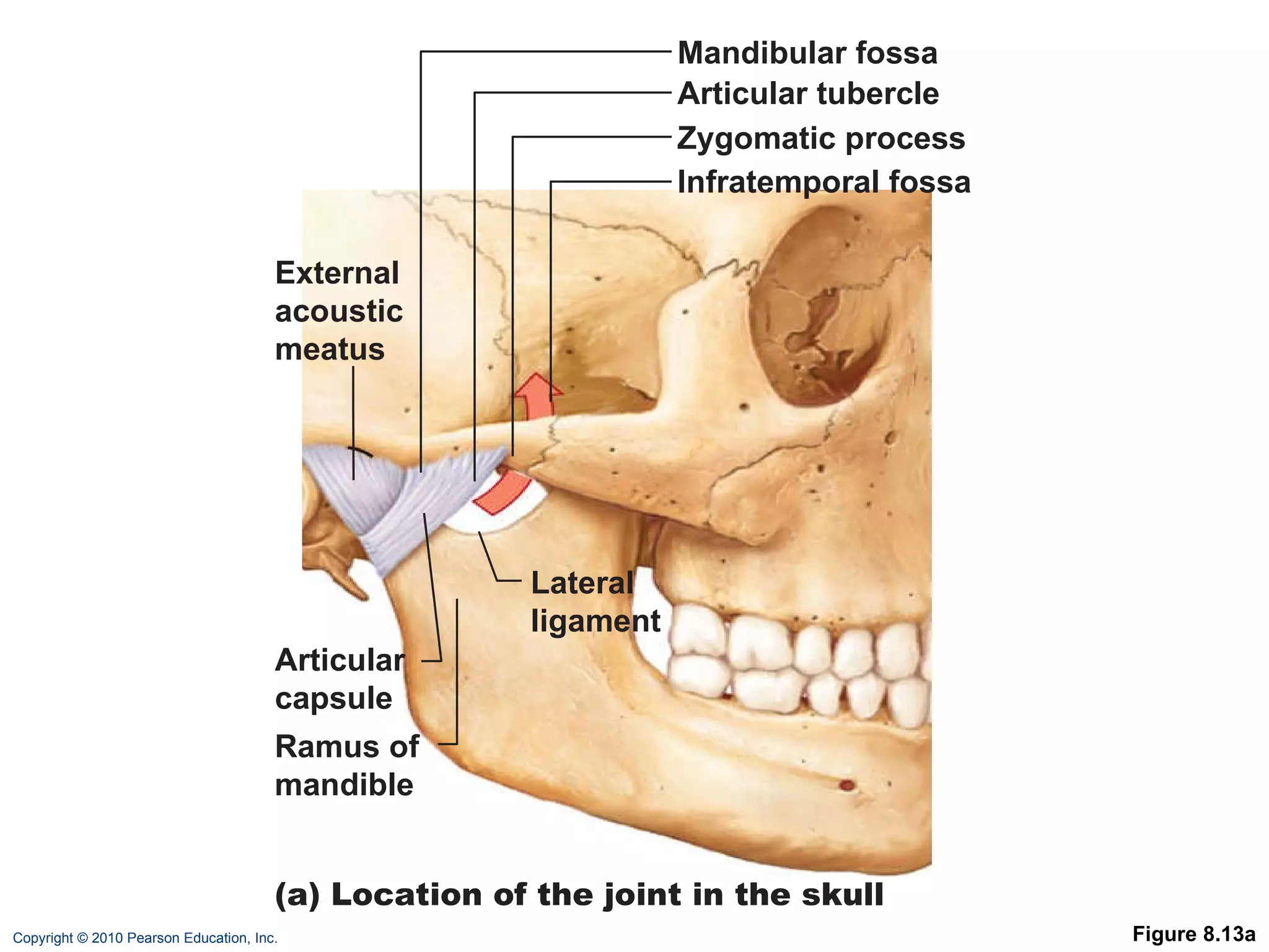
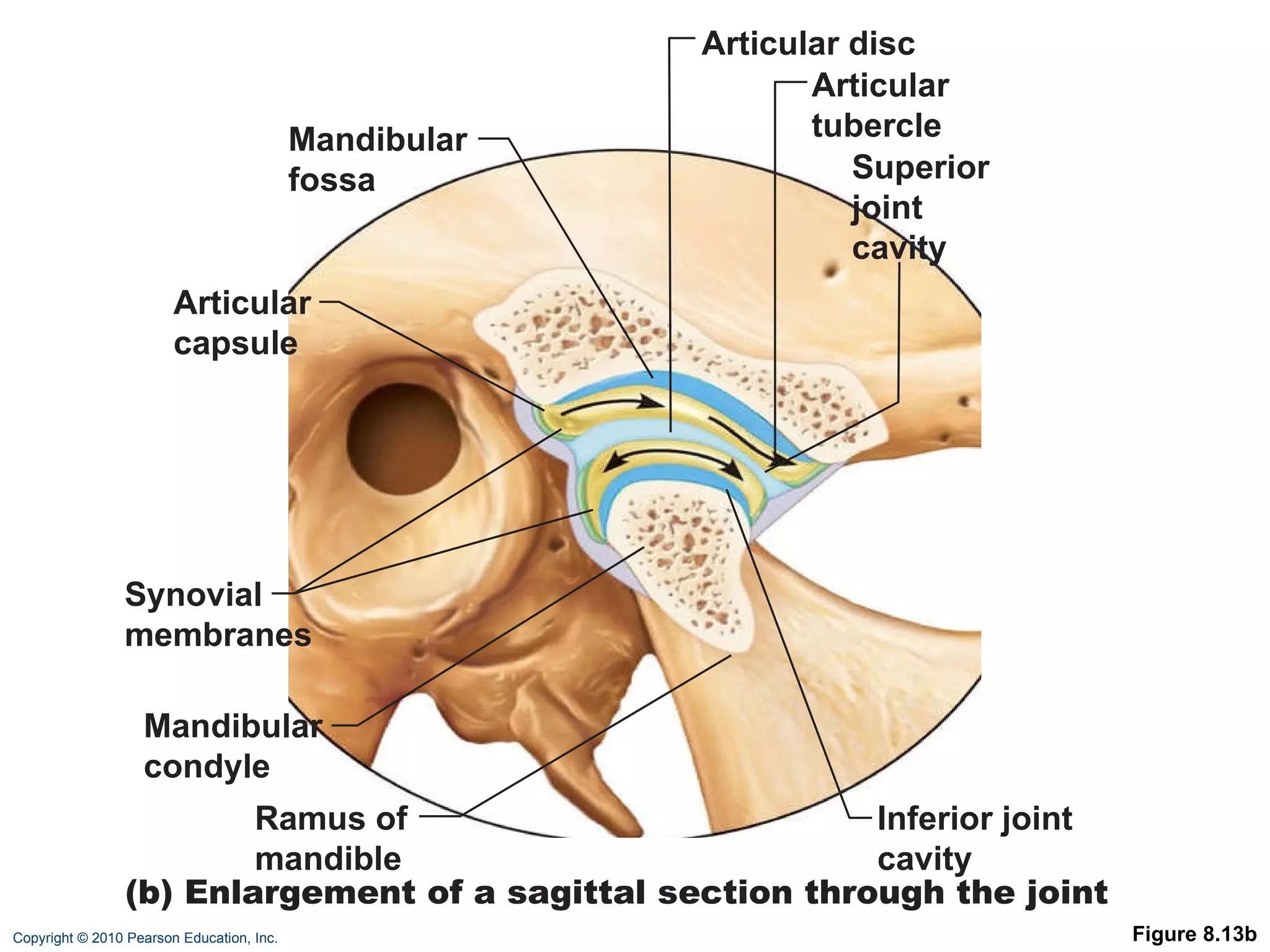
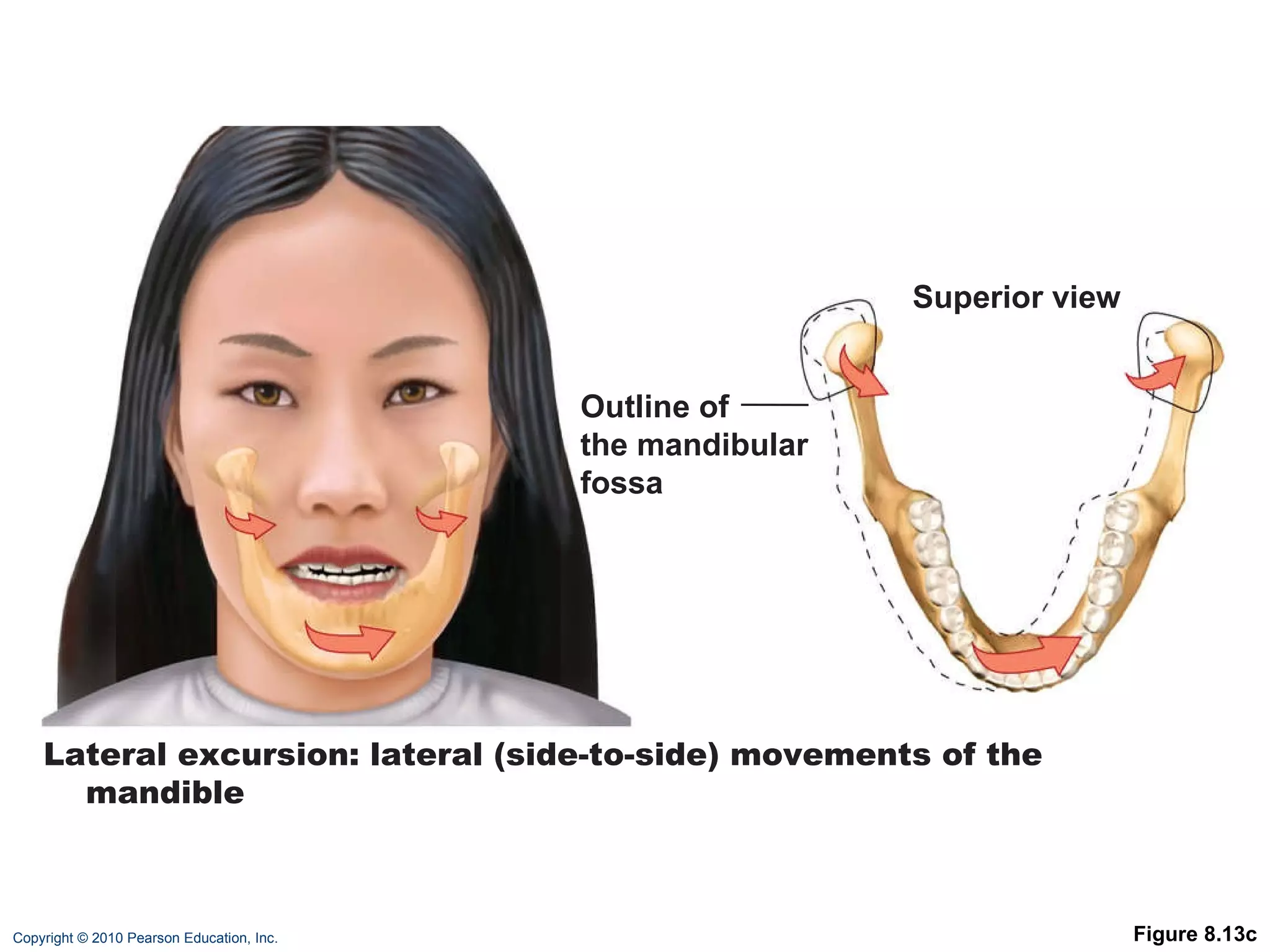
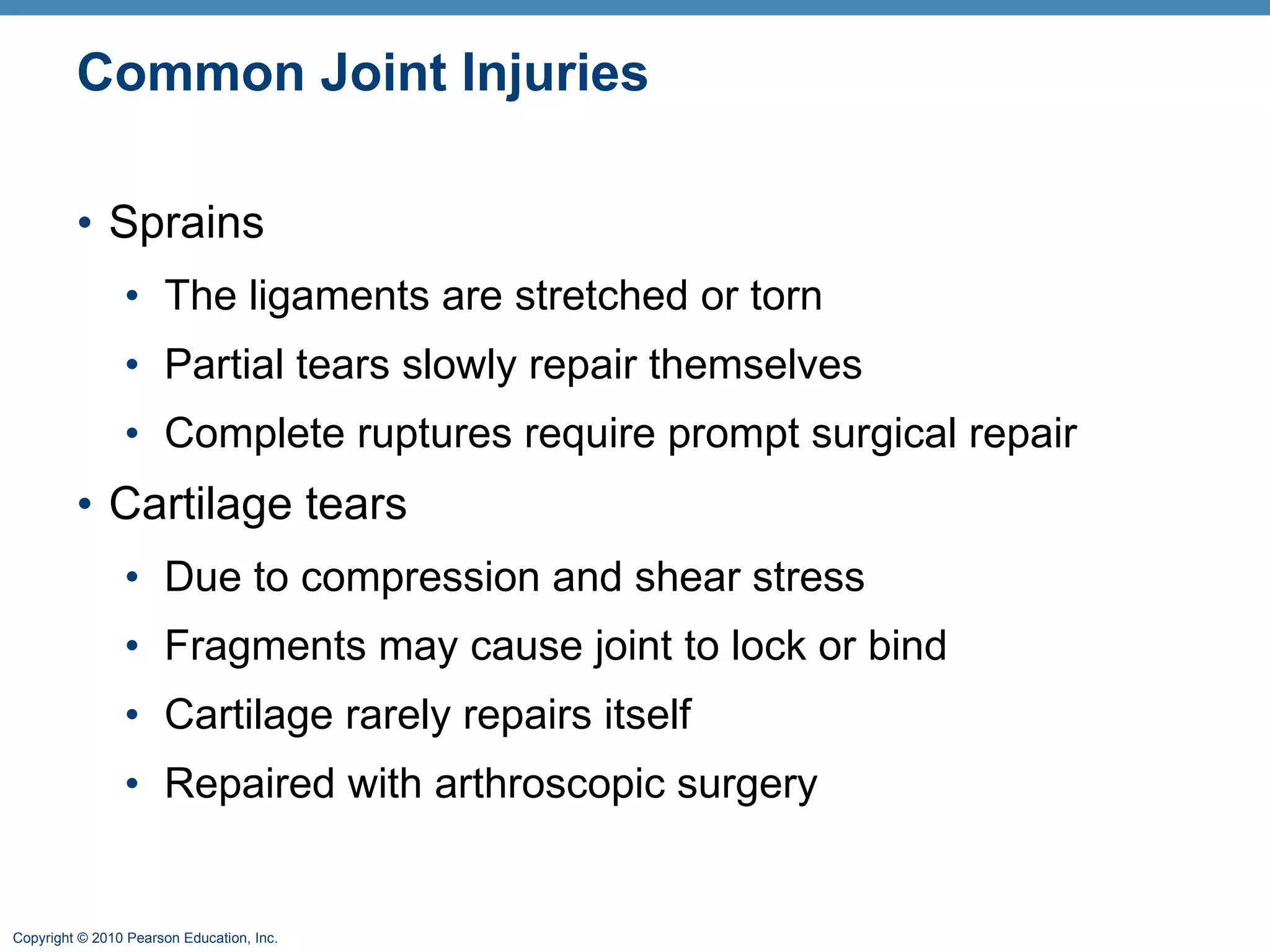
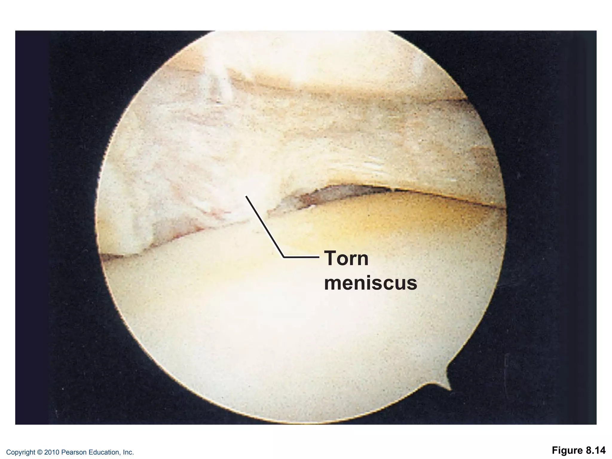
8  Joints: Part B
Classification of Synovial Joints Six types, based on shape of articular surfaces: Plane Hinge Pivot Condyloid Saddle Ball and socket
Plane Joints Nonaxial joints Flat articular surfaces Short gliding movements
Figure 8.7a a b c d e f Nonaxial Uniaxial Biaxial Multiaxial a Plane joint (intercarpal joint)
Hinge Joints Uniaxial joints  Motion along a single plane Flexion and extension only
Figure 8.7b b Hinge joint (elbow joint) a b c d e f Nonaxial Uniaxial Biaxial Multiaxial
Pivot Joints Rounded end of one bone conforms to a “sleeve,” or ring of another bone Uniaxial movement only
Figure 8.7c c Pivot joint (proximal radioulnar joint) a b c d e f Nonaxial Uniaxial Biaxial Multiaxial
Condyloid (Ellipsoidal) Joints Biaxial joints Both articular surfaces are oval Permit all angular movements
Figure 8.7d d Condyloid joint (metacarpophalangeal joint) a b c d e f Nonaxial Uniaxial Biaxial Multiaxial
Saddle Joints Biaxial Allow greater freedom of movement than condyloid joints Each articular surface has both concave and convex areas
Figure 8.7e e Saddle joint (carpometacarpal joint of thumb) a b c d e f Nonaxial Uniaxial Biaxial Multiaxial
Ball-and-Socket Joints Multiaxial joints The most freely moving synovial joints
Figure 8.7f f Ball-and-socket joint (shoulder joint) a b c d e f Nonaxial Uniaxial Biaxial Multiaxial
Knee Joint Largest, most complex joint of body Three joints surrounded by a single joint cavity: Femoropatellar joint: Plane joint Allows gliding motion during knee flexion Lateral and medial tibiofemoral joints between the femoral condyles and the C-shaped lateral and medial menisci (semilunar cartilages) of the tibia Allow flexion, extension, and some rotation when knee is partly flexed PLAY A&P Flix ™: Movement at the knee joint
Figure 8.8a (a) Sagittal section through the right knee joint Femur Tendon of quadriceps femoris Suprapatellar bursa Patella Subcutaneous prepatellar bursa Synovial cavity Lateral meniscus Posterior cruciate ligament Infrapatellar fat pad  Deep infrapatellar bursa Patellar ligament Articular capsule Lateral meniscus Anterior cruciate ligament Tibia
Figure 8.8b (b) Superior view of the right tibia in the knee joint, showing the menisci and cruciate ligaments Medial meniscus Articular cartilage on medial tibial condyle Anterior Anterior cruciate ligament Articular cartilage on lateral tibial condyle Lateral meniscus Posterior cruciate ligament
Knee Joint At least 12 associated bursae Capsule is reinforced by muscle tendons: E.g., quadriceps and semimembranosus tendons Joint capsule is thin and absent anteriorly Anteriorly, the quadriceps tendon gives rise to: Lateral and medial patellar retinacula Patellar ligament
Figure 8.8c Quadriceps femoris muscle Tendon of quadriceps femoris muscle Patella Lateral patellar retinaculum Medial patellar retinaculum Tibial collateral ligament Tibia Fibular collateral ligament Fibula (c) Anterior view of right knee Patellar ligament
Knee Joint Capsular and extracapsular ligaments Help prevent hyperextension Intracapsular ligaments:  Anterior and posterior cruciate ligaments Prevent anterior-posterior displacement Reside outside the synovial cavity
Figure 8.8d Articular capsule Oblique popliteal ligament Lateral head of gastrocnemius muscle Fibular collateral ligament Arcuate popliteal ligament Tibia Femur Medial head of gastrocnemius muscle Tendon of semimembranosus muscle (d) Posterior view of the joint capsule, including ligaments Popliteus muscle (cut) Tendon of adductor magnus  Bursa Tibial collateral ligament
PLAY Animation: Rotatable knee Figure 8.8e Fibular collateral ligament Posterior cruciate ligament Medial condyle Tibial collateral ligament Anterior cruciate ligament Medial meniscus Patellar ligament Patella Quadriceps tendon Lateral condyle of femur Lateral meniscus Fibula Tibia (e) Anterior view of flexed knee, showing the cruciate ligaments (articular capsule removed, and quadriceps tendon cut and reflected distally)
Figure 8.9 Lateral Medial Patella (outline) Tibial collateral ligament (torn) Medial meniscus (torn) Anterior cruciate ligament (torn) Hockey puck
Shoulder (Glenohumeral) Joint Ball-and-socket joint: head of humerus and glenoid fossa of the scapula Stability is sacrificed for greater freedom of movement
Figure 8.10a PLAY Animation: Rotatable shoulder Acromion of scapula Synovial membrane Fibrous capsule Hyaline cartilage Coracoacromial ligament Subacromial bursa Fibrous articular capsule Tendon sheath Tendon of long head of biceps brachii muscle Synovial cavity of the glenoid cavity containing synovial fluid Humerus (a) Frontal section through right shoulder joint
Shoulder Joint Reinforcing ligaments: Coracohumeral ligament—helps support the weight of the upper limb Three glenohumeral ligaments—somewhat weak anterior reinforcements
Shoulder joint Reinforcing muscle tendons: Tendon of the long head of biceps: Travels through the intertubercular groove  Secures the humerus to the glenoid cavity Four rotator cuff tendons encircle the shoulder joint: Subscapularis Supraspinatus Infraspinatus Teres minor PLAY A&P Flix ™: Rotator cuff muscles: An overview (a) PLAY A&P Flix ™: Rotator cuff muscles: An overview (b)
Figure 8.10c Acromion Coracoacromial ligament Subacromial bursa Coracohumeral ligament Greater tubercle of humerus Transverse humeral ligament Tendon sheath Tendon of long head of biceps brachii muscle Articular capsule reinforced by glenohumeral ligaments Subscapular bursa Tendon of the subscapularis muscle Scapula Coracoid process (c) Anterior view of right shoulder joint capsule
Figure 8.10d Acromion Coracoid process Articular capsule Glenoid cavity Glenoid labrum Tendon of long head of biceps brachii muscle  Glenohumeral ligaments Tendon of the subscapularis muscle  Scapula Posterior Anterior (d) Lateral view of socket of right shoulder joint, humerus removed
Elbow Joint Radius and ulna articulate with the humerus Hinge joint formed mainly by trochlear notch of ulna and trochlea of humerus Flexion and extension only PLAY A&P Flix ™: Movement at the elbow joint
Figure 8.11a Articular capsule Synovial membrane Synovial cavity Articular cartilage Coronoid process Tendon of brachialis muscle Ulna Humerus Fat pad Tendon of triceps muscle Bursa Trochlea Articular cartilage of the trochlear notch (a) Median sagittal section through right elbow (lateral view)
Elbow Joint Anular ligament—surrounds head of radius Two capsular ligaments restrict side-to-side movement: Ulnar collateral ligament Radial collateral ligament
Figure 8.11b Humerus Lateral epicondyle Articular capsule Radial collateral ligament Olecranon process Anular ligament Radius Ulna (b) Lateral view of right elbow joint
PLAY Animation: Rotatable elbow Figure 8.11d Articular capsule Anular ligament Coronoid process (d) Medial view of right elbow Radius Humerus Medial epicondyle Ulnar collateral ligament Ulna
Hip (Coxal) Joint Ball-and-socket joint Head of the femur articulates with the acetabulum Good range of motion, but limited by the deep socket  Acetabular labrum—enhances depth of socket PLAY A&P Flix ™: Movement at the hip joint: An overview
Figure 8.12a Articular cartilage Coxal (hip) bone Ligament of the head of  the femur  (ligamentum  teres) Synovial cavity Articular capsule Acetabular labrum  Femur (a) Frontal section through the right hip joint
Hip Joint Reinforcing ligaments: Iliofemoral ligament Pubofemoral ligament Ischiofemoral ligament Ligamentum teres
PLAY Animation: Rotatable hip Figure 8.12c Ischium Iliofemoral ligament Ischiofemoral ligament Greater trochanter of femur (c) Posterior view of right hip joint, capsule in place
Figure 8.12d Anterior inferior iliac spine Iliofemoral ligament Pubofemoral ligament Greater trochanter (d) Anterior view of right hip joint, capsule in place
Temporomandibular Joint (TMJ) Mandibular condyle articulates with the temporal bone Two types of movement Hinge—depression and elevation of mandible Gliding—e.g. side-to-side (lateral excursion) grinding of teeth Most easily dislocated joint in the body
Figure 8.13a Zygomatic process Mandibular fossa Articular tubercle Infratemporal fossa External acoustic meatus Articular capsule Ramus of mandible  Lateral ligament  (a) Location of the joint in the skull
Figure 8.13b Articular capsule Mandibular fossa Articular disc Articular tubercle Superior joint cavity Inferior joint cavity Mandibular condyle Ramus of mandible  Synovial membranes (b) Enlargement of a sagittal section through the joint
Figure 8.13c Lateral excursion: lateral (side-to-side) movements of the mandible Outline of the mandibular fossa Superior view
Common Joint Injuries Sprains The ligaments are stretched or torn Partial tears slowly repair themselves Complete ruptures require prompt surgical repair Cartilage tears Due to compression and shear stress Fragments may cause joint to lock or bind Cartilage rarely repairs itself Repaired with arthroscopic surgery
Figure 8.14 Torn meniscus
Common Joint Injuries Dislocations (luxations) Occur when bones are forced out of alignment Accompanied by sprains, inflammation, and joint immobilization Caused by serious falls or playing sports  Subluxation—partial dislocation of a joint
Inflammatory and Degenerative Conditions Bursitis An inflammation of a bursa, usually caused by a blow or friction Treated with rest and ice and, if severe, anti-inflammatory drugs Tendonitis Inflammation of tendon sheaths typically caused by overuse Symptoms and treatment similar to bursitis
Arthritis >100 different types of inflammatory or degenerative diseases that damage joints Most widespread crippling disease in the U.S. Symptoms; pain, stiffness, and swelling of a joint Acute forms: caused by bacteria, treated with antibiotics Chronic forms: osteoarthritis, rheumatoid arthritis, and gouty arthritis
Osteoarthritis (OA) Common, irreversible, degenerative (“wear-and-tear”) arthritis 85% of all Americans develop OA, more women than men Probably related to the normal aging process
Osteoarthritis (OA) More cartilage is destroyed than replaced in badly aligned or overworked joints Exposed bone ends thicken, enlarge, form bone spurs, and restrict movement Treatment: moderate activity, mild pain relievers, capsaicin creams, glucosamine and chondroitin sulfate
Rheumatoid Arthritis (RA) Chronic, inflammatory, autoimmune disease of unknown cause  Usually arises between age 40 and 50, but may occur at any age; affects 3 times as many women as men Signs and symptoms include joint pain and swelling (usually bilateral), anemia, osteoporosis, muscle weakness, and cardiovascular problems
Rheumatoid Arthritis  RA begins with synovitis of the affected joint Inflammatory blood cells migrate to the joint, release inflammatory chemicals  Inflamed synovial membrane thickens into a pannus Pannus erodes cartilage, scar tissue forms, articulating bone ends connect (ankylosis)
Figure 8.15
Rheumatoid Arthritis: Treatment Conservative therapy: aspirin, long-term use of antibiotics, and physical therapy Progressive treatment: anti-inflammatory drugs or immunosuppressants  New biological response modifier drugs neutralize inflammatory chemicals
Gouty Arthritis Deposition of uric acid crystals in joints and soft tissues, followed by inflammation More common in men  Typically affects the joint at the base of the great toe In untreated gouty arthritis, the bone ends fuse and immobilize the joint Treatment: drugs, plenty of water, avoidance of alcohol
Lyme Disease Caused by bacteria transmitted by the bites of ticks Symptoms: skin rash, flu-like symptoms, and foggy thinking May lead to joint pain and arthritis Treatment: antibiotics
Developmental Aspects of Joints By embryonic week 8, synovial joints resemble adult joints A joint’s size, shape, and flexibility are modified by use  Advancing years take their toll on joints: Ligaments and tendons shorten and weaken Intervertebral discs become more likely to herniate Most people in their 70s have some degree of OA Exercise that coaxes joints through their full range of motion is key to postponing joint problems

Ch 08 lecture_outline_b

  • 1.
    8 Joints:Part B
  • 2.
    Classification of SynovialJoints Six types, based on shape of articular surfaces: Plane Hinge Pivot Condyloid Saddle Ball and socket
  • 3.
    Plane Joints Nonaxialjoints Flat articular surfaces Short gliding movements
  • 4.
    Figure 8.7a ab c d e f Nonaxial Uniaxial Biaxial Multiaxial a Plane joint (intercarpal joint)
  • 5.
    Hinge Joints Uniaxialjoints Motion along a single plane Flexion and extension only
  • 6.
    Figure 8.7b bHinge joint (elbow joint) a b c d e f Nonaxial Uniaxial Biaxial Multiaxial
  • 7.
    Pivot Joints Roundedend of one bone conforms to a “sleeve,” or ring of another bone Uniaxial movement only
  • 8.
    Figure 8.7c cPivot joint (proximal radioulnar joint) a b c d e f Nonaxial Uniaxial Biaxial Multiaxial
  • 9.
    Condyloid (Ellipsoidal) JointsBiaxial joints Both articular surfaces are oval Permit all angular movements
  • 10.
    Figure 8.7d dCondyloid joint (metacarpophalangeal joint) a b c d e f Nonaxial Uniaxial Biaxial Multiaxial
  • 11.
    Saddle Joints BiaxialAllow greater freedom of movement than condyloid joints Each articular surface has both concave and convex areas
  • 12.
    Figure 8.7e eSaddle joint (carpometacarpal joint of thumb) a b c d e f Nonaxial Uniaxial Biaxial Multiaxial
  • 13.
    Ball-and-Socket Joints Multiaxialjoints The most freely moving synovial joints
  • 14.
    Figure 8.7f fBall-and-socket joint (shoulder joint) a b c d e f Nonaxial Uniaxial Biaxial Multiaxial
  • 15.
    Knee Joint Largest,most complex joint of body Three joints surrounded by a single joint cavity: Femoropatellar joint: Plane joint Allows gliding motion during knee flexion Lateral and medial tibiofemoral joints between the femoral condyles and the C-shaped lateral and medial menisci (semilunar cartilages) of the tibia Allow flexion, extension, and some rotation when knee is partly flexed PLAY A&P Flix ™: Movement at the knee joint
  • 16.
    Figure 8.8a (a)Sagittal section through the right knee joint Femur Tendon of quadriceps femoris Suprapatellar bursa Patella Subcutaneous prepatellar bursa Synovial cavity Lateral meniscus Posterior cruciate ligament Infrapatellar fat pad Deep infrapatellar bursa Patellar ligament Articular capsule Lateral meniscus Anterior cruciate ligament Tibia
  • 17.
    Figure 8.8b (b)Superior view of the right tibia in the knee joint, showing the menisci and cruciate ligaments Medial meniscus Articular cartilage on medial tibial condyle Anterior Anterior cruciate ligament Articular cartilage on lateral tibial condyle Lateral meniscus Posterior cruciate ligament
  • 18.
    Knee Joint Atleast 12 associated bursae Capsule is reinforced by muscle tendons: E.g., quadriceps and semimembranosus tendons Joint capsule is thin and absent anteriorly Anteriorly, the quadriceps tendon gives rise to: Lateral and medial patellar retinacula Patellar ligament
  • 19.
    Figure 8.8c Quadricepsfemoris muscle Tendon of quadriceps femoris muscle Patella Lateral patellar retinaculum Medial patellar retinaculum Tibial collateral ligament Tibia Fibular collateral ligament Fibula (c) Anterior view of right knee Patellar ligament
  • 20.
    Knee Joint Capsularand extracapsular ligaments Help prevent hyperextension Intracapsular ligaments: Anterior and posterior cruciate ligaments Prevent anterior-posterior displacement Reside outside the synovial cavity
  • 21.
    Figure 8.8d Articularcapsule Oblique popliteal ligament Lateral head of gastrocnemius muscle Fibular collateral ligament Arcuate popliteal ligament Tibia Femur Medial head of gastrocnemius muscle Tendon of semimembranosus muscle (d) Posterior view of the joint capsule, including ligaments Popliteus muscle (cut) Tendon of adductor magnus Bursa Tibial collateral ligament
  • 22.
    PLAY Animation: Rotatableknee Figure 8.8e Fibular collateral ligament Posterior cruciate ligament Medial condyle Tibial collateral ligament Anterior cruciate ligament Medial meniscus Patellar ligament Patella Quadriceps tendon Lateral condyle of femur Lateral meniscus Fibula Tibia (e) Anterior view of flexed knee, showing the cruciate ligaments (articular capsule removed, and quadriceps tendon cut and reflected distally)
  • 23.
    Figure 8.9 LateralMedial Patella (outline) Tibial collateral ligament (torn) Medial meniscus (torn) Anterior cruciate ligament (torn) Hockey puck
  • 24.
    Shoulder (Glenohumeral) JointBall-and-socket joint: head of humerus and glenoid fossa of the scapula Stability is sacrificed for greater freedom of movement
  • 25.
    Figure 8.10a PLAYAnimation: Rotatable shoulder Acromion of scapula Synovial membrane Fibrous capsule Hyaline cartilage Coracoacromial ligament Subacromial bursa Fibrous articular capsule Tendon sheath Tendon of long head of biceps brachii muscle Synovial cavity of the glenoid cavity containing synovial fluid Humerus (a) Frontal section through right shoulder joint
  • 26.
    Shoulder Joint Reinforcingligaments: Coracohumeral ligament—helps support the weight of the upper limb Three glenohumeral ligaments—somewhat weak anterior reinforcements
  • 27.
    Shoulder joint Reinforcingmuscle tendons: Tendon of the long head of biceps: Travels through the intertubercular groove Secures the humerus to the glenoid cavity Four rotator cuff tendons encircle the shoulder joint: Subscapularis Supraspinatus Infraspinatus Teres minor PLAY A&P Flix ™: Rotator cuff muscles: An overview (a) PLAY A&P Flix ™: Rotator cuff muscles: An overview (b)
  • 28.
    Figure 8.10c AcromionCoracoacromial ligament Subacromial bursa Coracohumeral ligament Greater tubercle of humerus Transverse humeral ligament Tendon sheath Tendon of long head of biceps brachii muscle Articular capsule reinforced by glenohumeral ligaments Subscapular bursa Tendon of the subscapularis muscle Scapula Coracoid process (c) Anterior view of right shoulder joint capsule
  • 29.
    Figure 8.10d AcromionCoracoid process Articular capsule Glenoid cavity Glenoid labrum Tendon of long head of biceps brachii muscle Glenohumeral ligaments Tendon of the subscapularis muscle Scapula Posterior Anterior (d) Lateral view of socket of right shoulder joint, humerus removed
  • 30.
    Elbow Joint Radiusand ulna articulate with the humerus Hinge joint formed mainly by trochlear notch of ulna and trochlea of humerus Flexion and extension only PLAY A&P Flix ™: Movement at the elbow joint
  • 31.
    Figure 8.11a Articularcapsule Synovial membrane Synovial cavity Articular cartilage Coronoid process Tendon of brachialis muscle Ulna Humerus Fat pad Tendon of triceps muscle Bursa Trochlea Articular cartilage of the trochlear notch (a) Median sagittal section through right elbow (lateral view)
  • 32.
    Elbow Joint Anularligament—surrounds head of radius Two capsular ligaments restrict side-to-side movement: Ulnar collateral ligament Radial collateral ligament
  • 33.
    Figure 8.11b HumerusLateral epicondyle Articular capsule Radial collateral ligament Olecranon process Anular ligament Radius Ulna (b) Lateral view of right elbow joint
  • 34.
    PLAY Animation: Rotatableelbow Figure 8.11d Articular capsule Anular ligament Coronoid process (d) Medial view of right elbow Radius Humerus Medial epicondyle Ulnar collateral ligament Ulna
  • 35.
    Hip (Coxal) JointBall-and-socket joint Head of the femur articulates with the acetabulum Good range of motion, but limited by the deep socket Acetabular labrum—enhances depth of socket PLAY A&P Flix ™: Movement at the hip joint: An overview
  • 36.
    Figure 8.12a Articularcartilage Coxal (hip) bone Ligament of the head of the femur (ligamentum teres) Synovial cavity Articular capsule Acetabular labrum Femur (a) Frontal section through the right hip joint
  • 37.
    Hip Joint Reinforcingligaments: Iliofemoral ligament Pubofemoral ligament Ischiofemoral ligament Ligamentum teres
  • 38.
    PLAY Animation: Rotatablehip Figure 8.12c Ischium Iliofemoral ligament Ischiofemoral ligament Greater trochanter of femur (c) Posterior view of right hip joint, capsule in place
  • 39.
    Figure 8.12d Anteriorinferior iliac spine Iliofemoral ligament Pubofemoral ligament Greater trochanter (d) Anterior view of right hip joint, capsule in place
  • 40.
    Temporomandibular Joint (TMJ)Mandibular condyle articulates with the temporal bone Two types of movement Hinge—depression and elevation of mandible Gliding—e.g. side-to-side (lateral excursion) grinding of teeth Most easily dislocated joint in the body
  • 41.
    Figure 8.13a Zygomaticprocess Mandibular fossa Articular tubercle Infratemporal fossa External acoustic meatus Articular capsule Ramus of mandible Lateral ligament (a) Location of the joint in the skull
  • 42.
    Figure 8.13b Articularcapsule Mandibular fossa Articular disc Articular tubercle Superior joint cavity Inferior joint cavity Mandibular condyle Ramus of mandible Synovial membranes (b) Enlargement of a sagittal section through the joint
  • 43.
    Figure 8.13c Lateralexcursion: lateral (side-to-side) movements of the mandible Outline of the mandibular fossa Superior view
  • 44.
    Common Joint InjuriesSprains The ligaments are stretched or torn Partial tears slowly repair themselves Complete ruptures require prompt surgical repair Cartilage tears Due to compression and shear stress Fragments may cause joint to lock or bind Cartilage rarely repairs itself Repaired with arthroscopic surgery
  • 45.
  • 46.
    Common Joint InjuriesDislocations (luxations) Occur when bones are forced out of alignment Accompanied by sprains, inflammation, and joint immobilization Caused by serious falls or playing sports Subluxation—partial dislocation of a joint
  • 47.
    Inflammatory and DegenerativeConditions Bursitis An inflammation of a bursa, usually caused by a blow or friction Treated with rest and ice and, if severe, anti-inflammatory drugs Tendonitis Inflammation of tendon sheaths typically caused by overuse Symptoms and treatment similar to bursitis
  • 48.
    Arthritis >100 differenttypes of inflammatory or degenerative diseases that damage joints Most widespread crippling disease in the U.S. Symptoms; pain, stiffness, and swelling of a joint Acute forms: caused by bacteria, treated with antibiotics Chronic forms: osteoarthritis, rheumatoid arthritis, and gouty arthritis
  • 49.
    Osteoarthritis (OA) Common,irreversible, degenerative (“wear-and-tear”) arthritis 85% of all Americans develop OA, more women than men Probably related to the normal aging process
  • 50.
    Osteoarthritis (OA) Morecartilage is destroyed than replaced in badly aligned or overworked joints Exposed bone ends thicken, enlarge, form bone spurs, and restrict movement Treatment: moderate activity, mild pain relievers, capsaicin creams, glucosamine and chondroitin sulfate
  • 51.
    Rheumatoid Arthritis (RA)Chronic, inflammatory, autoimmune disease of unknown cause Usually arises between age 40 and 50, but may occur at any age; affects 3 times as many women as men Signs and symptoms include joint pain and swelling (usually bilateral), anemia, osteoporosis, muscle weakness, and cardiovascular problems
  • 52.
    Rheumatoid Arthritis RA begins with synovitis of the affected joint Inflammatory blood cells migrate to the joint, release inflammatory chemicals Inflamed synovial membrane thickens into a pannus Pannus erodes cartilage, scar tissue forms, articulating bone ends connect (ankylosis)
  • 53.
  • 54.
    Rheumatoid Arthritis: TreatmentConservative therapy: aspirin, long-term use of antibiotics, and physical therapy Progressive treatment: anti-inflammatory drugs or immunosuppressants New biological response modifier drugs neutralize inflammatory chemicals
  • 55.
    Gouty Arthritis Depositionof uric acid crystals in joints and soft tissues, followed by inflammation More common in men Typically affects the joint at the base of the great toe In untreated gouty arthritis, the bone ends fuse and immobilize the joint Treatment: drugs, plenty of water, avoidance of alcohol
  • 56.
    Lyme Disease Causedby bacteria transmitted by the bites of ticks Symptoms: skin rash, flu-like symptoms, and foggy thinking May lead to joint pain and arthritis Treatment: antibiotics
  • 57.
    Developmental Aspects ofJoints By embryonic week 8, synovial joints resemble adult joints A joint’s size, shape, and flexibility are modified by use Advancing years take their toll on joints: Ligaments and tendons shorten and weaken Intervertebral discs become more likely to herniate Most people in their 70s have some degree of OA Exercise that coaxes joints through their full range of motion is key to postponing joint problems