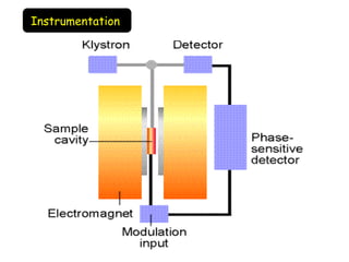
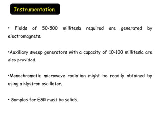
INSTRUMENTATION, APPLICATION, &
PRINCIPLE
• Electron Spin Resonance Spectroscopy
• Also called EPR Spectroscopy
– Electron Paramagnetic Resonance Spectroscopy
• Non-destructive technique
Electron Spin Resonance
• A chemical species with an odd number of electrons exhibits
characteristic magnetic properties much like the nucleus.
• The spinning action of an unpaired electron generates a magnetic moment
μ.
• If an intense magnetic field is applied, the electron assumes orientations
aligned with (lower energy –μHo
) or against (higher energy +μHo
) the field.
• An electron in a magnetic field is able to absorb energy of the proper
frequency ∆E =hυ which will catapult it from lower to higher energy level.
Principle
What causes the energy levels?
Resulting energy levels of an electron in a magnetic
field
• This phenomenon is known as electron resonance and the technique
employed to study this type of behavior is called as electron spin
resonance spectrometry.
•In a magnetic field of the order of 3400gauss, the appropriate quantum
of energy is obtained from radiation in the microwave region of
electromagnetic spectrum.
•It is common practice to subject the sample to differing magnetic
intensities keeping the microwave frequency constant. The magnetic field
is varied till resonance occurs.
•The area of ESR peak is directly proportional to the number of unpaired
electrons in the sample investigated and thus to the concentration of the
sample.
• In quantitative analysis, peak areas of the sample are compared to the
peak area of a standard which contains a known quantity of unpaired
electrons.
• ESR spectra do exhibit hyperfine splitting which is caused by interactions
between the spinning electrons and adjacent spinning magnetic nuclei.
• ESR spectra show no phenomenon like chemical shift as seen in NMR.
• Some proteins or enzymes contain intrinsic functional groups with
unpaired electrons however there are many which do not.
• Much information about these macromolecules can be gathered by
incorporating in them an extrinsic functional group with unpaired
electrons.
• Spin labeling refers to use of stable free radicals as reporter groups
or labels.
•Spin labels (stable free radicals) are usually molecules containing
nitroxide moiety that contains an unpaired electron localized on the
nitrogen and oxygen atoms.
SPIN LABELING
• A spin label (SL) is an organic molecule which possesses an
unpaired electron, usually on a nitrogen atom, and the ability to bind to
another molecule.
•Spin labels are normally used as tools for probing proteins or biological
membrane-local dynamics using electron paramagnetic
resonance spectroscopy.
•The site-directed spin labeling (SDSL) technique allows one to monitor a
specific region within a protein. In protein structure examinations, amino
acid-specific SLs can be used.
•The goal of spin labeling is somewhat similar to that of isotopic
substitution in NMR spectroscopy. There one replaces an atom lacking a
nuclear spin (and so is NMR-silent) with an isotope having a spin I = 0 (and
so is NMR-active). 
•Spin labelled fatty acids have been extensively used to understand
dynamic organization of lipids in bio-membranes and membrane biophysics.
•For example, stearic acid labelled with nitroxyl spin label moiety at
various carbons (5,7,9,12,13,14 and 16th) with respect to first carbon of
carbonyl group have been used to study the flexibility gradient of
membrane lipids to understand membrane fluidity conditions at different
depths of their lipid bilayer organization
Instrumentation
• Fields of 50-500 millitesla required are generated by
electromagnets.
•Auxillary sweep generators with a capacity of 10-100 millitesla are
also provided.
•Monochromatic microwave radiation might be readily obtained by
using a klystron oscillator.
• Samples for ESR must be solids.
Instrumentation
(1) KLYSTRONS
Klystron tube acts as the source of radiation. The frequency of the
monochromatic radiation is determined by the voltage applied to klystron.
 It is kept a fixed frequency by an automatic control circuit and provides a
power output of about 300 milli watts.
(2) WAVE GUIDE OR WAVEMETER
The wave meter is put in between the oscillator and attenuator to know the
frequency of microwaves produced by klystron oscillator. The wave meter
is usually calibrated in frequency unit (megahertz) instead of wavelength.
Wave guide is a hollow, rectangular brass tube. It is used to convey the
wave radiation to the sample and crystal.
Instrumentation
(3) ATTENUATORS
A calibrated attenuator is used to control the level of microwave power
from the source.
(4) SAMPLE CAVITIES
The heart of the ESR spectrometer is the resonant cavity containing
the sample. The sample is contained in a resonance cavity. In most of the
ESR spectrometers, dual sample cavities are generally used. This is done
for simultaneous observation of a sample and a reference material. Since
magnetic field interacts with the sample to cause spin resonance the
sample is placed where the intensity of magnetic field is greatest.
(5) CRYSTAL DETECTORS AND HOLDERS
A Silicon crystal detectors, which converts the radiation in D.C., has
widely been used as a detector of microwave radiation. Microwave Bridge
such as magic T and hybrid ring variety are most common.
(6) MODULATION COIL
The modulation of the signal at a frequency consistent with good signal
noise ratio in the crystal detector is accomplished by  a small alternating
variation of the magnetic field. The variation is produced by supplying an
A.C. signal to modulation coil oriented with respect to the sample in the
same direction as the magnetic field.
(7) MAGNET SYSTEM
The resonant cavity is placed between the pole pieces of an electromagnet.
An electro magnet capable of producing magnetic field of at least 5000
gauss is required for ESR. The field should be stable and uniform over the
sample volume. The stability of field is achieved by energizing the magnet
with a highly regulated power supply.
The ESR spectrum is recorded by slowly varying the magnetic field by
sweeping the current supplied to the magnet by the power supply. This
sweep is usually accomplished by with a variable speed motor drive. Both
the magnet as well as the power supply may require water cooling.
• ESR spectrometry is one of the main methods to study transition metal
containing metalloproteins.
• Biological macromolecules which lack unpaired electrons cannot be
studied by ESR because they do not resonate.
•Information obtained from ESR spectra:
•1] Rate of catalysis
•2] Active site geometry
•3] Denaturation and protein folding
•4] Enzyme-ligand interaction
APPLICATIONS
What compounds can you analyze?
• Applicable for species with one or more unpaired electrons
– Free radicals
– Transition metal compounds
• Useful for unstable paramagnetic compounds generated in situ
– Electrochemical oxidation or reduction
ESR
ESR
ESR

ESR

  • 1.
  • 2.
    • Electron SpinResonance Spectroscopy • Also called EPR Spectroscopy – Electron Paramagnetic Resonance Spectroscopy • Non-destructive technique Electron Spin Resonance
  • 4.
    • A chemicalspecies with an odd number of electrons exhibits characteristic magnetic properties much like the nucleus. • The spinning action of an unpaired electron generates a magnetic moment μ. • If an intense magnetic field is applied, the electron assumes orientations aligned with (lower energy –μHo ) or against (higher energy +μHo ) the field. • An electron in a magnetic field is able to absorb energy of the proper frequency ∆E =hυ which will catapult it from lower to higher energy level. Principle
  • 5.
    What causes theenergy levels? Resulting energy levels of an electron in a magnetic field
  • 6.
    • This phenomenonis known as electron resonance and the technique employed to study this type of behavior is called as electron spin resonance spectrometry. •In a magnetic field of the order of 3400gauss, the appropriate quantum of energy is obtained from radiation in the microwave region of electromagnetic spectrum. •It is common practice to subject the sample to differing magnetic intensities keeping the microwave frequency constant. The magnetic field is varied till resonance occurs. •The area of ESR peak is directly proportional to the number of unpaired electrons in the sample investigated and thus to the concentration of the sample.
  • 7.
    • In quantitativeanalysis, peak areas of the sample are compared to the peak area of a standard which contains a known quantity of unpaired electrons. • ESR spectra do exhibit hyperfine splitting which is caused by interactions between the spinning electrons and adjacent spinning magnetic nuclei. • ESR spectra show no phenomenon like chemical shift as seen in NMR.
  • 10.
    • Some proteinsor enzymes contain intrinsic functional groups with unpaired electrons however there are many which do not. • Much information about these macromolecules can be gathered by incorporating in them an extrinsic functional group with unpaired electrons. • Spin labeling refers to use of stable free radicals as reporter groups or labels. •Spin labels (stable free radicals) are usually molecules containing nitroxide moiety that contains an unpaired electron localized on the nitrogen and oxygen atoms. SPIN LABELING
  • 11.
    • A spin label (SL)is an organic molecule which possesses an unpaired electron, usually on a nitrogen atom, and the ability to bind to another molecule. •Spin labels are normally used as tools for probing proteins or biological membrane-local dynamics using electron paramagnetic resonance spectroscopy. •The site-directed spin labeling (SDSL) technique allows one to monitor a specific region within a protein. In protein structure examinations, amino acid-specific SLs can be used.
  • 12.
    •The goal ofspin labeling is somewhat similar to that of isotopic substitution in NMR spectroscopy. There one replaces an atom lacking a nuclear spin (and so is NMR-silent) with an isotope having a spin I = 0 (and so is NMR-active).  •Spin labelled fatty acids have been extensively used to understand dynamic organization of lipids in bio-membranes and membrane biophysics. •For example, stearic acid labelled with nitroxyl spin label moiety at various carbons (5,7,9,12,13,14 and 16th) with respect to first carbon of carbonyl group have been used to study the flexibility gradient of membrane lipids to understand membrane fluidity conditions at different depths of their lipid bilayer organization
  • 13.
  • 14.
    • Fields of50-500 millitesla required are generated by electromagnets. •Auxillary sweep generators with a capacity of 10-100 millitesla are also provided. •Monochromatic microwave radiation might be readily obtained by using a klystron oscillator. • Samples for ESR must be solids. Instrumentation
  • 16.
    (1) KLYSTRONS Klystron tubeacts as the source of radiation. The frequency of the monochromatic radiation is determined by the voltage applied to klystron.  It is kept a fixed frequency by an automatic control circuit and provides a power output of about 300 milli watts. (2) WAVE GUIDE OR WAVEMETER The wave meter is put in between the oscillator and attenuator to know the frequency of microwaves produced by klystron oscillator. The wave meter is usually calibrated in frequency unit (megahertz) instead of wavelength. Wave guide is a hollow, rectangular brass tube. It is used to convey the wave radiation to the sample and crystal. Instrumentation
  • 17.
    (3) ATTENUATORS A calibratedattenuator is used to control the level of microwave power from the source. (4) SAMPLE CAVITIES The heart of the ESR spectrometer is the resonant cavity containing the sample. The sample is contained in a resonance cavity. In most of the ESR spectrometers, dual sample cavities are generally used. This is done for simultaneous observation of a sample and a reference material. Since magnetic field interacts with the sample to cause spin resonance the sample is placed where the intensity of magnetic field is greatest.
  • 18.
    (5) CRYSTAL DETECTORS ANDHOLDERS A Silicon crystal detectors, which converts the radiation in D.C., has widely been used as a detector of microwave radiation. Microwave Bridge such as magic T and hybrid ring variety are most common. (6) MODULATION COIL The modulation of the signal at a frequency consistent with good signal noise ratio in the crystal detector is accomplished by  a small alternating variation of the magnetic field. The variation is produced by supplying an A.C. signal to modulation coil oriented with respect to the sample in the same direction as the magnetic field.
  • 19.
    (7) MAGNET SYSTEM Theresonant cavity is placed between the pole pieces of an electromagnet. An electro magnet capable of producing magnetic field of at least 5000 gauss is required for ESR. The field should be stable and uniform over the sample volume. The stability of field is achieved by energizing the magnet with a highly regulated power supply. The ESR spectrum is recorded by slowly varying the magnetic field by sweeping the current supplied to the magnet by the power supply. This sweep is usually accomplished by with a variable speed motor drive. Both the magnet as well as the power supply may require water cooling.
  • 20.
    • ESR spectrometryis one of the main methods to study transition metal containing metalloproteins. • Biological macromolecules which lack unpaired electrons cannot be studied by ESR because they do not resonate. •Information obtained from ESR spectra: •1] Rate of catalysis •2] Active site geometry •3] Denaturation and protein folding •4] Enzyme-ligand interaction APPLICATIONS
  • 21.
    What compounds canyou analyze? • Applicable for species with one or more unpaired electrons – Free radicals – Transition metal compounds • Useful for unstable paramagnetic compounds generated in situ – Electrochemical oxidation or reduction

Editor's Notes

  • #6 When an electron is placed within an applied magnetic field, Bo, the two possible spin states of the electron have different energies. This energy difference is a result of the Zeeman effect. The lower energy state occurs when the magnetic moment of the electron is aligned with the magnetic field and a higher energy state where m is aligned against the magnetic field. The two states are labeled by the projection of the electron spin, MS, on the direction of the magnetic field, where MS = -1/2 is the parallel state, and MS = +1/2 is the antiparallel state.
  • #22 However, this technique can only be applied to samples having one or more unpaired electrons.