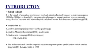
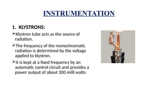
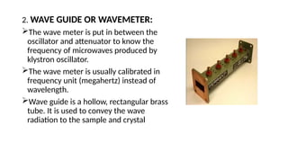
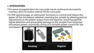
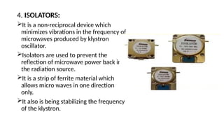
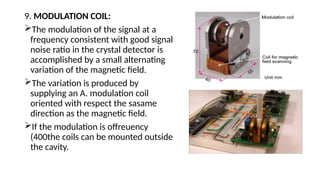
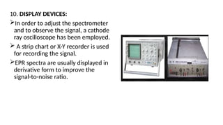
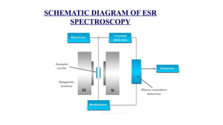
GURU GHASIDAS VISHWAVIDYALAYA, BILASPUR (CG)
Department of Biotechnology
ELECTRON SPIN RESONANACE SPECTROMETRY
Guided By:-
Dr. Santosh Kumar
Associate Professor
Department of Biotechnology,
GGV, Bilaspur, C.G
Presented By:
Roshni Dahariya
Khushboo Mahesh
Pratiksha Rajak
Priyanka Munda
CONTENTS :
1. Introduction
2. Hyperfine splitting &
hyperfine coupling
3. Instrumentation
4. Working
5. Application
ELECTRON SPIN RESONACE
SPECTROSCOPY
TERMS
Electron- Study of unpaired electron spins, and their interaction with their
environment study.
Spin- Electron spin is a quantum mechanical phenomenon. It is represented by ms,
ms can have one of only 2 values, + ½ and – ½
Paramagnetic- The general term paramagnetic is used to describe materials that are
attracted to a magnetic field.
Resonance-This may be the most important term in the EPR. The concept of
resonance is central to the power of magnetic resonance techniques.The general term
paramagnetic is used to describe materials that are attracted to a magnetic field erm
paramagnetic is used to describe materials that are attracted to a magnetic field
INTRODUCTON
• WHAT IS ESR?
It is the branch of absorption spectroscopy in which radiation having frequency in microwave region
(300Mhz-300Ghz) is absorbed by paramagnetic substances in induce transition between magnetic
energy level of electrons with unpaired spin is called as Electron Spin Resonance Spectroscopy(ESR)
• Also known as:-
1.Electron paramagnetic resonance (EPR) spectroscopy
2.Electron Magnetic Resonance (EMR) spectroscopy
3.Electron spin resonance (ESR) spectroscopy
EPR=EMR=ESR
• The molecules which contains unpaired electrons are paramagnetic species or free radical species
discovered by E.K. Zavoisky in 1944
ESR Phenomenon is shown by-
• Atoms having an odd number of electrons
• Atoms having partially filled inner electron shell
• Molecules that carry angular momentum of electronic origin
• Free radicals having unpaired electrons
PRINCIPLE of ESR
• ESR spectroscopy is based on the absorption of microwave radiation by an
unpaired electron when it is exposed to a strong magnetic field.
• The electronic energy levels of the atom or molecule will split into different
levels.
• The magnitude of the splitting is dependent on the strength of the applied
magnetic field.
• The atom or molecule can be excited from one split level to another in the
presence of an external radiation of frequency corresponding to the frequency
obtained from the difference in energy between the split levels. Such an
excitation is called a magnetic resonance absorption.
• The magnetic resonance frequency will hence be influenced by the local
environment of the atom or molecule.
Electron Spin Resonance
• The phenomenon of electron spin resonance (ESR) is based on the fact that
an electron is a charged particle.
• It spins around its axis and this causes it to act like a tiny bar magnet.
• Every electron has a magnetic moment and spin quantum number s = ½
with magnetic components ms = + ½ or ms = -1/2 .
• When a molecule or compound with an unpaired electron is placed in a
strong magnetic field, the spin of the unpaired electron can align in two
different ways creating two spin states ms = ± ½.
• The alignment can either be along the direction (parellel) to the magnetic
field which corresponds to the lower energy state ms = – ½ , and Opposite
(antiparallel) to the direction of the applied magnetic field ms = +½.
HYPERFINE SPLITTING AND
HYPERFINE COUPLING
• The interaction of magnetic moment of an unpaired electron with
these of neighboring magnetic active nuclei results in the splitting of
ESR signals . This type of splitting is known as the Hyperfine splitting.
• When the electron spin interacts with nuclear spin then each
electron spin state split into 2I+1 level this is known as
hyperfine splitting.
• If I = 0 , nuclear spin inactive ( does not Hyperfine Splitting).
• If I not equal 0 , nuclear spin active ( Hyperfine Splitting).
Rules to determine nuclear spin :-
1. A nucleus with an even number of proton and even no of
neutron has zero spin I= 0. For example 12, 16, 4,
𝑐 𝑂 𝐻𝑒
32, = 0
𝑆 𝐼
2. A Nucleus with an odd number of proton and odd no of
neutron has an integral spin.
For example 1, 14, = 1, 10, I = 3
𝐻 𝑁 𝐼 𝐵
3. A nucleus with an odd number of proton and even number of
neutron has half integral spin .
For example 1 , 13 , 31 , = 1 2 , 65 , = 3 2
𝐻 𝐶 𝑃 𝐼 𝐶𝑈 𝐼
Hyperfine Splitting Example :-
• Hydrogen Atom : Hydrogen atom contains one unpaired
electron with spin = 1/2 and one proton with nuclear spin I =
1/2. Hence, ms = 1/2 and mI = 1/2.
• In the absence of magnetic field, the electron spin energy
levels are degenerate (i.e. have same energy).
• When external magnetic field is applied, the degeneracy is
removed and the sublevel ms = + 1/2 goes up while sublevel
ms = 1/2 goes down.
• These two energy levels formed further interact with the
nucleus to give rise to four sublevels of different energy .
INSTRUMENTATION
1. KLYSTRONS:
Klystron tube acts as the source of
radiation.
The frequency of the monochromatic
radiation is determined by the voltage
applied to klystron.
It is kept at a fixed frequency by an
automatic control circuit and provides a
power output of about 300 milli watts
2. WAVE GUIDE OR WAVEMETER:
The wave meter is put in between the
oscillator and attenuator to know the
frequency of microwaves produced by
klystron oscillator.
The wave meter is usually calibrated in
frequency unit (megahertz) instead of
wavelength.
Wave guide is a hollow, rectangular brass
tube. It is used to convey the wave
radiation to the sample and crystal
3. ATTENUATORS:
The power propagated down the wave guide may be continuously decreased by
inserting a piece of resistive material into the wave guide.
In ESR spectroscopy, an attenuator functions to control and reduce the
power of the microwave radiation reaching the sample by allowing precise
adjustments to the power output from the klystron, ensuring optimal
signal detection and preventing damage to the sample from excessive
microwave power; essentially acting as a variable power control for the
microwave source.
4. ISOLATORS:
It is a non-reciprocal device which
minimizes vibrations in the frequency of
microwaves produced by klystron
oscillator.
Isolators are used to prevent the
reflection of microwave power back into
the radiation source.
It is a strip of ferrite material which
allows micro waves in one direction
only.
It also is being stabilizing the frequency
of the klystron.
5. SAMPLE CAVITIES:
The heart of the ESR spectrometer is the
resonant sample cavity.
In most of the ESR spectrometers, dual
sample cavities are generally used.
This is done for simultaneous observation
of a sample and a reference material.
Since magnetic field interact with sample
to cause spin resonance the sample is
placed where the intensity of magnetic
field is greatest.
6. COUPLERS AND MATCHING
SCREWS:
The various components of the
microwave assembly to be
coupled together by making use
of irises or slots of various sizes.
7. CRYSTAL DETECTORS AND HOLDERS:
A Silicon crystal detectors, which converts the radiation in D.C., has
widely been used as a detector of microwave radiation.
Microwave Bridge such as magic T and hybrid ring variety are most
common.
8. MAGNET SYSTEM:
The resonant cavity is placed between the poles pieces of an
electromagnet.
An electro magnet capable of producing magnetic field of at least
5000 gauss is required for ESR.
The field should be stable and uniform over the sample volume.
The stability of field is achieved by energizing the magnet with a
highly regulated power supply.
9. MODULATION COIL:
The modulation of the signal at a
frequency consistent with good signal
noise ratio in the crystal detector is
accomplished by a small alternating
variation of the magnetic field.
The variation is produced by
supplying an A. modulation coil
oriented with respect the sasame
direction as the magnetic field.
If the modulation is offreuency
(400the coils can be mounted outside
the cavity.
10. DISPLAY DEVICES:
In order to adjust the spectrometer
and to observe the signal, a cathode
ray oscilloscope has been employed.
 A strip chart or X-Y recorder is used
for recording the signal.
EPR spectra are usually displayed in
derivative form to improve the
signal-to-noise ratio.
SCHEMATIC DIAGRAM OF ESR
SPECTROSCOPY
APPLICATION
• ESR spectroscopy is one of the main methods used to study metalloproteins,
particularly those containing molybdenum, copper, iron, etc.
• Both copper and non-haem iron do not absorb radiation in visible and ultra
violet range, posses ESR absorbance peak in one of their oxidation state.
• Hence their appearance and disappearance of their ESR signal are used to
monitor their activity in multi enzyme system.
• In metalloproteins, the metal atom has characteristic number of ligands
coordinated to it in a definite geometrical arrangement. Studies using ESR
have shown that their geometry is frequently distorted.
Continue..
• The technique ESR has been extended by spin labeling. Thus spin
labeling glycerophosphatides with a suitable nitrox free radical the
laterate diffusion of the labelled molecules can be studied.
• Electron spin resonance is also extensively used to study the free
radicals caused by irradiation of biological material. The concentration
of free radicals in samples can also be determined.
Electron Spin Resonance (ESR) Spectroscopy

Electron Spin Resonance (ESR) Spectroscopy

  • 1.
    GURU GHASIDAS VISHWAVIDYALAYA,BILASPUR (CG) Department of Biotechnology ELECTRON SPIN RESONANACE SPECTROMETRY Guided By:- Dr. Santosh Kumar Associate Professor Department of Biotechnology, GGV, Bilaspur, C.G Presented By: Roshni Dahariya Khushboo Mahesh Pratiksha Rajak Priyanka Munda
  • 2.
    CONTENTS : 1. Introduction 2.Hyperfine splitting & hyperfine coupling 3. Instrumentation 4. Working 5. Application
  • 3.
  • 4.
    TERMS Electron- Study ofunpaired electron spins, and their interaction with their environment study. Spin- Electron spin is a quantum mechanical phenomenon. It is represented by ms, ms can have one of only 2 values, + ½ and – ½ Paramagnetic- The general term paramagnetic is used to describe materials that are attracted to a magnetic field. Resonance-This may be the most important term in the EPR. The concept of resonance is central to the power of magnetic resonance techniques.The general term paramagnetic is used to describe materials that are attracted to a magnetic field erm paramagnetic is used to describe materials that are attracted to a magnetic field
  • 5.
    INTRODUCTON • WHAT ISESR? It is the branch of absorption spectroscopy in which radiation having frequency in microwave region (300Mhz-300Ghz) is absorbed by paramagnetic substances in induce transition between magnetic energy level of electrons with unpaired spin is called as Electron Spin Resonance Spectroscopy(ESR) • Also known as:- 1.Electron paramagnetic resonance (EPR) spectroscopy 2.Electron Magnetic Resonance (EMR) spectroscopy 3.Electron spin resonance (ESR) spectroscopy EPR=EMR=ESR • The molecules which contains unpaired electrons are paramagnetic species or free radical species discovered by E.K. Zavoisky in 1944
  • 6.
    ESR Phenomenon isshown by- • Atoms having an odd number of electrons • Atoms having partially filled inner electron shell • Molecules that carry angular momentum of electronic origin • Free radicals having unpaired electrons
  • 7.
    PRINCIPLE of ESR •ESR spectroscopy is based on the absorption of microwave radiation by an unpaired electron when it is exposed to a strong magnetic field. • The electronic energy levels of the atom or molecule will split into different levels. • The magnitude of the splitting is dependent on the strength of the applied magnetic field. • The atom or molecule can be excited from one split level to another in the presence of an external radiation of frequency corresponding to the frequency obtained from the difference in energy between the split levels. Such an excitation is called a magnetic resonance absorption. • The magnetic resonance frequency will hence be influenced by the local environment of the atom or molecule.
  • 8.
    Electron Spin Resonance •The phenomenon of electron spin resonance (ESR) is based on the fact that an electron is a charged particle. • It spins around its axis and this causes it to act like a tiny bar magnet. • Every electron has a magnetic moment and spin quantum number s = ½ with magnetic components ms = + ½ or ms = -1/2 . • When a molecule or compound with an unpaired electron is placed in a strong magnetic field, the spin of the unpaired electron can align in two different ways creating two spin states ms = ± ½. • The alignment can either be along the direction (parellel) to the magnetic field which corresponds to the lower energy state ms = – ½ , and Opposite (antiparallel) to the direction of the applied magnetic field ms = +½.
  • 10.
    HYPERFINE SPLITTING AND HYPERFINECOUPLING • The interaction of magnetic moment of an unpaired electron with these of neighboring magnetic active nuclei results in the splitting of ESR signals . This type of splitting is known as the Hyperfine splitting. • When the electron spin interacts with nuclear spin then each electron spin state split into 2I+1 level this is known as hyperfine splitting. • If I = 0 , nuclear spin inactive ( does not Hyperfine Splitting). • If I not equal 0 , nuclear spin active ( Hyperfine Splitting).
  • 11.
    Rules to determinenuclear spin :- 1. A nucleus with an even number of proton and even no of neutron has zero spin I= 0. For example 12, 16, 4, 𝑐 𝑂 𝐻𝑒 32, = 0 𝑆 𝐼 2. A Nucleus with an odd number of proton and odd no of neutron has an integral spin. For example 1, 14, = 1, 10, I = 3 𝐻 𝑁 𝐼 𝐵 3. A nucleus with an odd number of proton and even number of neutron has half integral spin . For example 1 , 13 , 31 , = 1 2 , 65 , = 3 2 𝐻 𝐶 𝑃 𝐼 𝐶𝑈 𝐼
  • 12.
    Hyperfine Splitting Example:- • Hydrogen Atom : Hydrogen atom contains one unpaired electron with spin = 1/2 and one proton with nuclear spin I = 1/2. Hence, ms = 1/2 and mI = 1/2. • In the absence of magnetic field, the electron spin energy levels are degenerate (i.e. have same energy). • When external magnetic field is applied, the degeneracy is removed and the sublevel ms = + 1/2 goes up while sublevel ms = 1/2 goes down. • These two energy levels formed further interact with the nucleus to give rise to four sublevels of different energy .
  • 14.
    INSTRUMENTATION 1. KLYSTRONS: Klystron tubeacts as the source of radiation. The frequency of the monochromatic radiation is determined by the voltage applied to klystron. It is kept at a fixed frequency by an automatic control circuit and provides a power output of about 300 milli watts
  • 15.
    2. WAVE GUIDEOR WAVEMETER: The wave meter is put in between the oscillator and attenuator to know the frequency of microwaves produced by klystron oscillator. The wave meter is usually calibrated in frequency unit (megahertz) instead of wavelength. Wave guide is a hollow, rectangular brass tube. It is used to convey the wave radiation to the sample and crystal
  • 16.
    3. ATTENUATORS: The powerpropagated down the wave guide may be continuously decreased by inserting a piece of resistive material into the wave guide. In ESR spectroscopy, an attenuator functions to control and reduce the power of the microwave radiation reaching the sample by allowing precise adjustments to the power output from the klystron, ensuring optimal signal detection and preventing damage to the sample from excessive microwave power; essentially acting as a variable power control for the microwave source.
  • 17.
    4. ISOLATORS: It isa non-reciprocal device which minimizes vibrations in the frequency of microwaves produced by klystron oscillator. Isolators are used to prevent the reflection of microwave power back into the radiation source. It is a strip of ferrite material which allows micro waves in one direction only. It also is being stabilizing the frequency of the klystron.
  • 18.
    5. SAMPLE CAVITIES: Theheart of the ESR spectrometer is the resonant sample cavity. In most of the ESR spectrometers, dual sample cavities are generally used. This is done for simultaneous observation of a sample and a reference material. Since magnetic field interact with sample to cause spin resonance the sample is placed where the intensity of magnetic field is greatest.
  • 19.
    6. COUPLERS ANDMATCHING SCREWS: The various components of the microwave assembly to be coupled together by making use of irises or slots of various sizes.
  • 20.
    7. CRYSTAL DETECTORSAND HOLDERS: A Silicon crystal detectors, which converts the radiation in D.C., has widely been used as a detector of microwave radiation. Microwave Bridge such as magic T and hybrid ring variety are most common.
  • 21.
    8. MAGNET SYSTEM: Theresonant cavity is placed between the poles pieces of an electromagnet. An electro magnet capable of producing magnetic field of at least 5000 gauss is required for ESR. The field should be stable and uniform over the sample volume. The stability of field is achieved by energizing the magnet with a highly regulated power supply.
  • 22.
    9. MODULATION COIL: Themodulation of the signal at a frequency consistent with good signal noise ratio in the crystal detector is accomplished by a small alternating variation of the magnetic field. The variation is produced by supplying an A. modulation coil oriented with respect the sasame direction as the magnetic field. If the modulation is offreuency (400the coils can be mounted outside the cavity.
  • 23.
    10. DISPLAY DEVICES: Inorder to adjust the spectrometer and to observe the signal, a cathode ray oscilloscope has been employed.  A strip chart or X-Y recorder is used for recording the signal. EPR spectra are usually displayed in derivative form to improve the signal-to-noise ratio.
  • 24.
    SCHEMATIC DIAGRAM OFESR SPECTROSCOPY
  • 25.
    APPLICATION • ESR spectroscopyis one of the main methods used to study metalloproteins, particularly those containing molybdenum, copper, iron, etc. • Both copper and non-haem iron do not absorb radiation in visible and ultra violet range, posses ESR absorbance peak in one of their oxidation state. • Hence their appearance and disappearance of their ESR signal are used to monitor their activity in multi enzyme system. • In metalloproteins, the metal atom has characteristic number of ligands coordinated to it in a definite geometrical arrangement. Studies using ESR have shown that their geometry is frequently distorted.
  • 26.
    Continue.. • The techniqueESR has been extended by spin labeling. Thus spin labeling glycerophosphatides with a suitable nitrox free radical the laterate diffusion of the labelled molecules can be studied. • Electron spin resonance is also extensively used to study the free radicals caused by irradiation of biological material. The concentration of free radicals in samples can also be determined.