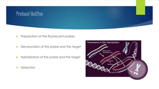
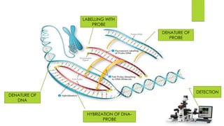
FISH FLOURESCENT IN-SITU HYBRIDIZATION
NARENDRA YADAV
PAPER 3 UNIT 2
INTRODUCTION
 FISH is a cytogenetic technique that uses fluorescent probes that bind to
only those parts of the chormosomes with a high degree of sequences
complementarity.
 Fluorescent probe-DNA complex can be studied under fluroscent
microscope.
 Developed by the biomedical researchers in early’ 1980s.
Protocol Outline
 Preparation of the fluorescent probes
 Denaturation of the probe and the target
 Hybridization of the probe and the target
 Detection
DENATURE OF
DNA
DENATURE OF
PROBE
LABELLING WITH
PROBE
HYBRIZATION OF DNA-
PROBE
DETECTION
Flourescent labled organism
MicroRNA(green) and mRNA(red) visualization in
differentiating C1C12 cells
Probes
 Complementary sequences of target nucleic acids
 Designed against the sequence of interest.
 Probes are tagged with fluorescent dyes like biotin, fluorescein,
Digoxigenin
 Size ranges from 20-40 bp tO1000bp.
PROBES
 Stellaris(R) RNA FISH probes.
 Fiber FISH.
 Q-FISH.
 Flow-FISH.
APPLICATION
 Use to detect and localize the presence and absence of the specific DNA
sequences on chormosomes.
 FISH often used for finding specific features in DNA for use in genetic
counselling, medicine, and species identification.
 FISH can also be used to detect and localize specific RNA target (mRNA,
IncRNA, miRNA) in cells.
 Also used to detect circulating tumor cells, and tissues samples.
Continue…
 Diseases that are diagnosed using FISH include Angelman syndrome,
22q13 deletion syndrome,, acute lymphoblastic leukemia, Cri-du-
chat, and Down syndrome.
 FISH can also be used to compare the genomes of two biological
species, to deduce evolutionary relationships.
BIBLIOGRAPHY
 GOOGLE-IMAGES
 WIKIPEDIA
THANK YOU

Fish(flourescent in-situ hybridization)

  • 1.
    FISH FLOURESCENT IN-SITUHYBRIDIZATION NARENDRA YADAV PAPER 3 UNIT 2
  • 2.
    INTRODUCTION  FISH isa cytogenetic technique that uses fluorescent probes that bind to only those parts of the chormosomes with a high degree of sequences complementarity.  Fluorescent probe-DNA complex can be studied under fluroscent microscope.  Developed by the biomedical researchers in early’ 1980s.
  • 3.
    Protocol Outline  Preparationof the fluorescent probes  Denaturation of the probe and the target  Hybridization of the probe and the target  Detection
  • 4.
    DENATURE OF DNA DENATURE OF PROBE LABELLINGWITH PROBE HYBRIZATION OF DNA- PROBE DETECTION
  • 5.
  • 6.
    MicroRNA(green) and mRNA(red)visualization in differentiating C1C12 cells
  • 7.
    Probes  Complementary sequencesof target nucleic acids  Designed against the sequence of interest.  Probes are tagged with fluorescent dyes like biotin, fluorescein, Digoxigenin  Size ranges from 20-40 bp tO1000bp.
  • 8.
    PROBES  Stellaris(R) RNAFISH probes.  Fiber FISH.  Q-FISH.  Flow-FISH.
  • 9.
    APPLICATION  Use todetect and localize the presence and absence of the specific DNA sequences on chormosomes.  FISH often used for finding specific features in DNA for use in genetic counselling, medicine, and species identification.  FISH can also be used to detect and localize specific RNA target (mRNA, IncRNA, miRNA) in cells.  Also used to detect circulating tumor cells, and tissues samples.
  • 10.
    Continue…  Diseases thatare diagnosed using FISH include Angelman syndrome, 22q13 deletion syndrome,, acute lymphoblastic leukemia, Cri-du- chat, and Down syndrome.  FISH can also be used to compare the genomes of two biological species, to deduce evolutionary relationships.
  • 11.
  • 12.