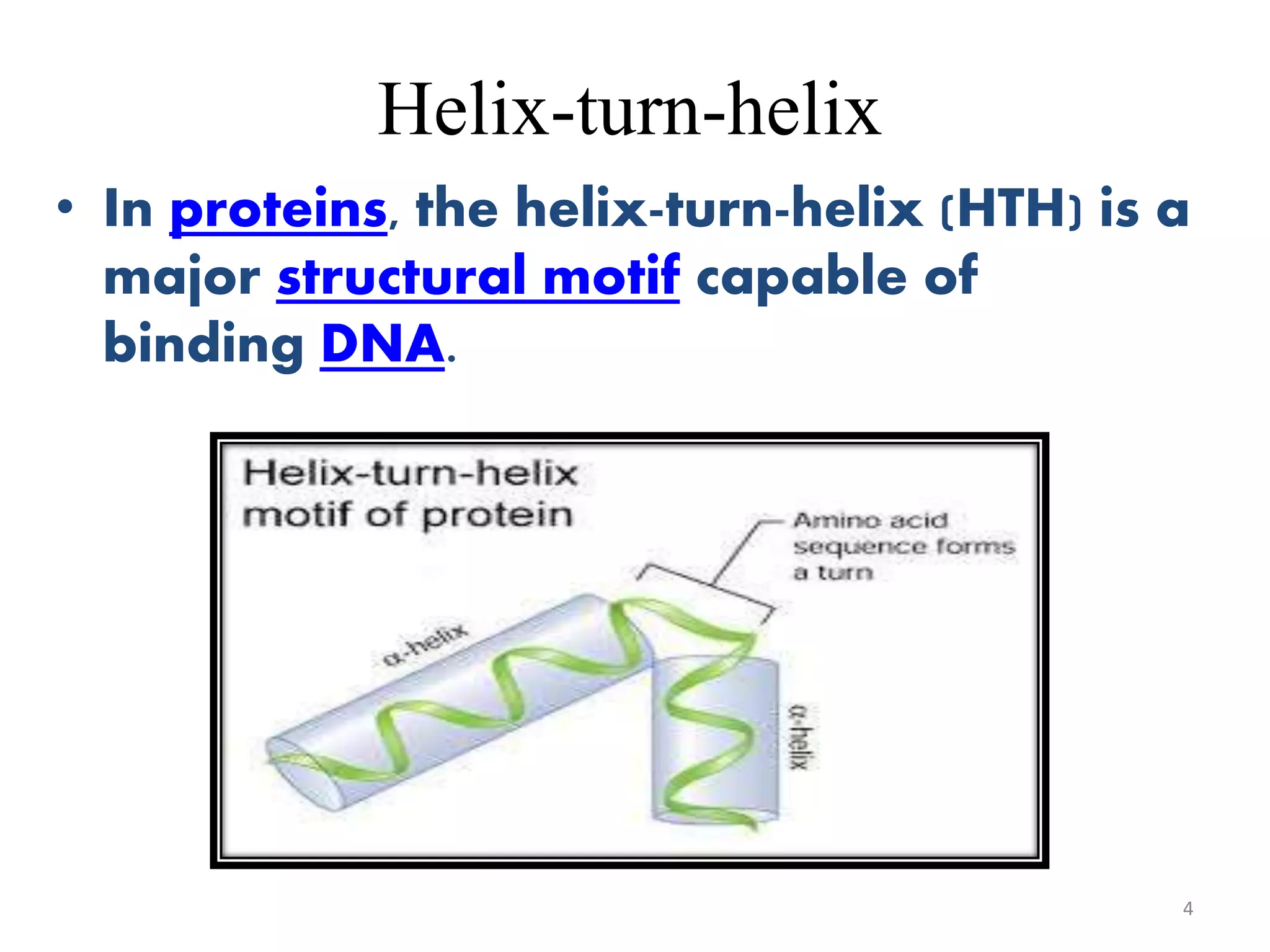
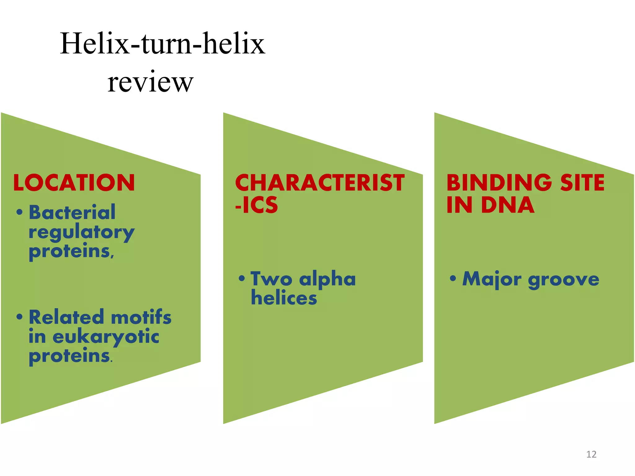
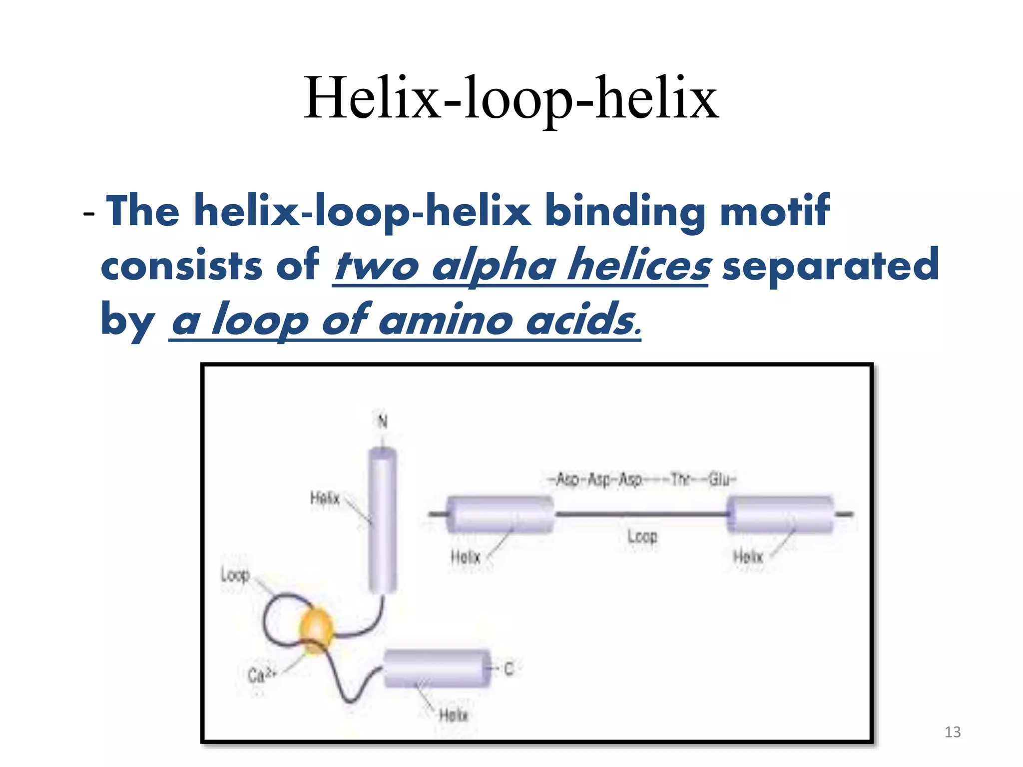
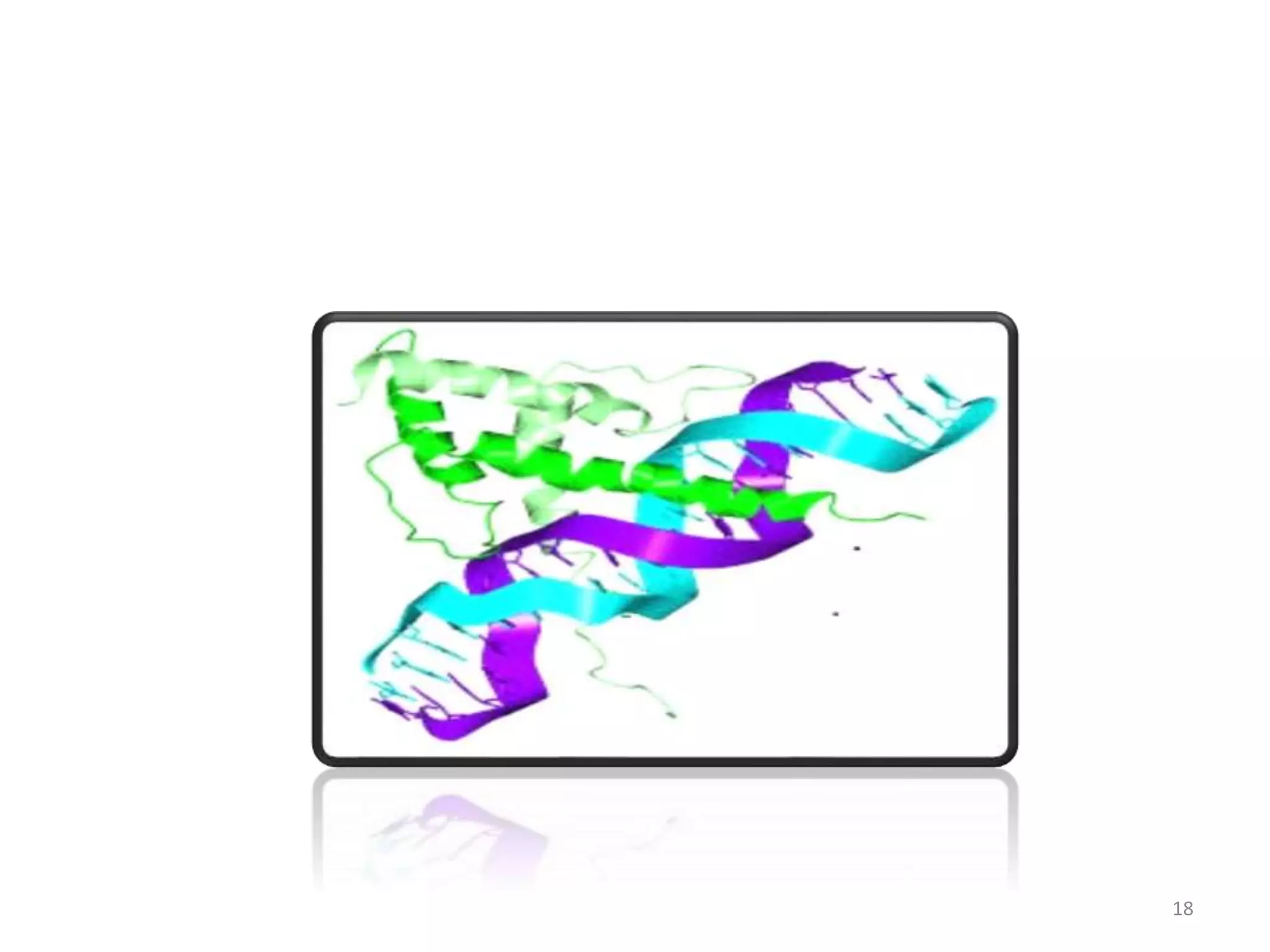
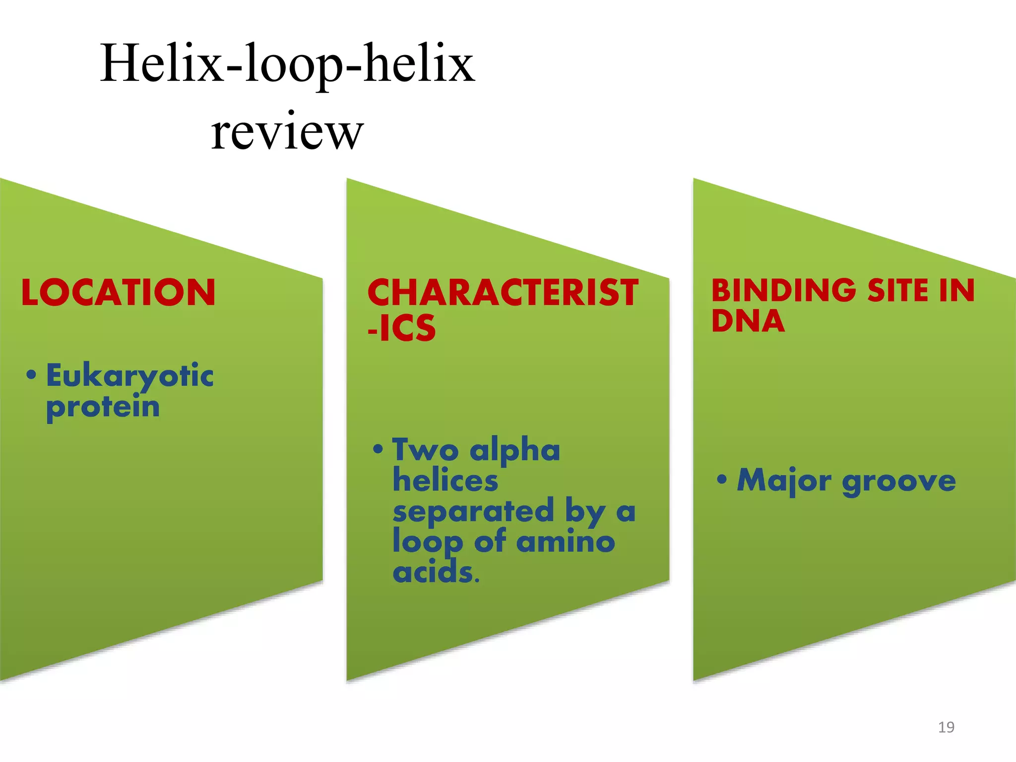
HELIX- TURN- HELIX
&
HELIX- LOOP- HELIX
BIOTECHNOLOGY
- BY- NAREN
YADAV
DNA – Binding Domain
A DNA-binding domain (DBD) is an
independently folded protein domain that
contains at least one motif that recognizes
double- or single-stranded DNA. A DBD can
recognize a specific DNA sequence
(a recognition sequence) or have a general
affinity to DNA.
2
Types of DNA-Binding Motifs
• 1. Steroid Receptor
• 2. Zinc Fingers
• 3. Leucine Zipper
• 4. Homeodomain
• 5.HELIX-TURN-HELIX
• 6.HELIX-LOOP-HELIX
3
Helix-turn-helix
• In proteins, the helix-turn-helix (HTH) is a
major structural motif capable of
binding DNA.
4
DISCOVERY
• The discovery of the helix-turn-helix
motif was based on similarities
between several genes
encoding transcription regulatory
proteins from bacteriophage
lambda and Escherichia coli: Cro, CAP,
and λ repressor, which were found to
share a common 20-25 amino
acid sequence that facilitates DNA
recognition. 5
EXAMPLE
6
The λ repressor of
bacteriophage
lambda employs a
helix-turn-helix (left;
green) to
bindDNA (right; blue
and red).
STRUCTURE
• The structure consist of two α helices joined
by a short strand of amino acid.
• The recognition and binding to DNA by
helix-turn-helix proteins is done by the
two α helices, one occupying the N-
terminal end of the motif, the other at
the C-terminus.
• In most cases, such as in the Cro
repressor, the second helix contributes
most to DNA recognition, and hence it is
often called the "recognition helix".
7
Helix-turn-helix
C-terminal binds to major groove, N-terminal
helps to position the complex
8
FUNCTION
 The helix-turn-helix (HTH) is a major structural
motif capable of binding DNA. It is found in
many proteins that regulate gene expression.
 It binds to the major groove of DNA through
a series of hydrogen bonds and various Van
der Waals interactions with exposed bases.
The other α helix stabilizes the interaction
between protein and DNA, but does not play
a particularly strong role in its recognition.
 The recognition helix and its preceding helix
always have the same relative orientation.
9
10
CLASSIFICATION OF HELIX
TURN HELIX
Di-helical
Tri-helical
Tetra-helical
Winged helix-turn-helix
Other modified helix-turn-helix motifs
11
Helix-turn-helix
review
LOCATION
•Bacterial
regulatory
proteins,
•Related motifs
in eukaryotic
proteins.
CHARACTERIST
-ICS
•Two alpha
helices
BINDING SITE
IN DNA
•Major groove
12
Helix-loop-helix
- The helix-loop-helix binding motif
consists of two alpha helices separated
by a loop of amino acids.
13
STRUCTURE
-Two polypeptide chains with the motif
join to form a functional DNA-binding
protein.
- A highly basic set of amino acids in one
of the helices binds to the DNA.
- Transcription factor including this
domain are in dimeric form.
14
CONTINUE…
• In general, one helix is smaller, and,
due to the flexibility of the loop, allows
dimerization by folding and packing
against another helix.
• The larger helix typically contains the
DNA-binding regions.
• bHLH proteins typically bind to
a consensus sequence called an E-box,
CANNTG.
15
FUNCTION
16
 bHLH transcription factors are often
important in development or cell
activity. BMAL1-Clock is a core
transcription complex in the
molecular circadian clock.
 Other genes, like c-Myc and HIF-1,
have been linked to cancer due to
their effects on cell growth and
metabolism.
REGULATION
• Since many bHLH transcription
factors are heterodimeric, their activity
is often highly regulated by the
dimerization of the subunits.
• One subunit's expression or availability
is often controlled, whereas the other
subunit is constitutively expressed.
17
18
Helix-loop-helix
review
LOCATION
•Eukaryotic
protein
CHARACTERIST
-ICS
•Two alpha
helices
separated by a
loop of amino
acids.
BINDING SITE IN
DNA
•Major groove
19
EXAMPLES
• AhR,BMAL-1-CLOCK,C-Myc,
• N-Myc,MyoD,Myf5,Scleraxis,
• Neurogenis, etc…
20
•THANK YOU
21

HELIX-LOOP-HELIX, HELIX-TURN-HELIX

  • 1.
    HELIX- TURN- HELIX & HELIX-LOOP- HELIX BIOTECHNOLOGY - BY- NAREN YADAV
  • 2.
    DNA – BindingDomain A DNA-binding domain (DBD) is an independently folded protein domain that contains at least one motif that recognizes double- or single-stranded DNA. A DBD can recognize a specific DNA sequence (a recognition sequence) or have a general affinity to DNA. 2
  • 3.
    Types of DNA-BindingMotifs • 1. Steroid Receptor • 2. Zinc Fingers • 3. Leucine Zipper • 4. Homeodomain • 5.HELIX-TURN-HELIX • 6.HELIX-LOOP-HELIX 3
  • 4.
    Helix-turn-helix • In proteins,the helix-turn-helix (HTH) is a major structural motif capable of binding DNA. 4
  • 5.
    DISCOVERY • The discoveryof the helix-turn-helix motif was based on similarities between several genes encoding transcription regulatory proteins from bacteriophage lambda and Escherichia coli: Cro, CAP, and λ repressor, which were found to share a common 20-25 amino acid sequence that facilitates DNA recognition. 5
  • 6.
    EXAMPLE 6 The λ repressorof bacteriophage lambda employs a helix-turn-helix (left; green) to bindDNA (right; blue and red).
  • 7.
    STRUCTURE • The structureconsist of two α helices joined by a short strand of amino acid. • The recognition and binding to DNA by helix-turn-helix proteins is done by the two α helices, one occupying the N- terminal end of the motif, the other at the C-terminus. • In most cases, such as in the Cro repressor, the second helix contributes most to DNA recognition, and hence it is often called the "recognition helix". 7
  • 8.
    Helix-turn-helix C-terminal binds tomajor groove, N-terminal helps to position the complex 8
  • 9.
    FUNCTION  The helix-turn-helix(HTH) is a major structural motif capable of binding DNA. It is found in many proteins that regulate gene expression.  It binds to the major groove of DNA through a series of hydrogen bonds and various Van der Waals interactions with exposed bases. The other α helix stabilizes the interaction between protein and DNA, but does not play a particularly strong role in its recognition.  The recognition helix and its preceding helix always have the same relative orientation. 9
  • 10.
  • 11.
    CLASSIFICATION OF HELIX TURNHELIX Di-helical Tri-helical Tetra-helical Winged helix-turn-helix Other modified helix-turn-helix motifs 11
  • 12.
  • 13.
    Helix-loop-helix - The helix-loop-helixbinding motif consists of two alpha helices separated by a loop of amino acids. 13
  • 14.
    STRUCTURE -Two polypeptide chainswith the motif join to form a functional DNA-binding protein. - A highly basic set of amino acids in one of the helices binds to the DNA. - Transcription factor including this domain are in dimeric form. 14
  • 15.
    CONTINUE… • In general,one helix is smaller, and, due to the flexibility of the loop, allows dimerization by folding and packing against another helix. • The larger helix typically contains the DNA-binding regions. • bHLH proteins typically bind to a consensus sequence called an E-box, CANNTG. 15
  • 16.
    FUNCTION 16  bHLH transcriptionfactors are often important in development or cell activity. BMAL1-Clock is a core transcription complex in the molecular circadian clock.  Other genes, like c-Myc and HIF-1, have been linked to cancer due to their effects on cell growth and metabolism.
  • 17.
    REGULATION • Since manybHLH transcription factors are heterodimeric, their activity is often highly regulated by the dimerization of the subunits. • One subunit's expression or availability is often controlled, whereas the other subunit is constitutively expressed. 17
  • 18.
  • 19.
  • 20.
  • 21.