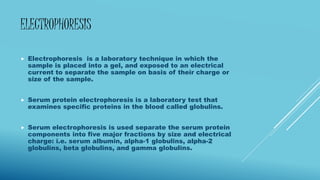
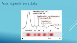
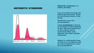
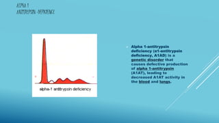
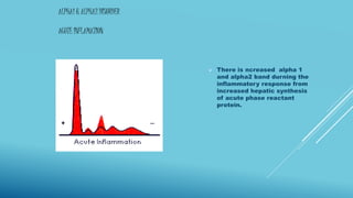
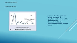
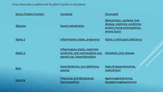
NARENDRA YADAV
PAPER 4
SERUM ELECTROPHORESIS
INTRODUCTION
 Serum is the component that is neither a blood cell nor a
clotting factor; it is the blood plasma not including the
fibrinogens.
 Serum includes all the electrolytes, antibodies, antigens,
hormones, and any exogenous substances (e.g., drugs
and microorganisms).
 Serum is used in numerous diagnostic tests, as well as
blood typing.
COMPONENT OF SERUM
 Serum contains two major protein groups: albumin and
globulin.
 Using protein electrophoresis, these two groups can be
separated into five smaller groups (fractions):
1. albumin fraction
2. alpha 1 fraction
3. alpha 2 fraction
4. beta fraction
5.gamma fraction.
ALBUMIN
 Produce by liver.
 Largest protein component of serum.
 Moves toward anode durning electrophoresis.
 Usually a single band is seen.
 Heterozygous individuals may produce bisalbuminemia
ALPHA 1 FRACTION &
ALPHA 2 FRACTION
 Alpha 1 fraction component:
1.anti-trypsin=protease inhibitor.
2.thyroid binding protein= binding to thyroid hormones.
3.transcortin=Transcortin, transporting cortisol hormones.
 Alpha 2 fraction component:
1.ceruloplamin=copper-carrying protein.
2. haptoglobins= binds free hemoglobin released with
high affinity and thereby inhibits its oxidative activity.
BETA AND GAMMA FRACTION
 Beta fractions consist of:
1. transferrin=iron-binding glycoproteins.
2.lipoproteins= LDL& HDL.
 Gamma fraction cosist of:
 1. immunoglobulins.
ELECTROPHORESIS
 Electrophoresis is a laboratory technique in which the
sample is placed into a gel, and exposed to an electrical
current to separate the sample on basis of their charge or
size of the sample.
 Serum protein electrophoresis is a laboratory test that
examines specific proteins in the blood called globulins.
 Serum electrophoresis is used separate the serum protein
components into five major fractions by size and electrical
charge: i.e. serum albumin, alpha-1 globulins, alpha-2
globulins, beta globulins, and gamma globulins.
PRINCIPLE
 A charged particle placed in an electrical field migrates towards
the anode or cathode depending on the net charge carried by the
particle.
 Serum proteins carry a negative charge at pH 8.6 and move
toward the anode.
 Serum is placed into a sample within low EEO agarose gel in an
alkaline buffer.
 A standardized voltage is applied and upon staining, five distinct
protein bands (albumin, α1, α2, β, and γ) are seen.
 The membrane is then run through a densitometer and the light
absorbance by the protein bands is recorded.
 The absorbance of light is proportional to the relative protein
concentration. The protein concentration of each band is then
calculated.
PROTOCOL
 Preparation of agarose gel.
 Sample preparations.
 Electrophoresis.
 Staining.
 Destaining.
 Visualizing.
INTERPRETATION
 The densest band are of albumin.
Which run towards the anode durning
the electrophoresis.
 Alpha 1 fraction
 Alpha 2 fraction
 Beta fraction
 Gamma fraction which move towards
the cathode durning the
electrophoresis. The band is
observed back of the origin.
Normal Graph after interpretation
PATHOLOGICAL CONDITION ASSOCIATED WITH A CHARACTERISTIC
SERUM PROTEIN ELECTROPHORESIS PATTERN.
ALBUMIN DISORDER
(HEPATIC CIRRHOSIS)
 Abnormal function of
liver.
 Protein synthesis is
compromised and the
concentration of albumin
and protein in the alpha
and band are decreased.
 Additional common
finding is beta-gamma
bridging due to
increased IgA.
NEPHROTIC SYNDROME
 Nephrotic syndrome is a
kidney disorder.
 Loss of protein through the
kidneys(proteinuria) leads
to low protein levels in the
blood.
 Synthesis cause its
accumulation.
 Large proteinuria is due to
an increase in permeability
of the "filtering membrane"
of the kidney, which
selectively filter only
albumin and other smaller
molecular.
 Alpha2 is sufficiently large
so that it is not filtered and
increased Synthesis cause
its accumulation.
ALPHA 1
ANTITRYPSIN -DEFICIENCY
 Alpha 1-antitrypsin
deficiency (α1-antitrypsin
deficiency, A1AD) is a
genetic disorder that
causes defective production
of alpha 1-antitrypsin
(A1AT), leading to
decreased A1AT activity in
the blood and lungs.
ALPHA1 & ALPHA2 DISORDER
ACUTE INFLAMATION
 There is ncreased alpha 1
and alpha2 band durning the
inflammatory response from
increased hepatic synthesis
of acute phase reactant
protein.
GAMA FRACTION DISORDER
CHRONIC INFLAMATION
 Immunoglobulin synthesis
by the activated B
lymphocytes transformed to
plasma cells is
demonstrated by the
increased polyclonal gamma
band.
IMMUNOGLOBULIN DEFICIENCY
 Deficient
immunoglobulins
synthesis is
demonstrated by
deminsihed gamma
band.
MONOCLONAL GAMMOPATHY
 Usually sharp band in the
gamma region suggest the
presence of a homogenous
immunoglobulins.
 Homogenous
immunoglobulins are also
found in waldenstrom’s
macroglobulinemia
Serum Protein Fraction Increased Decreased
Albumin Severe dehydration
Malnutrition, cachexia, liver
disease, nephrotic syndrome,
protein-losing enteropathies,
severe burns
Alpha-1 Inflammatory states, pregnancy Alpha-1 antitrypsin deficiency
Alpha-2
Inflammatory states, nephrotic
syndrome, oral contraceptive use,
steroid use, hyperthyroidism
Hemolysis, liver disease
Beta
Hyperlipidemia, iron-deficiency
anemia
Hypo-B-lipoproteinemia,
malnutrition
Gamma
Polyclonal and Monoclonal
Gammopathies
Agammaglobulinemia,
hypogammaglobulinemia
Various disease states or conditions alter the pattern of proteins in electrophoresis
“
”
Thank you
“
”
REFERENCE
GOOGLE
WIKIPEDIA
BIOTECHNOLOGY JOURNAL

SERUM ELECTROPHORESIS

  • 1.
  • 2.
    INTRODUCTION  Serum isthe component that is neither a blood cell nor a clotting factor; it is the blood plasma not including the fibrinogens.  Serum includes all the electrolytes, antibodies, antigens, hormones, and any exogenous substances (e.g., drugs and microorganisms).  Serum is used in numerous diagnostic tests, as well as blood typing.
  • 3.
    COMPONENT OF SERUM Serum contains two major protein groups: albumin and globulin.  Using protein electrophoresis, these two groups can be separated into five smaller groups (fractions): 1. albumin fraction 2. alpha 1 fraction 3. alpha 2 fraction 4. beta fraction 5.gamma fraction.
  • 4.
    ALBUMIN  Produce byliver.  Largest protein component of serum.  Moves toward anode durning electrophoresis.  Usually a single band is seen.  Heterozygous individuals may produce bisalbuminemia
  • 5.
    ALPHA 1 FRACTION& ALPHA 2 FRACTION  Alpha 1 fraction component: 1.anti-trypsin=protease inhibitor. 2.thyroid binding protein= binding to thyroid hormones. 3.transcortin=Transcortin, transporting cortisol hormones.  Alpha 2 fraction component: 1.ceruloplamin=copper-carrying protein. 2. haptoglobins= binds free hemoglobin released with high affinity and thereby inhibits its oxidative activity.
  • 6.
    BETA AND GAMMAFRACTION  Beta fractions consist of: 1. transferrin=iron-binding glycoproteins. 2.lipoproteins= LDL& HDL.  Gamma fraction cosist of:  1. immunoglobulins.
  • 7.
    ELECTROPHORESIS  Electrophoresis isa laboratory technique in which the sample is placed into a gel, and exposed to an electrical current to separate the sample on basis of their charge or size of the sample.  Serum protein electrophoresis is a laboratory test that examines specific proteins in the blood called globulins.  Serum electrophoresis is used separate the serum protein components into five major fractions by size and electrical charge: i.e. serum albumin, alpha-1 globulins, alpha-2 globulins, beta globulins, and gamma globulins.
  • 8.
    PRINCIPLE  A chargedparticle placed in an electrical field migrates towards the anode or cathode depending on the net charge carried by the particle.  Serum proteins carry a negative charge at pH 8.6 and move toward the anode.  Serum is placed into a sample within low EEO agarose gel in an alkaline buffer.  A standardized voltage is applied and upon staining, five distinct protein bands (albumin, α1, α2, β, and γ) are seen.  The membrane is then run through a densitometer and the light absorbance by the protein bands is recorded.  The absorbance of light is proportional to the relative protein concentration. The protein concentration of each band is then calculated.
  • 9.
    PROTOCOL  Preparation ofagarose gel.  Sample preparations.  Electrophoresis.  Staining.  Destaining.  Visualizing.
  • 10.
    INTERPRETATION  The densestband are of albumin. Which run towards the anode durning the electrophoresis.  Alpha 1 fraction  Alpha 2 fraction  Beta fraction  Gamma fraction which move towards the cathode durning the electrophoresis. The band is observed back of the origin.
  • 11.
    Normal Graph afterinterpretation
  • 12.
    PATHOLOGICAL CONDITION ASSOCIATEDWITH A CHARACTERISTIC SERUM PROTEIN ELECTROPHORESIS PATTERN.
  • 13.
    ALBUMIN DISORDER (HEPATIC CIRRHOSIS) Abnormal function of liver.  Protein synthesis is compromised and the concentration of albumin and protein in the alpha and band are decreased.  Additional common finding is beta-gamma bridging due to increased IgA.
  • 14.
    NEPHROTIC SYNDROME  Nephroticsyndrome is a kidney disorder.  Loss of protein through the kidneys(proteinuria) leads to low protein levels in the blood.  Synthesis cause its accumulation.  Large proteinuria is due to an increase in permeability of the "filtering membrane" of the kidney, which selectively filter only albumin and other smaller molecular.  Alpha2 is sufficiently large so that it is not filtered and increased Synthesis cause its accumulation.
  • 15.
    ALPHA 1 ANTITRYPSIN -DEFICIENCY Alpha 1-antitrypsin deficiency (α1-antitrypsin deficiency, A1AD) is a genetic disorder that causes defective production of alpha 1-antitrypsin (A1AT), leading to decreased A1AT activity in the blood and lungs.
  • 16.
    ALPHA1 & ALPHA2DISORDER ACUTE INFLAMATION  There is ncreased alpha 1 and alpha2 band durning the inflammatory response from increased hepatic synthesis of acute phase reactant protein.
  • 17.
    GAMA FRACTION DISORDER CHRONICINFLAMATION  Immunoglobulin synthesis by the activated B lymphocytes transformed to plasma cells is demonstrated by the increased polyclonal gamma band.
  • 18.
  • 19.
    MONOCLONAL GAMMOPATHY  Usuallysharp band in the gamma region suggest the presence of a homogenous immunoglobulins.  Homogenous immunoglobulins are also found in waldenstrom’s macroglobulinemia
  • 20.
    Serum Protein FractionIncreased Decreased Albumin Severe dehydration Malnutrition, cachexia, liver disease, nephrotic syndrome, protein-losing enteropathies, severe burns Alpha-1 Inflammatory states, pregnancy Alpha-1 antitrypsin deficiency Alpha-2 Inflammatory states, nephrotic syndrome, oral contraceptive use, steroid use, hyperthyroidism Hemolysis, liver disease Beta Hyperlipidemia, iron-deficiency anemia Hypo-B-lipoproteinemia, malnutrition Gamma Polyclonal and Monoclonal Gammopathies Agammaglobulinemia, hypogammaglobulinemia Various disease states or conditions alter the pattern of proteins in electrophoresis
  • 21.
  • 22.