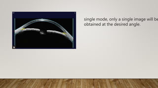
SlideShare a Scribd company logo
ROLE OF ASOCT & UBM
DR.M.DINESH
UBM (ultrasound biomicroscope), ASOCT (anterior segment
optical coherence tomography) provide detailed
• cross-sectional images of the
• cornea,
• anterior chamber,
• angle,
• iris, and
• in case of UBM the lens.
• Optical coherence tomography (OCT) is a
• cross-sectional,
• 3D,
• high-resolution imaging modality that uses
• low coherence interferometry to achieve
• axial resolution in the range of 3–20 μm.
• Anterior segment imaging using OCT was first demonstrated in 1994 by
Izatt, et al. using light with a wavelength of 830 nm
principle of OCT
1. The principle of OCT is based on Michelson’s interferometry
2. Posterior segment OCT uses a lower wavelength of light at 830 nm
3. ASOCT uses a higher wavelength 1310 nm
The anterior segment OCT (ASOCT) at 1310 nm wavelength of light is
better suited for AC angle imaging due
1. Reduced scattering
1. ensures better penetration through ocular structures like the
sclera and the iris and hence a more detailed AC angle
morphology
2. More dissipation/absorption
1. The higher(1310 nm) wavelength light is strongly absorbed by
water in ocular media(vitreous) and therefore, only 10% of the
light incident on the cornea reaches the retina causing no damage
to the retina
2. The improved retinal protection allows for the use of high power
illumination which enables highspeed imaging
The high-speed imaging helps in various ways:
1. Reduces examination time
2. Eliminates motion artifacts
3. Enables imaging of dynamic ocular events
4. Allows for rapid survey of relatively large areas.
The ASOCT has 6 imaging modes:
1. High-resolution cornea single
2. High-resolution cornea quad
3. Pachymetry
4. Anterior segment single
5. Anterior segment double
6. Anterior segment quad
single mode, only a single image will be
obtained at the desired angle.
1. Double mode takes images at the
preset angles of 20 to 200, &160 to
340
2. 1800 preset angle separation will
remain the same i.e.,when manually
changing the angle of image
capture, all angles will rotate
together
1. quad mode of both high-
resolution cornea and AS
2. 4 cross-sectional images are taken
at 0 to 180, 45 to 225, 90 to 270,
and 135 to 315 degrees,
respectively
3. If the operator adjusts the angle by
15 degrees, the new images will be
captured at 15 to 195, 60 to 240,
105 to 285, and 150 to 330.
4. All images have been rotated by
15 degrees and are still separated
by
90 degrees.
Two resolution modes of imaging
Standard resolution imaging High/enhanced resolution
imaging
Broader view of AS
16 mm width /6 mm depth
More detailed imaging
10 mm width/3 mm depth
Full overview of AS:
Cornea, anterior chamber, iris
and both angles
Cornea and any segment
needing detailed evaluation
256 scans 0.125 seconds 512 scans 0.250 seconds
Measurement tools
The different tools available are:
1. Calipers
2. Flap tool
3. Angle tools—ICAT (Iridocorneal angle tool) and ACA (Anterior
chamber angle) tools
4. Chamber tools for anterior chamber depth and width
measurements
5. Anterior segment refractive tool set.
(A) The caliper tool;
(B) The flap tool;
(C) The iridocorneal angle tool;
(D) The chamber tools:
1. Central corneal thickness;
2. Anterior chamber depth;
3. Angle to angle distance; and
4. Crystalline lens rise
Applications of ASOCT
can be broadly grouped into applications in:
• Cornea
• Biometry and phakic IOL
• Glaucoma
Limitations
ASOCT
a. Cannot penetrate through pigmented tissue
b. Cannot image anything past the posterior pigmented epithelium of
the iris.
Applications in Cornea
1. LASIK
2. DSAEK
3. Dystrophies and degenerations
4. Corneal inflammatory and infltrative disorders
5. Keratoplasty
6. Keratoconus
7. Intacs
8. Descemets detachment
1. ASOCT can assist in diagnosis and documentation of corneal
conditions such as dystrophies and degenerations, as well as
assorted inflammatory pathologies.
2. It can be used to diagnose and manage corneal infiltrates, ulcers,
dellen or scars.
3. The depth and extent as obtained by ASOCT is superior to that of
anterior segment photography.
4. Monitoring of corneal infection is possible especially when deep,
and aggressive infection occurs.
5. Hypopyon and hyphema can also be monitored as a result of
severe infection or trauma.
6. ASOCT has the capability to determine the depth of a foreign
body or
the presence of residue once removed
7. Corneal dystrophies affect one or more layers of the cornea.
8. High definition images obtained by ASOCT allow the practitioner to
visualize the epithelium, stroma and endothelium with clear
differentiation.
9. ASOCT can document corneal topography pre and post Descemet’s
stripping automated endothelial keratoplasty (DSAEK) in the
treatment of pseudophakic bullous keratopathy (PBK)
10. ASOCT has the capability to image keratoconus and other ectastias
to determine and document the severity of thinning and scarring.
11. ASOCT pachymetry maps scan areas from the central cornea to 10
mm, calculating a global thickness measurement in both clear and
opacified corneas.
12. Pachymetry maps allow the surgeon to create accurate and
repeatable flap thicknesses in LASIK surgical
planning.Postoperatively, residual bed thickness can be imaged
utilizing these thickness maps
Biometry in Postrefractive Surgery Cases
1. To measure the posterior corneal curvature accurately,
2. To develop an IOL power formula based on the ASOCT corneal
power measurement.
Phakic Intraocular Lens
1. The ASOCT is looked upon with a great deal of optimism regarding
its
potential to guide the sizing of IOLs
Some of the Phakic IOL tools. It shows the “RAINBOW”
(0.5 mm, 1 mm and 1.5 mm from the cornea), “SAFETY” (distance from
cornea) at 180°, center and 0° and “VAULT” (distance between Phakic
IOL and crystalline lens) at 180°, center and 0°)
The use of the “flap tool” in post-LASIK cases to measure the precise
thickness;
(B) The global pachymetry map, which is formed from 16 modifed high
sresolution scans
Applications in Glaucoma
ASOCT is used:
• to study the normal anatomy and physiology
• for screening of the spectrum of angle-closure glaucoma
• to study plateau iris syndrome
• to study mechanism of malignant glaucoma
• to test the efficacy of laser peripheral iridotomy, and
• to test the patency of glaucoma drainage device.
ASOCT of the angle Gonioscopy
It is a non-technical device It requires technical skill
Objective test Subjective test
Comfortable procedure May be discomfortable
Non-contact procedure Contact procedure
No light artifact
Light , indentation, and
accommodation
artifacts may be found
Control for focus is easy More difcult procedure technically
Able to document landmark Documentation is difficult
It is not a standardized
procedure
Standardized procedure. Different
grading systems and lenses are
required
expensive inexpensive
Two photographs of the same angle
A.The photo on the left was taken with the room lights on and it shows a
clearly open angle.
B.The one on the right was taken after turning the lights off, which
demonstrates iris-cornea apposition anterior to the scleral spur which is
marked by the arrow
i.e.,oppositional angle closure
1.Angle-closure
case of synechiae angle closure
subtle case of synechial
closure in an eye with a
plateau iris
Laser Iridotomy
ASOCT images of an eye: (A) Before and; (B) After undergoing laser
iridotomy
The angle appears occludable before procedure, but it clearly
demonstrates that the iridotomy has increased the angle’s width
(A) The angle appears occludable before procedure; (B) The iridotomy
has increased the angle’s width
• Iridotomy procedures have shown dramatic improvement of the
angle
structure when pupillary block mechanisms are present.
• Certain patients’ angles do not improve as expected following
iridotomy, nonpupillary block mechanisms (i.e., plateau iris, lens-
related anterior rotation of the iris) can be easily identified.
• A limitation of the ASOCT during angle imaging
• inability to visualize pathology causing primary or secondary
glaucoma including trabecular pigment or narrow bands of
peripheral synechiae.
Defnitions
Angle Opening Distance
Angle opening distance (AOD500) is calculated as the perpendicular
distance measured from the TM at 500 µm anterior to the scleral spur
to the anterior iris surface.
AOD 750 is calculated as the perpendicular distance measured from the
TM at 750 µm anterior to the scleral spur to the anterior iris surface.
Angle Recess Area
ARA is measured as described by Ishikawa, et al.
1. The defining boundaries of this triangular area are the AOD 500 or
AOD 750 (the base), the angle recess (the apex), and the iris surface
and inner corneoscleral wall form sides of the triangle
2. The ARA is a better measurement parameter than the AOD because it
takes into account the whole contour of the iris surface rather than
measuring at a single point on the iris as is the case with the AOD.
3. ARA may be less sensitive in identifying a narrow angle in eyes with a
relatively deep angle recess.
B) Angle recess area
Trabecular Iris Space Area (TISA):
The defining boundaries of TISA a trapezoidal are as follows:
1. Anteriorly -the AOD 500 or AOD 750;
2. Posteriorly -a line drawn from the scleral spur perpendicular to the
plane of the inner scleral wall to the opposing iris;
3. Superiorly -the inner corneoscleral wall;
4. inferiorly -the iris surface
5. this parameter represents the actual
filtering area more accurately when
compared with the ARA because the
TISA excludes the nonfiltering region
behind the scleral spur.
Trabecular iris contact length (TICL)
defined as the linear distance of iris contact with the corneoscleral
surface beginning at the scleral spur and extending anteriorly in an
anatomically
apposed or synechially closed-angle.
Trabecular iris angle
defined as an angle measured with the apex in the iris recess and the
arms of the angle passing through a point on the TM 500 µm from
the scleral spur and a point on the iris perpendicularly
The composite image of all the parameters AOD, ARA and
TISA
Scleral Spur Angle
defined as an angle measured with the apex at the sclera spur and the
arms of the angle passing through a point on the TM 500 µm from the
scleral spur
and a point on the iris perpendicular.
Plateau Iris :
Plateau iris configuration (PIC)
1. is characterized by an appositional angle with a flat iris configuration, in
contrast to an anterior bowing of the iris seen in a “typical” angle
closure glaucoma in which there is a more crowded anterior chamber
due to a hyperopic/short eye.
2. Classically with PIC
1. the iris configuration is planar and the depth of the AC is N
2. the iris root is often short and inserted anteriorly on the ciliary face,
causing a shallow and narrow angle.
3. With plateau iris configuration there is a relative pupil block
mechanism.
A)figurative images of a normal eye
B)eye with plateau iris syndrome
ASOCT image of an eye with plateau iris
Plateau iris syndrome (PIS)
1. d/to an abnormal anterior position of the ciliary body which
alters the position of the peripheral iris in relation to the
trabecular meshwork resulting in obstruction to aqueous
outflow.
2. PIS may be triggered by spontaneous pupillary dilation (for
example, in darkness), or in response to mydriatic agents.
3. Slit lamp evaluation of patients with plateau iris configuration
usually shows a normal ACD(ant chamb depth) and a flat iris
plane.
4. on indentation gonioscopy a “double hump sign” is chr of PIS .
1. peripheral “hump” - ciliary body propping up the iris root
2. central “hump” - the central third of the iris resting over the
surface of the lens
5. Though, plateau iris is believed to have a deep central chamber
unlike a typical narrow angled individual who is hyperopic, UBM
studies have found patients with plateau iris to have shallower
anterior chamber.
Malignant glaucoma :
1. following filtration surgery it remains one of the most challenging
problems.
2. It refers to a shallow or flat central and peripheral anterior chamber
caused by the forward movement of the lens-iris and iris-hyaloid
diaphragm accompanied by elevated IOP, in the presence of a patent
peripheral iridectomy.
3. ASOCT findings include
1. irido-corneal touch,
2. anterior displacement of the iris root, and
3. appositional angle-closure.
4. The imaging of anterior rotation of the ciliary body with apposition to
the iris is found with UBM but not with ASOCT because of absorption
of infrared light by deeper scleral tissues and the pigment epithelium
of the iris & thereby the visualization of lens, ciliary body, or anterior
vitreous structures was limited .
5. However, the UBM method requires a coupling medium in an
immersion bath, which restricts examinations in the immediate
postoperative period.
5. The goal of the treatment for malignant glaucoma has been directed to
restore
the normal aqueous flow into the anterior chamber with resultant
deepening. ASOCT has revealed to be helpful for the objective
assessment of the therapeutic effects, and to recognize eyes that are
unresponsive to medical or surgical management with a recurrent flat
anterior chamber postoperatively
6. An important risk factor to develop malignant glaucoma is a chronic
glaucoma with a narrow irido-corneal angle and an increased scleral
thickness, which is recognised by ASOCT in the early stagesof a
ciliolenticular block monitoring central and peripheral shallowing of the
anterior chamber to improve therapeutic management and prevent
progression
Postoperative flat chamber: malignant
glaucoma
Bleb morphology
1. it is an indicator of bleb function and a predictor of bleb-related
complications such as bleb leak, blebitis, and bleb-related
endophthalmitis.
2. Bleb morphology indicates the function of the filtration shunt created
by the trabeculectomy procedure and guides the ophthalmologist in
performing interventions such as needling and suture lysis in order to
optimize shunt function.
3. ASOCT has been used to image trabeculectomy bleb to provide
information about internal structure that is not available at the slit lamp.
4. It is able to provide clear images of the blebwall, cavity, flap and ostium
as displayed below.
5. Successful blebs display conjunctival thickening as a hallmark of
success,
regardless of degree of bleb elevation. This reflects facility of
transconjunctival aqueous flow.
5. Highly elevated blebs sometimes display marked conjunctival
thickening and only a small cavity.
6. In failed blebs, ASOCT is particularly useful in imaging failed blebs to
demonstrate the level of failure.
7. Ostial closure, flap fibrosis and presumed episcleral fibrosis in the
absence of the former two situations are all clearly demonstrated.
8. In the early postoperative period, a failing bleb with a closely apposed
scleral flap may be resuscitated by suture lysis, resulting in a more
expanded bleb.
Hence ASOCT is a useful tool to image trabeculectomy
blebs and may aid the clinician in postoperative bleb management.
9. It can also image the intrascleral lake and implant used in non-
penetrating glaucoma surgery (deep sclerectomy) and glaucoma
drainage devices
Trabeculectomy bleb
Ultrasound biomicroscopy (UBM)
is a method of high frequency ultrasound imaging used to generate
images approaching light microscopic resolution up to a depth of 4–5
mm from the surface.
instruments
3 main components of the system are
1. the transducer
2. Signal processor and
3. an articulated arm to steady the scanning head and provide precise
motion control
The system is controlled and synchronized by a computer for further
analysis
instruments frequencies between 35 and 100 MHz,
resolution of 60 microns and a depth penetration up to about 4 mm.
The UBM machine from Zeiss-Humphrey with a 50 MHz probe
The technique of performing UBM with the probe immersed
in a coupling solution placed over the area of interest.
The technique :
1. Technique used for examination is the immersion technique using a fluid
standoff
2. An eye cup permits evaluation avoiding distortion caused by the eyelids
and permits the use of a coupling solution.
3. The procedure is done in the supine position under topical anesthesia
4. The eyecup is used to separate the lids and is filled with 1%
methylcellulose or normal saline.
5. The transducer is immersed in the solution and placed directly over the
part to be scanned.
6. As the arm can be tilted as well as rotated in the horizontal plane, various
sections of the same area can be scanned.
7. The cornea and anterior segment can be easily studied, and the
conjunctiva, sclera and peripheral retina up to the limits of rotation of the
eye in various directions.
Ultrasound Biomicroscopy of Normal Ocular Structures
1. The cornea is seen as a multilayered structure, with a highly
reflective epithelium, a high reflective Bowman’s membrane, and a
high reflective line consisting of the endothelium and Descemet’s
membrane.
2. UBM can measure
1. ACD
2. anterior chamber angle
3. Iris thickness
4. iridolenticular contact distance.
3. The ciliary body, ciliary processes and the ciliary sulcus are well
distinguished on UBM. The ciliary processes show a
variable configuration. The anterior lens surface can be seen, as
well as the anterior zonules. The posterior zonule and vitreous
face may be seen in very shallow chambered eyes.
Uses of UBM :
Corneal and Scleral Diseases
1. The UBM performed in opaque corneas can be helpful in deciding the
prognosis and planning surgery in eyes undergoing keratoplasty.
2. “Virtual gonioscopy” in these instances gives an idea of angle details.
The lens status can also be assessed.
3. Depth of involvement in scleral and episcleral inflammation can help in
diagnosis and evaluating response to treatment.
4. In cases of necrotizing scleritis there may be a spontaneous bleb
formation and the communication can be picked up on the UBM.
Ocular Surface Tumors :
1. Conjunctival and corneal surface tumors can be assessed
2. Information regarding the depth of tumor ,involvement of adjacent
and intraocular structures and tumor residue/recurrence following
surgical excision
The UBM image of the same eye shows , mass
is
homogenous, low reflective, conjunctival and
does not involve the sclera
Image showing a nasal limbal mass
Glaucoma :
1. UBM can image all the angle components, iris, and ciliary body
complex.
2. It is also possible to study the behavior of the iris and angle under
various conditions of illumination
3. Angle-closure can occur at four anatomic levels;
1. the iris (pupillary block),
2. the ciliary body (plateau iris),
3. the lens (phacomorphic glaucoma, and
4. The anterior vitreous face (malignant glaucoma).
Narrow angle with plateau iris Opening up of the angle following a laser
iridotomy
Poor response to peripheral iridotomy may be due to the plateau iris
configuration, where the anteriorly located ciliary processes block the
ciliary sulcus preventing the peripheral iris from falling back after iridotomy
Malignant glaucoma :
1. UBM - able to demonstrate anterior rotation of the ciliary processes to
make a diagnosis of malignant glaucoma
2. It has allowed classification of malignant glaucoma into two groups;
one with supraciliary effusion, which responds to medical management
better and another without effusion that usually requires surgical
management.
3. UBM helps in deciding the course of management at an early stage
1. Extremely shallow anterior chamber.
2. Occluded angle.
3. Forward rotation of the ciliary body.
In pigment dispersion syndrome there is reverse pupillary block where the
posterior bowing of the iris causes an increase in iris-lens contact and
sometimes iris zonular contact as well.
Widely opened angle
Typical Posterior bowing of the peripheral iris
Peripheral Anterior Synechiae
• Reveal the extent of irido corneal adhesions, even if the cornea is hazy or
opaque
Functional Status of Filtering Surgery
• Whether the sclerostomy aperture is patent or blocked internally.
• whether the peripheral iridectomy is patent.
• whether the filtering bleb is flat, shallow or deep
Cataract surgery :
1. Preoperative phacodonesis
2. Area of zonular loss
3. the integrity of the posterior capsule especially in cases of
traumatic cataract can be assessed & plan surgery accordingly.
IOL Complications
1. The UBM is useful in studying various IOL complications.
2. Optic and haptic locations can be assessed accurately by looking for
a strong echo at their interface plane.
3. This ability to image the IOL helps in determining whether structural
changes induced by the IOL are responsible for postoperative
complications.
4. These can be due to retained cortex or contact of the IOL haptic
with the iris or ciliary body.
5. Poor quality of vision due to tilt or decentration of the IOL may
necessitate repositioning or exchange.
6. Demonstration of malpositioned haptics, zonular and capsular
dehiscence, haptic erosion into ocular tissues, decentration and tilt
of IOL can be helpful in assessing these IOL related complications
and planning management strategies
Anterior Segment Cystic Lesions and Tumors
1. Cystic lesions of the cornea, iris and ciliary body
Iris tumors and nevi
Small ciliary bodytumors as well
as cysts, which can mimic tumors are best assessed on UBM
2. The UBM is useful in defining the posterior extent of ciliary body
tumors as well as the anterior extent of peripheral choroidal
tumors
Trauma :
UBM can demonstrate anterior segment foreign bodies, cyclodialysis
clefts, zonular damage and angle recession
The most common clinical presentation of an irido-ciliary
cyst is a peripheral iris elevation - the typical UBM finding
of a thin walled structure with no internal reflectivity is
diagnostic.
Angle recession is imaged as a tear into the face of the
ciliary body. Ciliary body tissue is still imaged attached
to
the scleral spur.
Zonular rupture
Vitreoretinal Surgery
1. UBM clearly images structural changes at the sclerotomies in
post pars plana vitrectomy (PPV) & it distinguishes healing
patterns
2. UBM is helpful in evaluating PPV related complications such as
anterior hyaloidal fibrovascular proliferation and retinal break
formation and planning management.
Anterior segment Inflammation and Hypotony
1. Uveitic eyes very often have miotic pupils and hazy media making it
difficult to evaluate the posterior segment.
2. Conventional ultrasound is able to image the posterior segment but
the UBM helps in studying the ciliary body and pars plana in detail,
identifying edema, thickening, atrophy of ciliary processes, cyclitic
membranes, pars plana exudates and membranes as well as ciliary
body traction
3. In cases of unexplained hypotony, supraciliary effusion, ciliary body
membranes, causing traction or atrophic ciliary processes can be
demonstrated
AS-OCT VS UBM
• Optical VS Ultrasound
• Resolution : 15m VS 50m
• Procedure : noncontact vs contact
• Scan Dimension : 16*6mm VS 5*5mm
• Image posterior iris : No vs Yes
THAN Q
Ref :
Diagnostic Procedures in OPHTHALMOLOGY 3rd ed HV Nema
Uses of ASOCT in LASIK cases:
1. Comprehensive pachymetry map of the entire cornea can be derived
2. The thickness of the cornea from the individual scans as well as the
global pachymetry map can be used to determine ablation rates
3. One of the major factors causing variation in this parameter is
stromal hydration.The ASOCT can measure this hydration minimizing
the differences between planned and actual ablation depths
4. Superior resolution of morphological features
5. Detection of normal from abnormally thin corneas in which
subtractive refractive surgery like LASIK may need to be avoided
6. ASOCT has been utilized in monitoring the epithelial healing progression
under therapeutic contact lenses (TCL) following lamellar keratoplasty
and epiLASIK.
7. Visualizing the epithelium postoperatively allows the surgeon to remove
the TCL at the appropriate time without traumatizing the new epithelium
or risking a secondary infection by leaving the TCL longer than necessary.
Schematic diagram showing the processes involved in
obtaining scans using the UBM
Optical Principles
• Imaging with OCT is based on Michelson interferometer and
includes complex analysis of reflections of low coherence light from
the ocular tissue (low coherence interferometry).
Michelson Interferometer
• A beam of light passes through a semi-transparent mirror that
splits the beam into two.
• These two beams of light are then thrown on two equidistant
mirrors;
reflected light from these mirrors is then picked up and summed up
by a detector.
• The equidistant mirrors reflect the light wave in same phase
• If one of the mirrors is moved by a distance less than the
wavelength of the incident light, the reflected lights from the two
mirrors will then possess a phase difference.
• This phase difference then produces an interference pattern at
the level of the detector
Anterior segment OCT & UBM

More Related Content

What's hot

Soft Contact Lens Fitting
Soft Contact Lens FittingSoft Contact Lens Fitting
Soft Contact Lens Fitting
Vishakh Nair
 

What's hot (20)

Contact lens in keratoconus
Contact lens in keratoconusContact lens in keratoconus
Contact lens in keratoconus
 
Test for stereopsis
Test for stereopsisTest for stereopsis
Test for stereopsis
 
Humphrey visual field analyser (HVFA)
Humphrey visual field analyser (HVFA)Humphrey visual field analyser (HVFA)
Humphrey visual field analyser (HVFA)
 
Surgical induced astigmatism
Surgical induced astigmatismSurgical induced astigmatism
Surgical induced astigmatism
 
Acaratio
AcaratioAcaratio
Acaratio
 
Pentacam
Pentacam Pentacam
Pentacam
 
JCC -Jackson Cross Cylinder
JCC -Jackson Cross CylinderJCC -Jackson Cross Cylinder
JCC -Jackson Cross Cylinder
 
Contact lens fitting in keratoconus copy
Contact lens fitting in keratoconus   copyContact lens fitting in keratoconus   copy
Contact lens fitting in keratoconus copy
 
Iol power calculation in pediatric patients
Iol power calculation in pediatric patientsIol power calculation in pediatric patients
Iol power calculation in pediatric patients
 
AS-OCT
AS-OCTAS-OCT
AS-OCT
 
Biometry
BiometryBiometry
Biometry
 
keratoprosthesis
keratoprosthesiskeratoprosthesis
keratoprosthesis
 
fitting RGP lenses
fitting RGP lensesfitting RGP lenses
fitting RGP lenses
 
Vitreous substitutes
Vitreous substitutesVitreous substitutes
Vitreous substitutes
 
Role of oct in glaucoma
Role of oct in glaucomaRole of oct in glaucoma
Role of oct in glaucoma
 
Ac/a ratio
Ac/a ratio Ac/a ratio
Ac/a ratio
 
Soft Contact Lens Fitting
Soft Contact Lens FittingSoft Contact Lens Fitting
Soft Contact Lens Fitting
 
My ppt
My pptMy ppt
My ppt
 
AS OCT & UBM - Dr Shylesh B Dabke
AS OCT & UBM - Dr Shylesh B DabkeAS OCT & UBM - Dr Shylesh B Dabke
AS OCT & UBM - Dr Shylesh B Dabke
 
Binocular balancing
Binocular balancing Binocular balancing
Binocular balancing
 

Similar to Anterior segment OCT & UBM

OCULAR BIOMETRY AND IOL.pptx
OCULAR BIOMETRY AND IOL.pptxOCULAR BIOMETRY AND IOL.pptx
OCULAR BIOMETRY AND IOL.pptx
Ayienwi Wilson
 
Optical coherence tomography
Optical coherence tomographyOptical coherence tomography
Optical coherence tomography
Samuel Ponraj
 

Similar to Anterior segment OCT & UBM (20)

Pentacam and Corneal topography
Pentacam and Corneal topographyPentacam and Corneal topography
Pentacam and Corneal topography
 
OCULAR BIOMETRY AND IOL.pptx
OCULAR BIOMETRY AND IOL.pptxOCULAR BIOMETRY AND IOL.pptx
OCULAR BIOMETRY AND IOL.pptx
 
Ultrasound biomicrosopy in glaucoma
Ultrasound biomicrosopy in glaucomaUltrasound biomicrosopy in glaucoma
Ultrasound biomicrosopy in glaucoma
 
ultrasound biomicroscopy
ultrasound biomicroscopyultrasound biomicroscopy
ultrasound biomicroscopy
 
CORNEAL TOPOGRAPHY by Florina Deka & Dhanjit Borah
CORNEAL TOPOGRAPHY by Florina Deka & Dhanjit BorahCORNEAL TOPOGRAPHY by Florina Deka & Dhanjit Borah
CORNEAL TOPOGRAPHY by Florina Deka & Dhanjit Borah
 
Biometry.pptx
Biometry.pptxBiometry.pptx
Biometry.pptx
 
biometry for ON.ppt
biometry for ON.pptbiometry for ON.ppt
biometry for ON.ppt
 
Optical coherence tomography
Optical coherence tomographyOptical coherence tomography
Optical coherence tomography
 
Biometry instruments & equipment
Biometry instruments & equipmentBiometry instruments & equipment
Biometry instruments & equipment
 
Slide Deck For Cope Course 10 11 2010
Slide Deck For Cope Course 10 11 2010Slide Deck For Cope Course 10 11 2010
Slide Deck For Cope Course 10 11 2010
 
National Ocular Biometry Course (NOBC) 2015 An echoslide presentation
National Ocular Biometry Course (NOBC) 2015 An echoslide presentation National Ocular Biometry Course (NOBC) 2015 An echoslide presentation
National Ocular Biometry Course (NOBC) 2015 An echoslide presentation
 
introduction into Biometry and it's formulas.pptx
introduction into Biometry and it's formulas.pptxintroduction into Biometry and it's formulas.pptx
introduction into Biometry and it's formulas.pptx
 
A SCAN, B SCAN & BIOMETRY---19.pptx
A SCAN, B SCAN & BIOMETRY---19.pptxA SCAN, B SCAN & BIOMETRY---19.pptx
A SCAN, B SCAN & BIOMETRY---19.pptx
 
Ocular Biometry- IOL calculation methods
Ocular Biometry- IOL calculation methodsOcular Biometry- IOL calculation methods
Ocular Biometry- IOL calculation methods
 
Advances in presbyopia treatment
Advances in presbyopia treatmentAdvances in presbyopia treatment
Advances in presbyopia treatment
 
oct-ujjval solanki
oct-ujjval solankioct-ujjval solanki
oct-ujjval solanki
 
High Precision Biometry
High Precision BiometryHigh Precision Biometry
High Precision Biometry
 
ASOCT NEW SLIDES.pptx
ASOCT NEW SLIDES.pptxASOCT NEW SLIDES.pptx
ASOCT NEW SLIDES.pptx
 
OCT , Laser therapy for DR , Vitrectomy
OCT , Laser therapy for DR , VitrectomyOCT , Laser therapy for DR , Vitrectomy
OCT , Laser therapy for DR , Vitrectomy
 
CSO MS-39
CSO MS-39CSO MS-39
CSO MS-39
 

More from Dinesh Madduri (16)

corneo scleral wound healing in sics
corneo scleral wound healing in sics corneo scleral wound healing in sics
corneo scleral wound healing in sics
 
ocular Toxoplasmosis
ocular Toxoplasmosisocular Toxoplasmosis
ocular Toxoplasmosis
 
Sics steps
Sics stepsSics steps
Sics steps
 
Oct in post seg disorders
Oct in post seg disordersOct in post seg disorders
Oct in post seg disorders
 
Glaucoma drainage devices
Glaucoma drainage devicesGlaucoma drainage devices
Glaucoma drainage devices
 
Recent advances in dcr
Recent advances in dcrRecent advances in dcr
Recent advances in dcr
 
Slitlampexamination dinesh
Slitlampexamination dineshSlitlampexamination dinesh
Slitlampexamination dinesh
 
Neurotrophic keratopathy
Neurotrophic keratopathyNeurotrophic keratopathy
Neurotrophic keratopathy
 
Keratoconus f dinesh
Keratoconus f dineshKeratoconus f dinesh
Keratoconus f dinesh
 
Confocal microscopy dinesh
Confocal microscopy dineshConfocal microscopy dinesh
Confocal microscopy dinesh
 
Evaluation and initial management of patients with ocular and adnexal trauma
Evaluation and initial management of patients with ocular and adnexal traumaEvaluation and initial management of patients with ocular and adnexal trauma
Evaluation and initial management of patients with ocular and adnexal trauma
 
Cmv retinitis
Cmv retinitisCmv retinitis
Cmv retinitis
 
Biologicals in uveitis
Biologicals in uveitisBiologicals in uveitis
Biologicals in uveitis
 
Botox
BotoxBotox
Botox
 
Bacterial keratitis
Bacterial keratitisBacterial keratitis
Bacterial keratitis
 
Artificial anterior chamber
Artificial anterior chamberArtificial anterior chamber
Artificial anterior chamber
 

Recently uploaded

Industrial Training Report- AKTU Industrial Training Report
Industrial Training Report- AKTU Industrial Training ReportIndustrial Training Report- AKTU Industrial Training Report
Industrial Training Report- AKTU Industrial Training Report
Avinash Rai
 
Additional Benefits for Employee Website.pdf
Additional Benefits for Employee Website.pdfAdditional Benefits for Employee Website.pdf
Additional Benefits for Employee Website.pdf
joachimlavalley1
 

Recently uploaded (20)

Danh sách HSG Bộ môn cấp trường - Cấp THPT.pdf
Danh sách HSG Bộ môn cấp trường - Cấp THPT.pdfDanh sách HSG Bộ môn cấp trường - Cấp THPT.pdf
Danh sách HSG Bộ môn cấp trường - Cấp THPT.pdf
 
Basic Civil Engineering Notes of Chapter-6, Topic- Ecosystem, Biodiversity G...
Basic Civil Engineering Notes of Chapter-6,  Topic- Ecosystem, Biodiversity G...Basic Civil Engineering Notes of Chapter-6,  Topic- Ecosystem, Biodiversity G...
Basic Civil Engineering Notes of Chapter-6, Topic- Ecosystem, Biodiversity G...
 
Mattingly "AI & Prompt Design: Limitations and Solutions with LLMs"
Mattingly "AI & Prompt Design: Limitations and Solutions with LLMs"Mattingly "AI & Prompt Design: Limitations and Solutions with LLMs"
Mattingly "AI & Prompt Design: Limitations and Solutions with LLMs"
 
2024_Student Session 2_ Set Plan Preparation.pptx
2024_Student Session 2_ Set Plan Preparation.pptx2024_Student Session 2_ Set Plan Preparation.pptx
2024_Student Session 2_ Set Plan Preparation.pptx
 
Open Educational Resources Primer PowerPoint
Open Educational Resources Primer PowerPointOpen Educational Resources Primer PowerPoint
Open Educational Resources Primer PowerPoint
 
The Benefits and Challenges of Open Educational Resources
The Benefits and Challenges of Open Educational ResourcesThe Benefits and Challenges of Open Educational Resources
The Benefits and Challenges of Open Educational Resources
 
Benefits and Challenges of Using Open Educational Resources
Benefits and Challenges of Using Open Educational ResourcesBenefits and Challenges of Using Open Educational Resources
Benefits and Challenges of Using Open Educational Resources
 
Basic_QTL_Marker-assisted_Selection_Sourabh.ppt
Basic_QTL_Marker-assisted_Selection_Sourabh.pptBasic_QTL_Marker-assisted_Selection_Sourabh.ppt
Basic_QTL_Marker-assisted_Selection_Sourabh.ppt
 
Industrial Training Report- AKTU Industrial Training Report
Industrial Training Report- AKTU Industrial Training ReportIndustrial Training Report- AKTU Industrial Training Report
Industrial Training Report- AKTU Industrial Training Report
 
MARUTI SUZUKI- A Successful Joint Venture in India.pptx
MARUTI SUZUKI- A Successful Joint Venture in India.pptxMARUTI SUZUKI- A Successful Joint Venture in India.pptx
MARUTI SUZUKI- A Successful Joint Venture in India.pptx
 
Introduction to Quality Improvement Essentials
Introduction to Quality Improvement EssentialsIntroduction to Quality Improvement Essentials
Introduction to Quality Improvement Essentials
 
Operations Management - Book1.p - Dr. Abdulfatah A. Salem
Operations Management - Book1.p  - Dr. Abdulfatah A. SalemOperations Management - Book1.p  - Dr. Abdulfatah A. Salem
Operations Management - Book1.p - Dr. Abdulfatah A. Salem
 
The Last Leaf, a short story by O. Henry
The Last Leaf, a short story by O. HenryThe Last Leaf, a short story by O. Henry
The Last Leaf, a short story by O. Henry
 
Salient features of Environment protection Act 1986.pptx
Salient features of Environment protection Act 1986.pptxSalient features of Environment protection Act 1986.pptx
Salient features of Environment protection Act 1986.pptx
 
50 ĐỀ LUYỆN THI IOE LỚP 9 - NĂM HỌC 2022-2023 (CÓ LINK HÌNH, FILE AUDIO VÀ ĐÁ...
50 ĐỀ LUYỆN THI IOE LỚP 9 - NĂM HỌC 2022-2023 (CÓ LINK HÌNH, FILE AUDIO VÀ ĐÁ...50 ĐỀ LUYỆN THI IOE LỚP 9 - NĂM HỌC 2022-2023 (CÓ LINK HÌNH, FILE AUDIO VÀ ĐÁ...
50 ĐỀ LUYỆN THI IOE LỚP 9 - NĂM HỌC 2022-2023 (CÓ LINK HÌNH, FILE AUDIO VÀ ĐÁ...
 
[GDSC YCCE] Build with AI Online Presentation
[GDSC YCCE] Build with AI Online Presentation[GDSC YCCE] Build with AI Online Presentation
[GDSC YCCE] Build with AI Online Presentation
 
The impact of social media on mental health and well-being has been a topic o...
The impact of social media on mental health and well-being has been a topic o...The impact of social media on mental health and well-being has been a topic o...
The impact of social media on mental health and well-being has been a topic o...
 
Basic phrases for greeting and assisting costumers
Basic phrases for greeting and assisting costumersBasic phrases for greeting and assisting costumers
Basic phrases for greeting and assisting costumers
 
Keeping Your Information Safe with Centralized Security Services
Keeping Your Information Safe with Centralized Security ServicesKeeping Your Information Safe with Centralized Security Services
Keeping Your Information Safe with Centralized Security Services
 
Additional Benefits for Employee Website.pdf
Additional Benefits for Employee Website.pdfAdditional Benefits for Employee Website.pdf
Additional Benefits for Employee Website.pdf
 

Anterior segment OCT & UBM

  • 1. ROLE OF ASOCT & UBM DR.M.DINESH
  • 2. UBM (ultrasound biomicroscope), ASOCT (anterior segment optical coherence tomography) provide detailed • cross-sectional images of the • cornea, • anterior chamber, • angle, • iris, and • in case of UBM the lens.
  • 3. • Optical coherence tomography (OCT) is a • cross-sectional, • 3D, • high-resolution imaging modality that uses • low coherence interferometry to achieve • axial resolution in the range of 3–20 μm. • Anterior segment imaging using OCT was first demonstrated in 1994 by Izatt, et al. using light with a wavelength of 830 nm
  • 4. principle of OCT 1. The principle of OCT is based on Michelson’s interferometry 2. Posterior segment OCT uses a lower wavelength of light at 830 nm 3. ASOCT uses a higher wavelength 1310 nm
  • 5. The anterior segment OCT (ASOCT) at 1310 nm wavelength of light is better suited for AC angle imaging due 1. Reduced scattering 1. ensures better penetration through ocular structures like the sclera and the iris and hence a more detailed AC angle morphology 2. More dissipation/absorption 1. The higher(1310 nm) wavelength light is strongly absorbed by water in ocular media(vitreous) and therefore, only 10% of the light incident on the cornea reaches the retina causing no damage to the retina 2. The improved retinal protection allows for the use of high power illumination which enables highspeed imaging
  • 6. The high-speed imaging helps in various ways: 1. Reduces examination time 2. Eliminates motion artifacts 3. Enables imaging of dynamic ocular events 4. Allows for rapid survey of relatively large areas.
  • 7. The ASOCT has 6 imaging modes: 1. High-resolution cornea single 2. High-resolution cornea quad 3. Pachymetry 4. Anterior segment single 5. Anterior segment double 6. Anterior segment quad
  • 8. single mode, only a single image will be obtained at the desired angle.
  • 9. 1. Double mode takes images at the preset angles of 20 to 200, &160 to 340 2. 1800 preset angle separation will remain the same i.e.,when manually changing the angle of image capture, all angles will rotate together
  • 10. 1. quad mode of both high- resolution cornea and AS 2. 4 cross-sectional images are taken at 0 to 180, 45 to 225, 90 to 270, and 135 to 315 degrees, respectively 3. If the operator adjusts the angle by 15 degrees, the new images will be captured at 15 to 195, 60 to 240, 105 to 285, and 150 to 330. 4. All images have been rotated by 15 degrees and are still separated by 90 degrees.
  • 11. Two resolution modes of imaging Standard resolution imaging High/enhanced resolution imaging Broader view of AS 16 mm width /6 mm depth More detailed imaging 10 mm width/3 mm depth Full overview of AS: Cornea, anterior chamber, iris and both angles Cornea and any segment needing detailed evaluation 256 scans 0.125 seconds 512 scans 0.250 seconds
  • 12. Measurement tools The different tools available are: 1. Calipers 2. Flap tool 3. Angle tools—ICAT (Iridocorneal angle tool) and ACA (Anterior chamber angle) tools 4. Chamber tools for anterior chamber depth and width measurements 5. Anterior segment refractive tool set.
  • 13. (A) The caliper tool; (B) The flap tool;
  • 14. (C) The iridocorneal angle tool; (D) The chamber tools: 1. Central corneal thickness; 2. Anterior chamber depth; 3. Angle to angle distance; and 4. Crystalline lens rise
  • 15.
  • 16. Applications of ASOCT can be broadly grouped into applications in: • Cornea • Biometry and phakic IOL • Glaucoma Limitations ASOCT a. Cannot penetrate through pigmented tissue b. Cannot image anything past the posterior pigmented epithelium of the iris.
  • 17. Applications in Cornea 1. LASIK 2. DSAEK 3. Dystrophies and degenerations 4. Corneal inflammatory and infltrative disorders 5. Keratoplasty 6. Keratoconus 7. Intacs 8. Descemets detachment
  • 18. 1. ASOCT can assist in diagnosis and documentation of corneal conditions such as dystrophies and degenerations, as well as assorted inflammatory pathologies. 2. It can be used to diagnose and manage corneal infiltrates, ulcers, dellen or scars. 3. The depth and extent as obtained by ASOCT is superior to that of anterior segment photography. 4. Monitoring of corneal infection is possible especially when deep, and aggressive infection occurs. 5. Hypopyon and hyphema can also be monitored as a result of severe infection or trauma. 6. ASOCT has the capability to determine the depth of a foreign body or the presence of residue once removed
  • 19. 7. Corneal dystrophies affect one or more layers of the cornea. 8. High definition images obtained by ASOCT allow the practitioner to visualize the epithelium, stroma and endothelium with clear differentiation. 9. ASOCT can document corneal topography pre and post Descemet’s stripping automated endothelial keratoplasty (DSAEK) in the treatment of pseudophakic bullous keratopathy (PBK) 10. ASOCT has the capability to image keratoconus and other ectastias to determine and document the severity of thinning and scarring. 11. ASOCT pachymetry maps scan areas from the central cornea to 10 mm, calculating a global thickness measurement in both clear and opacified corneas. 12. Pachymetry maps allow the surgeon to create accurate and repeatable flap thicknesses in LASIK surgical planning.Postoperatively, residual bed thickness can be imaged utilizing these thickness maps
  • 20. Biometry in Postrefractive Surgery Cases 1. To measure the posterior corneal curvature accurately, 2. To develop an IOL power formula based on the ASOCT corneal power measurement. Phakic Intraocular Lens 1. The ASOCT is looked upon with a great deal of optimism regarding its potential to guide the sizing of IOLs
  • 21. Some of the Phakic IOL tools. It shows the “RAINBOW” (0.5 mm, 1 mm and 1.5 mm from the cornea), “SAFETY” (distance from cornea) at 180°, center and 0° and “VAULT” (distance between Phakic IOL and crystalline lens) at 180°, center and 0°)
  • 22. The use of the “flap tool” in post-LASIK cases to measure the precise thickness;
  • 23. (B) The global pachymetry map, which is formed from 16 modifed high sresolution scans
  • 24. Applications in Glaucoma ASOCT is used: • to study the normal anatomy and physiology • for screening of the spectrum of angle-closure glaucoma • to study plateau iris syndrome • to study mechanism of malignant glaucoma • to test the efficacy of laser peripheral iridotomy, and • to test the patency of glaucoma drainage device.
  • 25. ASOCT of the angle Gonioscopy It is a non-technical device It requires technical skill Objective test Subjective test Comfortable procedure May be discomfortable Non-contact procedure Contact procedure No light artifact Light , indentation, and accommodation artifacts may be found Control for focus is easy More difcult procedure technically Able to document landmark Documentation is difficult It is not a standardized procedure Standardized procedure. Different grading systems and lenses are required expensive inexpensive
  • 26. Two photographs of the same angle A.The photo on the left was taken with the room lights on and it shows a clearly open angle. B.The one on the right was taken after turning the lights off, which demonstrates iris-cornea apposition anterior to the scleral spur which is marked by the arrow i.e.,oppositional angle closure 1.Angle-closure
  • 27. case of synechiae angle closure subtle case of synechial closure in an eye with a plateau iris
  • 28. Laser Iridotomy ASOCT images of an eye: (A) Before and; (B) After undergoing laser iridotomy The angle appears occludable before procedure, but it clearly demonstrates that the iridotomy has increased the angle’s width
  • 29. (A) The angle appears occludable before procedure; (B) The iridotomy has increased the angle’s width
  • 30. • Iridotomy procedures have shown dramatic improvement of the angle structure when pupillary block mechanisms are present. • Certain patients’ angles do not improve as expected following iridotomy, nonpupillary block mechanisms (i.e., plateau iris, lens- related anterior rotation of the iris) can be easily identified. • A limitation of the ASOCT during angle imaging • inability to visualize pathology causing primary or secondary glaucoma including trabecular pigment or narrow bands of peripheral synechiae.
  • 31. Defnitions Angle Opening Distance Angle opening distance (AOD500) is calculated as the perpendicular distance measured from the TM at 500 µm anterior to the scleral spur to the anterior iris surface. AOD 750 is calculated as the perpendicular distance measured from the TM at 750 µm anterior to the scleral spur to the anterior iris surface.
  • 32. Angle Recess Area ARA is measured as described by Ishikawa, et al. 1. The defining boundaries of this triangular area are the AOD 500 or AOD 750 (the base), the angle recess (the apex), and the iris surface and inner corneoscleral wall form sides of the triangle 2. The ARA is a better measurement parameter than the AOD because it takes into account the whole contour of the iris surface rather than measuring at a single point on the iris as is the case with the AOD. 3. ARA may be less sensitive in identifying a narrow angle in eyes with a relatively deep angle recess.
  • 34. Trabecular Iris Space Area (TISA): The defining boundaries of TISA a trapezoidal are as follows: 1. Anteriorly -the AOD 500 or AOD 750; 2. Posteriorly -a line drawn from the scleral spur perpendicular to the plane of the inner scleral wall to the opposing iris; 3. Superiorly -the inner corneoscleral wall; 4. inferiorly -the iris surface 5. this parameter represents the actual filtering area more accurately when compared with the ARA because the TISA excludes the nonfiltering region behind the scleral spur.
  • 35. Trabecular iris contact length (TICL) defined as the linear distance of iris contact with the corneoscleral surface beginning at the scleral spur and extending anteriorly in an anatomically apposed or synechially closed-angle.
  • 36. Trabecular iris angle defined as an angle measured with the apex in the iris recess and the arms of the angle passing through a point on the TM 500 µm from the scleral spur and a point on the iris perpendicularly
  • 37. The composite image of all the parameters AOD, ARA and TISA
  • 38. Scleral Spur Angle defined as an angle measured with the apex at the sclera spur and the arms of the angle passing through a point on the TM 500 µm from the scleral spur and a point on the iris perpendicular.
  • 39. Plateau Iris : Plateau iris configuration (PIC) 1. is characterized by an appositional angle with a flat iris configuration, in contrast to an anterior bowing of the iris seen in a “typical” angle closure glaucoma in which there is a more crowded anterior chamber due to a hyperopic/short eye. 2. Classically with PIC 1. the iris configuration is planar and the depth of the AC is N 2. the iris root is often short and inserted anteriorly on the ciliary face, causing a shallow and narrow angle. 3. With plateau iris configuration there is a relative pupil block mechanism.
  • 40. A)figurative images of a normal eye B)eye with plateau iris syndrome
  • 41. ASOCT image of an eye with plateau iris
  • 42. Plateau iris syndrome (PIS) 1. d/to an abnormal anterior position of the ciliary body which alters the position of the peripheral iris in relation to the trabecular meshwork resulting in obstruction to aqueous outflow. 2. PIS may be triggered by spontaneous pupillary dilation (for example, in darkness), or in response to mydriatic agents. 3. Slit lamp evaluation of patients with plateau iris configuration usually shows a normal ACD(ant chamb depth) and a flat iris plane. 4. on indentation gonioscopy a “double hump sign” is chr of PIS . 1. peripheral “hump” - ciliary body propping up the iris root 2. central “hump” - the central third of the iris resting over the surface of the lens
  • 43. 5. Though, plateau iris is believed to have a deep central chamber unlike a typical narrow angled individual who is hyperopic, UBM studies have found patients with plateau iris to have shallower anterior chamber.
  • 44. Malignant glaucoma : 1. following filtration surgery it remains one of the most challenging problems. 2. It refers to a shallow or flat central and peripheral anterior chamber caused by the forward movement of the lens-iris and iris-hyaloid diaphragm accompanied by elevated IOP, in the presence of a patent peripheral iridectomy. 3. ASOCT findings include 1. irido-corneal touch, 2. anterior displacement of the iris root, and 3. appositional angle-closure.
  • 45. 4. The imaging of anterior rotation of the ciliary body with apposition to the iris is found with UBM but not with ASOCT because of absorption of infrared light by deeper scleral tissues and the pigment epithelium of the iris & thereby the visualization of lens, ciliary body, or anterior vitreous structures was limited . 5. However, the UBM method requires a coupling medium in an immersion bath, which restricts examinations in the immediate postoperative period.
  • 46. 5. The goal of the treatment for malignant glaucoma has been directed to restore the normal aqueous flow into the anterior chamber with resultant deepening. ASOCT has revealed to be helpful for the objective assessment of the therapeutic effects, and to recognize eyes that are unresponsive to medical or surgical management with a recurrent flat anterior chamber postoperatively 6. An important risk factor to develop malignant glaucoma is a chronic glaucoma with a narrow irido-corneal angle and an increased scleral thickness, which is recognised by ASOCT in the early stagesof a ciliolenticular block monitoring central and peripheral shallowing of the anterior chamber to improve therapeutic management and prevent progression
  • 47. Postoperative flat chamber: malignant glaucoma
  • 48. Bleb morphology 1. it is an indicator of bleb function and a predictor of bleb-related complications such as bleb leak, blebitis, and bleb-related endophthalmitis. 2. Bleb morphology indicates the function of the filtration shunt created by the trabeculectomy procedure and guides the ophthalmologist in performing interventions such as needling and suture lysis in order to optimize shunt function. 3. ASOCT has been used to image trabeculectomy bleb to provide information about internal structure that is not available at the slit lamp. 4. It is able to provide clear images of the blebwall, cavity, flap and ostium as displayed below. 5. Successful blebs display conjunctival thickening as a hallmark of success, regardless of degree of bleb elevation. This reflects facility of transconjunctival aqueous flow.
  • 49. 5. Highly elevated blebs sometimes display marked conjunctival thickening and only a small cavity. 6. In failed blebs, ASOCT is particularly useful in imaging failed blebs to demonstrate the level of failure. 7. Ostial closure, flap fibrosis and presumed episcleral fibrosis in the absence of the former two situations are all clearly demonstrated. 8. In the early postoperative period, a failing bleb with a closely apposed scleral flap may be resuscitated by suture lysis, resulting in a more expanded bleb. Hence ASOCT is a useful tool to image trabeculectomy blebs and may aid the clinician in postoperative bleb management. 9. It can also image the intrascleral lake and implant used in non- penetrating glaucoma surgery (deep sclerectomy) and glaucoma drainage devices
  • 51. Ultrasound biomicroscopy (UBM) is a method of high frequency ultrasound imaging used to generate images approaching light microscopic resolution up to a depth of 4–5 mm from the surface. instruments 3 main components of the system are 1. the transducer 2. Signal processor and 3. an articulated arm to steady the scanning head and provide precise motion control The system is controlled and synchronized by a computer for further analysis instruments frequencies between 35 and 100 MHz, resolution of 60 microns and a depth penetration up to about 4 mm.
  • 52. The UBM machine from Zeiss-Humphrey with a 50 MHz probe
  • 53. The technique of performing UBM with the probe immersed in a coupling solution placed over the area of interest.
  • 54.
  • 55. The technique : 1. Technique used for examination is the immersion technique using a fluid standoff 2. An eye cup permits evaluation avoiding distortion caused by the eyelids and permits the use of a coupling solution. 3. The procedure is done in the supine position under topical anesthesia 4. The eyecup is used to separate the lids and is filled with 1% methylcellulose or normal saline. 5. The transducer is immersed in the solution and placed directly over the part to be scanned. 6. As the arm can be tilted as well as rotated in the horizontal plane, various sections of the same area can be scanned. 7. The cornea and anterior segment can be easily studied, and the conjunctiva, sclera and peripheral retina up to the limits of rotation of the eye in various directions.
  • 56. Ultrasound Biomicroscopy of Normal Ocular Structures 1. The cornea is seen as a multilayered structure, with a highly reflective epithelium, a high reflective Bowman’s membrane, and a high reflective line consisting of the endothelium and Descemet’s membrane. 2. UBM can measure 1. ACD 2. anterior chamber angle 3. Iris thickness 4. iridolenticular contact distance. 3. The ciliary body, ciliary processes and the ciliary sulcus are well distinguished on UBM. The ciliary processes show a variable configuration. The anterior lens surface can be seen, as well as the anterior zonules. The posterior zonule and vitreous face may be seen in very shallow chambered eyes.
  • 57. Uses of UBM : Corneal and Scleral Diseases 1. The UBM performed in opaque corneas can be helpful in deciding the prognosis and planning surgery in eyes undergoing keratoplasty. 2. “Virtual gonioscopy” in these instances gives an idea of angle details. The lens status can also be assessed. 3. Depth of involvement in scleral and episcleral inflammation can help in diagnosis and evaluating response to treatment. 4. In cases of necrotizing scleritis there may be a spontaneous bleb formation and the communication can be picked up on the UBM.
  • 58.
  • 59. Ocular Surface Tumors : 1. Conjunctival and corneal surface tumors can be assessed 2. Information regarding the depth of tumor ,involvement of adjacent and intraocular structures and tumor residue/recurrence following surgical excision The UBM image of the same eye shows , mass is homogenous, low reflective, conjunctival and does not involve the sclera Image showing a nasal limbal mass
  • 60. Glaucoma : 1. UBM can image all the angle components, iris, and ciliary body complex. 2. It is also possible to study the behavior of the iris and angle under various conditions of illumination 3. Angle-closure can occur at four anatomic levels; 1. the iris (pupillary block), 2. the ciliary body (plateau iris), 3. the lens (phacomorphic glaucoma, and 4. The anterior vitreous face (malignant glaucoma).
  • 61. Narrow angle with plateau iris Opening up of the angle following a laser iridotomy
  • 62. Poor response to peripheral iridotomy may be due to the plateau iris configuration, where the anteriorly located ciliary processes block the ciliary sulcus preventing the peripheral iris from falling back after iridotomy
  • 63. Malignant glaucoma : 1. UBM - able to demonstrate anterior rotation of the ciliary processes to make a diagnosis of malignant glaucoma 2. It has allowed classification of malignant glaucoma into two groups; one with supraciliary effusion, which responds to medical management better and another without effusion that usually requires surgical management. 3. UBM helps in deciding the course of management at an early stage 1. Extremely shallow anterior chamber. 2. Occluded angle. 3. Forward rotation of the ciliary body.
  • 64. In pigment dispersion syndrome there is reverse pupillary block where the posterior bowing of the iris causes an increase in iris-lens contact and sometimes iris zonular contact as well. Widely opened angle Typical Posterior bowing of the peripheral iris
  • 65. Peripheral Anterior Synechiae • Reveal the extent of irido corneal adhesions, even if the cornea is hazy or opaque
  • 66. Functional Status of Filtering Surgery • Whether the sclerostomy aperture is patent or blocked internally. • whether the peripheral iridectomy is patent. • whether the filtering bleb is flat, shallow or deep
  • 67. Cataract surgery : 1. Preoperative phacodonesis 2. Area of zonular loss 3. the integrity of the posterior capsule especially in cases of traumatic cataract can be assessed & plan surgery accordingly.
  • 68. IOL Complications 1. The UBM is useful in studying various IOL complications. 2. Optic and haptic locations can be assessed accurately by looking for a strong echo at their interface plane. 3. This ability to image the IOL helps in determining whether structural changes induced by the IOL are responsible for postoperative complications. 4. These can be due to retained cortex or contact of the IOL haptic with the iris or ciliary body. 5. Poor quality of vision due to tilt or decentration of the IOL may necessitate repositioning or exchange. 6. Demonstration of malpositioned haptics, zonular and capsular dehiscence, haptic erosion into ocular tissues, decentration and tilt of IOL can be helpful in assessing these IOL related complications and planning management strategies
  • 69. Anterior Segment Cystic Lesions and Tumors 1. Cystic lesions of the cornea, iris and ciliary body Iris tumors and nevi Small ciliary bodytumors as well as cysts, which can mimic tumors are best assessed on UBM 2. The UBM is useful in defining the posterior extent of ciliary body tumors as well as the anterior extent of peripheral choroidal tumors
  • 70. Trauma : UBM can demonstrate anterior segment foreign bodies, cyclodialysis clefts, zonular damage and angle recession
  • 71. The most common clinical presentation of an irido-ciliary cyst is a peripheral iris elevation - the typical UBM finding of a thin walled structure with no internal reflectivity is diagnostic.
  • 72. Angle recession is imaged as a tear into the face of the ciliary body. Ciliary body tissue is still imaged attached to the scleral spur. Zonular rupture
  • 73. Vitreoretinal Surgery 1. UBM clearly images structural changes at the sclerotomies in post pars plana vitrectomy (PPV) & it distinguishes healing patterns 2. UBM is helpful in evaluating PPV related complications such as anterior hyaloidal fibrovascular proliferation and retinal break formation and planning management.
  • 74. Anterior segment Inflammation and Hypotony 1. Uveitic eyes very often have miotic pupils and hazy media making it difficult to evaluate the posterior segment. 2. Conventional ultrasound is able to image the posterior segment but the UBM helps in studying the ciliary body and pars plana in detail, identifying edema, thickening, atrophy of ciliary processes, cyclitic membranes, pars plana exudates and membranes as well as ciliary body traction 3. In cases of unexplained hypotony, supraciliary effusion, ciliary body membranes, causing traction or atrophic ciliary processes can be demonstrated
  • 75. AS-OCT VS UBM • Optical VS Ultrasound • Resolution : 15m VS 50m • Procedure : noncontact vs contact • Scan Dimension : 16*6mm VS 5*5mm • Image posterior iris : No vs Yes
  • 76. THAN Q Ref : Diagnostic Procedures in OPHTHALMOLOGY 3rd ed HV Nema
  • 77.
  • 78. Uses of ASOCT in LASIK cases: 1. Comprehensive pachymetry map of the entire cornea can be derived 2. The thickness of the cornea from the individual scans as well as the global pachymetry map can be used to determine ablation rates 3. One of the major factors causing variation in this parameter is stromal hydration.The ASOCT can measure this hydration minimizing the differences between planned and actual ablation depths 4. Superior resolution of morphological features 5. Detection of normal from abnormally thin corneas in which subtractive refractive surgery like LASIK may need to be avoided
  • 79. 6. ASOCT has been utilized in monitoring the epithelial healing progression under therapeutic contact lenses (TCL) following lamellar keratoplasty and epiLASIK. 7. Visualizing the epithelium postoperatively allows the surgeon to remove the TCL at the appropriate time without traumatizing the new epithelium or risking a secondary infection by leaving the TCL longer than necessary.
  • 80. Schematic diagram showing the processes involved in obtaining scans using the UBM
  • 81. Optical Principles • Imaging with OCT is based on Michelson interferometer and includes complex analysis of reflections of low coherence light from the ocular tissue (low coherence interferometry). Michelson Interferometer • A beam of light passes through a semi-transparent mirror that splits the beam into two. • These two beams of light are then thrown on two equidistant mirrors; reflected light from these mirrors is then picked up and summed up by a detector. • The equidistant mirrors reflect the light wave in same phase
  • 82. • If one of the mirrors is moved by a distance less than the wavelength of the incident light, the reflected lights from the two mirrors will then possess a phase difference. • This phase difference then produces an interference pattern at the level of the detector