ULTRASOUND BIOMICROSCOPY
SIVATEJA CHALLA
LAYOUT
• INTRODUCTION
• PRINCIPLE
• TECHNIQUE
• NORMAL ANATOMICAL STRUCTURES
• APPLICATIONS
• UBM VS AS OCT
• LIMITATIONS
INTRODUCTION
INTRODUCTION
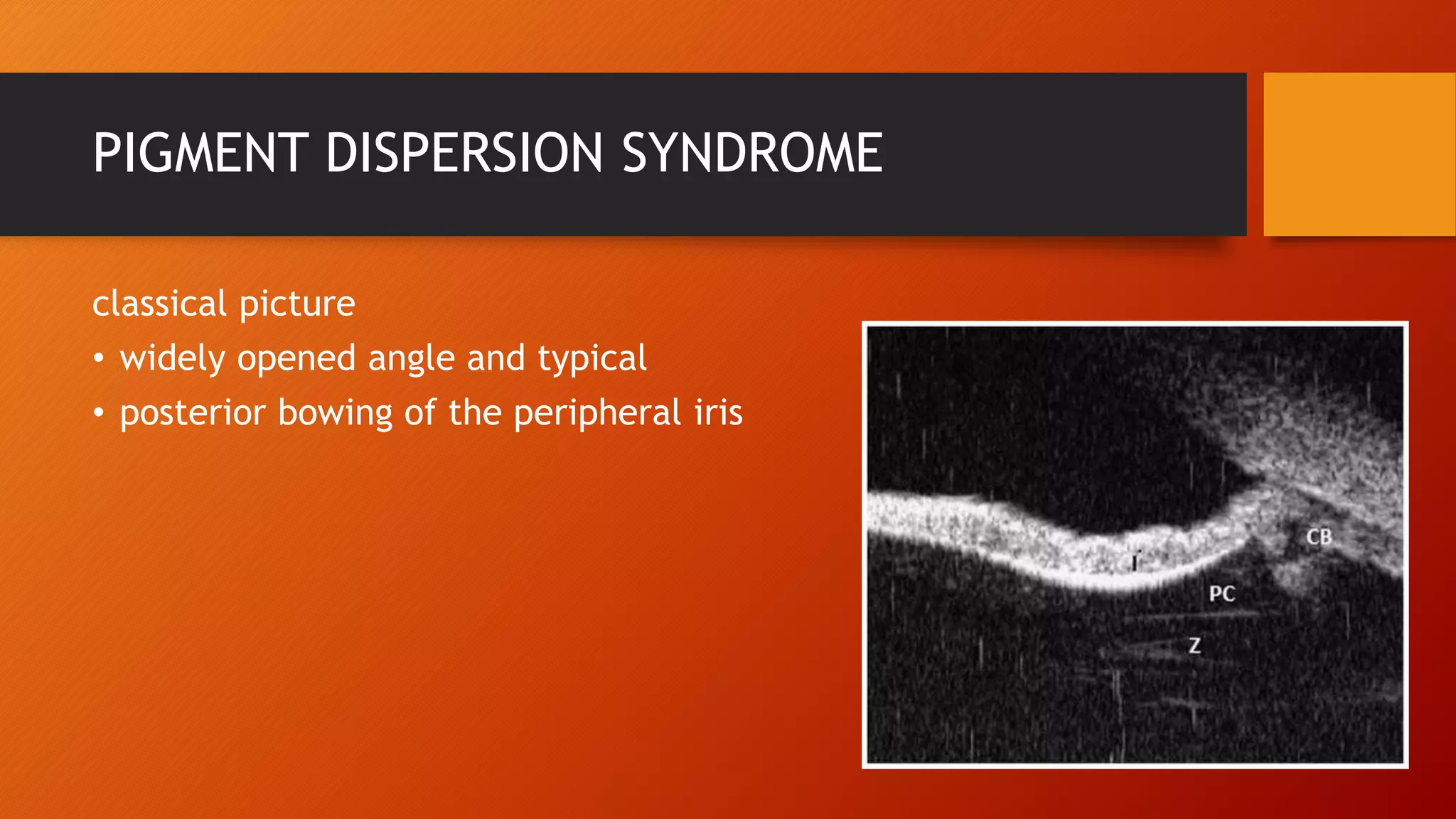
• Ultrasound biomicroscopy (UBM) provides high-resolution imaging
of ocular structures anterior to the pars plana region of the eye
• Developed by Pavlin, Sherar and Foster in Toronto in the late
1980s
• Provides exceptionally detailed two-dimensional gray-scale
images of the various anterior segment structures and evaluates
them both quantitatively and qualitatively
PRINCIPLE
PRINCIPLE
• It acts on a principle similar to that of the B-scan (10Mhz)
• Frequency 50Mhz
• More frequency, less penetration(5mm) and more resolution
• Limited depth of penetration is also associated with a smaller
angular field (4x4 mm)
• addition to the tissues easily seen using conventional methods (ie,
slit lamp), such as the cornea, iris, and sclera, structures including
the ciliary body and zonules, hidden from clinical observation,
can be imaged and their morphology assessed
TECHNIQUE
TECHNIQUE
There are three main components of the UBM machine.
1.Transducer
2. High-frequency signal processing.
3. Video monitor
Transducer 50 MHz
This radiofrequency travels the body tissue and is
reflected back to the transducer. The reflected
radio frequency is processed by the signal
processing unit.
Signal processing unit
 The signal processing unit in UBM is specially
designed to handle high frequency signals.
 Subtle movements.
 Special motion control device.
 Mounted on a pulley with the piezoelectric
crystal fixed on a large handle
Patient in supine position
Local anesthetic
Eye cup (plastic or silicone) which is
used to create a small water bath
Methyl cellulose or normal saline can
be used as coupling solution
The reflected signal is best detected
when the transducer is oriented so
that the ultrasound beam strikes the
targeted surface perpendicularly
The crystal of the transducer is placed
in saline approximately 2 mm from
the eye surface.
(This distance of 2 mm prevents injury
to the cornea and also helps as a fluid
standoff.)
NORMAL OCULAR STRUCTURES
Epithelium
Bowmanns membrane
Stroma
Endothelium and DM
The Scleral Spur Can Be Identified In The Region Where The Radiopaque Shadow Of The Sclera
Merges With The Relatively Radiolucent Shadow Of The Cornea And Is Often Visible As A Change In
The Configuration When The Inner Border As The Sclera Merges Into The Cornea.
APPLICATIONS
QUALITATIVE STUDIES AND QUANTITATIVE STUDIES
Quantitative Ultrasound Biomicroscopy
• Quantitative studies includes Simple measurements methods such as
a) Distance
b) Angle measurement
Biometry of the Anterior Segment
Corneal thickness
Anterior chamber depth
Posterior chamber depth
IOL thickness
Iris thickness
Ciliary body thickness
Scleral thickness .
Cannot determine lens thickness
(cannot penetrate till post capsule of lens)
Measurement of AC angle
Qualitative Ultrasound Biomicroscopy
• Glaucoma
• Cornea
• Tumors
• Trauma
• sclera
• Intraocular lenses
• Ocular adnexa
GLAUCOMA
open-angle glaucoma
• can be used to measure the anterior
chamber angle in degrees
• assess the configuration of the
peripheral iris
• evaluate the iris insertion in relation to
the trabecular meshwork
• see if there is an anterior insertion of
the iris or an anteriorly displaced
ciliary body
Narrow angles
• UBM shows the extent of angle
closure
• depth of the anterior and posterior
chambers
• identifies pathologic processes
pushing the lens and iris forward.
determine the mechanism of elevated intraocular pressure
(angle-closure versus open-angle) by showing the
relationship between the peripheral iris and trabecular
meshwork.
PUPIL BLOCK GLAUCOMA
(A) The angle shows appositional closure owing to anterior
bowing of the iris.
(B) The angle is open with a flattened iris after laser peripheral
iridotomy.
PLATEAU IRIS
Plateau iris has been defined based on UBM by Kumar
et al if all criteria fulfilled in at least 2 quadrants:
1.The ciliary process was anteriorly directed, supporting
the peripheral iris so that it was parallel to the
trabecular meshwork.
2. The iris root had a steep rise from its point of
insertion, followed by a downward angulation from the
corneoscleral wall.
3. Presence of a central flat iris plane.
4. An absent ciliary sulcus.
5. Irido-angle contact (above the level of the scleral
spur) in the same quadrant.
Kumar, et al. Prevalence of plateau iris in primary angle closure
suspects, a UBM study. Ophthalmology 2008;115:430-34.
To Determine Occludability of the Angle
• perform dark room provocative testing with the UBM, to study the
spontaneous occlusion of the angle under conditions of decreased
illumination
• Helps to identify “at risk” population which can then be subjected to a
laser iridotomy
• better than dark room gonioscopy because the latter is time consuming
and standardization of slit-lamp illumination is difficult
Congenital glaucoma
Common features-
• thin stretched out ciliary body,
• abnormal tissue at the irido corneal
angle
• abnormal insertion of the ciliary body
PIGMENT DISPERSION SYNDROME
classical picture
• widely opened angle and typical
• posterior bowing of the peripheral iris
Peripheral anterior synechiae
• reveal the extent of irido corneal
adhesions, even if the cornea is hazy or
opaque
Malignant glaucoma
• Extremely shallow anterior chamber,
• occluded angle,
• forward rotation of the ciliary body
• with or without fluid in the suprachoroidal
space.
Functional Status of a Filtering Surgery
• whether the sclerostomy aperture is patent
or blocked internally,
• whether the peripheral iridectomy is patent
• whether the filtering bleb is flat, shallow or
deep
CORNEA
Graft host junction Epithelial bullae in oedema Corneal dystrophy
Lamellar keratoplasty Stromal scarring Adherent leucoma
Granular dystrophy
TUMOURS
• Iris tumors, ciliary body tumors and anterior choroidal tumors can
be imaged
• Borders of the tumor are usually detectable by the change in
reflectivity from surrounding structures
CHOROIDAL TUMOUR Irido ciliary cyst
The most common
clinical presentation of
an irido-ciliary cyst is a
peripheral iris elevation -
the typical UBM finding of
a thin walled structure
with no internal
reflectivity is diagnostic.
TRAUMA
CYCLODIALYSIS
Angle recession is imaged as a tear into the face of
the ciliary body. Ciliary body tissue is still imaged
attached to the scleral spur.
Zonular rupture
Hemorrhagic cyst within the iris
SCLERA
EPISCLERITIS
INTRAOCULAR LENSES
• Can be helpful in analyzing intraocular lens position and determining
the source of the problem if all does not go well
• Anterior chamber depth after surgery can be measured with a high
degree of accuracy.
• margins of the optic can be easily imaged and decentration analysed
• Haptic location in relationship to surrounding structures can be
determined and, in the case of posterior chamber lenses, one can
usually determine if the haptic is in the capsular bag
iris isadherent to a membrane
on the surface of the lens
Conjunctival and Adnexal Disease
UBM AS-OCT
• contact technique • Non contact technique
• Coupling media used • Not used
• Sound energy used • Light energy used
• Scleral spur less distinct • More distinct
• Posterior lens capsule not seen • Seen
• patient supine, positioning theoretically causes the
iris diaphragm to fall back deepens the AC and opens
the angle.
• Patient sitting,so no alterations
• pressure on the eyecup used while scanning can
influence angle configuration
• Nil
• Only 1 quadrant can be imaged at a time • 4 quadrants can be scanned at once.
UBM AS-OCT
• there is no fixed reference point and the angle region
measured is located subjectively as nasal, temporal,
superior, inferior, and so forth, not in exact degrees of
an arc
• With AS-OCT, keeping the fixation angle (the angle
between the instrument’s optical axis and the eye’s
line of sight) at 0 degree, finding the exact location of
the measured angle in degrees of an arc is possible.
• UBM procedureis more time consuming and requires a
highly skilled operator to obtain high-quality precision
images.
• No skills required
Limitations
• risk for infection or corneal abrasion because of the
contact nature of the examination
• it is contraindicated in suspected open-globe injuries.
Limitations
• AS-OCT are that it cannot obtain clear images through
opaque media and is obstructed by the eyelids,
making imaging of the superior and inferior angles
difficult
• also provides limited visualization of the ciliary body.
LIMITATIONS
• The most important limitation of UBM is depth.UBM cannot
visualize structures deeper more that4 mm from the surface.
• The other limitation is that UBM cannot be performed in presence
of an open corneal or scleral wound.
THANK YOU

ultrasound biomicroscopy

  • 1.
  • 2.
    LAYOUT • INTRODUCTION • PRINCIPLE •TECHNIQUE • NORMAL ANATOMICAL STRUCTURES • APPLICATIONS • UBM VS AS OCT • LIMITATIONS
  • 3.
  • 4.
    INTRODUCTION • Ultrasound biomicroscopy(UBM) provides high-resolution imaging of ocular structures anterior to the pars plana region of the eye • Developed by Pavlin, Sherar and Foster in Toronto in the late 1980s • Provides exceptionally detailed two-dimensional gray-scale images of the various anterior segment structures and evaluates them both quantitatively and qualitatively
  • 5.
  • 6.
    PRINCIPLE • It actson a principle similar to that of the B-scan (10Mhz) • Frequency 50Mhz • More frequency, less penetration(5mm) and more resolution • Limited depth of penetration is also associated with a smaller angular field (4x4 mm) • addition to the tissues easily seen using conventional methods (ie, slit lamp), such as the cornea, iris, and sclera, structures including the ciliary body and zonules, hidden from clinical observation, can be imaged and their morphology assessed
  • 8.
  • 9.
    TECHNIQUE There are threemain components of the UBM machine. 1.Transducer 2. High-frequency signal processing. 3. Video monitor
  • 10.
    Transducer 50 MHz Thisradiofrequency travels the body tissue and is reflected back to the transducer. The reflected radio frequency is processed by the signal processing unit. Signal processing unit  The signal processing unit in UBM is specially designed to handle high frequency signals.  Subtle movements.  Special motion control device.  Mounted on a pulley with the piezoelectric crystal fixed on a large handle
  • 11.
    Patient in supineposition Local anesthetic Eye cup (plastic or silicone) which is used to create a small water bath Methyl cellulose or normal saline can be used as coupling solution The reflected signal is best detected when the transducer is oriented so that the ultrasound beam strikes the targeted surface perpendicularly The crystal of the transducer is placed in saline approximately 2 mm from the eye surface. (This distance of 2 mm prevents injury to the cornea and also helps as a fluid standoff.)
  • 15.
  • 16.
    NORMAL OCULAR STRUCTURES Epithelium Bowmannsmembrane Stroma Endothelium and DM
  • 17.
    The Scleral SpurCan Be Identified In The Region Where The Radiopaque Shadow Of The Sclera Merges With The Relatively Radiolucent Shadow Of The Cornea And Is Often Visible As A Change In The Configuration When The Inner Border As The Sclera Merges Into The Cornea.
  • 18.
  • 19.
    Quantitative Ultrasound Biomicroscopy •Quantitative studies includes Simple measurements methods such as a) Distance b) Angle measurement
  • 20.
    Biometry of theAnterior Segment Corneal thickness Anterior chamber depth Posterior chamber depth IOL thickness Iris thickness Ciliary body thickness Scleral thickness . Cannot determine lens thickness (cannot penetrate till post capsule of lens)
  • 21.
  • 23.
    Qualitative Ultrasound Biomicroscopy •Glaucoma • Cornea • Tumors • Trauma • sclera • Intraocular lenses • Ocular adnexa
  • 24.
    GLAUCOMA open-angle glaucoma • canbe used to measure the anterior chamber angle in degrees • assess the configuration of the peripheral iris • evaluate the iris insertion in relation to the trabecular meshwork • see if there is an anterior insertion of the iris or an anteriorly displaced ciliary body Narrow angles • UBM shows the extent of angle closure • depth of the anterior and posterior chambers • identifies pathologic processes pushing the lens and iris forward. determine the mechanism of elevated intraocular pressure (angle-closure versus open-angle) by showing the relationship between the peripheral iris and trabecular meshwork.
  • 25.
    PUPIL BLOCK GLAUCOMA (A)The angle shows appositional closure owing to anterior bowing of the iris. (B) The angle is open with a flattened iris after laser peripheral iridotomy.
  • 26.
    PLATEAU IRIS Plateau irishas been defined based on UBM by Kumar et al if all criteria fulfilled in at least 2 quadrants: 1.The ciliary process was anteriorly directed, supporting the peripheral iris so that it was parallel to the trabecular meshwork. 2. The iris root had a steep rise from its point of insertion, followed by a downward angulation from the corneoscleral wall. 3. Presence of a central flat iris plane. 4. An absent ciliary sulcus. 5. Irido-angle contact (above the level of the scleral spur) in the same quadrant. Kumar, et al. Prevalence of plateau iris in primary angle closure suspects, a UBM study. Ophthalmology 2008;115:430-34.
  • 27.
    To Determine Occludabilityof the Angle • perform dark room provocative testing with the UBM, to study the spontaneous occlusion of the angle under conditions of decreased illumination • Helps to identify “at risk” population which can then be subjected to a laser iridotomy • better than dark room gonioscopy because the latter is time consuming and standardization of slit-lamp illumination is difficult
  • 28.
    Congenital glaucoma Common features- •thin stretched out ciliary body, • abnormal tissue at the irido corneal angle • abnormal insertion of the ciliary body
  • 29.
    PIGMENT DISPERSION SYNDROME classicalpicture • widely opened angle and typical • posterior bowing of the peripheral iris
  • 30.
    Peripheral anterior synechiae •reveal the extent of irido corneal adhesions, even if the cornea is hazy or opaque
  • 31.
    Malignant glaucoma • Extremelyshallow anterior chamber, • occluded angle, • forward rotation of the ciliary body • with or without fluid in the suprachoroidal space.
  • 32.
    Functional Status ofa Filtering Surgery • whether the sclerostomy aperture is patent or blocked internally, • whether the peripheral iridectomy is patent • whether the filtering bleb is flat, shallow or deep
  • 33.
  • 34.
    Graft host junctionEpithelial bullae in oedema Corneal dystrophy Lamellar keratoplasty Stromal scarring Adherent leucoma
  • 35.
  • 36.
    TUMOURS • Iris tumors,ciliary body tumors and anterior choroidal tumors can be imaged • Borders of the tumor are usually detectable by the change in reflectivity from surrounding structures
  • 37.
    CHOROIDAL TUMOUR Iridociliary cyst The most common clinical presentation of an irido-ciliary cyst is a peripheral iris elevation - the typical UBM finding of a thin walled structure with no internal reflectivity is diagnostic.
  • 38.
  • 39.
    Angle recession isimaged as a tear into the face of the ciliary body. Ciliary body tissue is still imaged attached to the scleral spur. Zonular rupture
  • 40.
  • 41.
  • 43.
    INTRAOCULAR LENSES • Canbe helpful in analyzing intraocular lens position and determining the source of the problem if all does not go well • Anterior chamber depth after surgery can be measured with a high degree of accuracy. • margins of the optic can be easily imaged and decentration analysed • Haptic location in relationship to surrounding structures can be determined and, in the case of posterior chamber lenses, one can usually determine if the haptic is in the capsular bag
  • 44.
    iris isadherent toa membrane on the surface of the lens
  • 45.
  • 47.
    UBM AS-OCT • contacttechnique • Non contact technique • Coupling media used • Not used • Sound energy used • Light energy used • Scleral spur less distinct • More distinct • Posterior lens capsule not seen • Seen • patient supine, positioning theoretically causes the iris diaphragm to fall back deepens the AC and opens the angle. • Patient sitting,so no alterations • pressure on the eyecup used while scanning can influence angle configuration • Nil • Only 1 quadrant can be imaged at a time • 4 quadrants can be scanned at once.
  • 48.
    UBM AS-OCT • thereis no fixed reference point and the angle region measured is located subjectively as nasal, temporal, superior, inferior, and so forth, not in exact degrees of an arc • With AS-OCT, keeping the fixation angle (the angle between the instrument’s optical axis and the eye’s line of sight) at 0 degree, finding the exact location of the measured angle in degrees of an arc is possible. • UBM procedureis more time consuming and requires a highly skilled operator to obtain high-quality precision images. • No skills required Limitations • risk for infection or corneal abrasion because of the contact nature of the examination • it is contraindicated in suspected open-globe injuries. Limitations • AS-OCT are that it cannot obtain clear images through opaque media and is obstructed by the eyelids, making imaging of the superior and inferior angles difficult • also provides limited visualization of the ciliary body.
  • 49.
    LIMITATIONS • The mostimportant limitation of UBM is depth.UBM cannot visualize structures deeper more that4 mm from the surface. • The other limitation is that UBM cannot be performed in presence of an open corneal or scleral wound.
  • 50.

Editor's Notes

  • #25 In addition, imaging of the anterior segment structures is possible even in eyes with corneal edema or corneal opacification that precludes gonioscopic assessment.
  • #26 PUPILLARY BLOCK AND FOLLOWING IRIDECTOMY. A space is present between the ciliary processes and poste­rior iris.
  • #30 irido-zonular contact
  • #33 The grading of the bleb is done according to intrableb reflectivity, visibility of route under the scleral flap, formation of cavernous fluid filled space, and bleb height. Following four grades have been described: • Low reflective L • High reflective H • Encapsulated E • Flat F Eyes with good IOP control mainly have ’L’ type blebs, these have low to moderate intrableb reflectivity, visible intrascleral route and higher intrableb height. Flat and encapsulated blebs generally denote a surgical failure.