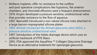
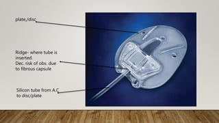
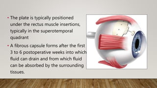
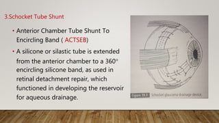
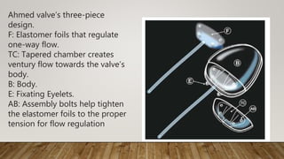
SlideShare a Scribd company logo
1 of 91
GLAUCOMA DRAINAGE DEVICES
DR.M.DINESH
Glaucoma drainage devices (GDDs)
Principle
• work by creating an alternate pathway for aqueous outflow by
• channeling aqueous from AC through a tube of implant
• towards sub-conjunctival space or suprachoroidal space
• where it is absorbed into systemic circulation by diffusion and
through scleral veins.
Drainage Devices
• Setons (latin -Bristle) –
• Refers to solid (non hollow )shaft prevents wound
apposition facilitating bulk flow by surface tension
• unsuccessful as they were unable to maintain fistula
patency
• Sources of Seton: Silk, Horse hair, gold, Tantalum wire,
platinum, gelatin, Silicon, PMMA.
• Stunt (Tubular structure) - a passive tubular structure
incapable of influencing either antegrade or retrograde flow.
• Valve - Tubular structure for unidirectional flow
History
• 1907 :The first attempt to implant a drainage device was made by
Rollet and Moreau ,when they performed a double paracentesis and
used horse hair through the corneal punctures to treat patients with
painful absolute glaucoma.
• 1912: The first translimbal glaucoma drainage device implanted by
Zorab ;
device used silk thread to drain fluid
• 1969 :Molteno – 1st scientifically explained the pathophysiology of
bleb resistance and designed a tube. All of the currently available
GDD are based on fundamentals of Molteno implants.
• Molteno implants, offer no resistance to the outflow
and post operative complications like hypotony, flat anterior
chambers, and choroidal effusions were a frequent phenomemon.
• 1976 :Krupin developed a pressure sensitive, unidirectional valve
that provides resistance to the flow of aqueous.
• 1992 :Baerveldt introduced a non-valved silicone tube attached to
a large barium-impregnated silicone platen
• 1993 :Ahmed introduced the Ahmed glaucoma valve (AGV), a
pressure sensitive unidirectional valve.
• 1997: Introduction of the Helies drainage device which uses an
artificial meshwork of PTFE fibers
• 2001: FDA approved the AquaFlow ™ Collagen Glaucoma Drainage
Device as an alternative treatment for openangle glaucoma
Implants can be studied under
1. Type of plate
2. Type of material
3. Type of opening
1.Type of plate – of the available rigid and flexible plates the later
causes less inflammation
2.Type of material
• Most commonly used materials are
• Silicon -Baerveldt, Krupin, Ahmed
• Polypropylene -Ahmed, Molteno
• Hydroxylapatite
• Expandable polytetrafluoroethylene
3.Types of opening
1.GDDs with no resistance
A)Non-valved/Non-restrictive implants/Open-Tube Drainage
devices
• Rely on resistance formed by fibrous capsule or bleb
• Bleb grows around the implant and creates a space for fluid to
drain and be absorbed by surrounding tissue
1. Molteno
2. Bareveldt
3. Schocket
4. Express miniature glaucoma shunt R 50
5. Istent from glaukos
B)Valved/Flow Restrictive implants:
• Only drains fluid at a certain IOP
• Valve opens and fluid is drained into a reservoir where it is
absorbed by surrounding tissues
1. AGV
2. Krupin slit valve
3. Others Joseph , Optimed &White GDD
2.GDD with variable resistance:
1. Molteno dual ridge device
2. Baerveldt bioseal (is a flap that overhangs the silicone tube as it
opens on the endplate )
3. Deep light gold micro shunt from SOLX (suprachoroidal shunt)
Ref :35 Glaucoma drainage devices DOS times vol .13 issue no 8 Feb 2008
Indications :
• One or more filtering procedures that have failed
• Refractory infantile glaucoma
• Neovascular glaucoma
• Traumatic glaucoma
• Uveitic glaucoma
• Glaucoma in aphakia or pseudophakia
• Glaucoma following corneal transplantation
• Glaucoma with Penetrating keratoplasty
• Glaucoma with Retinal detachment surgery
• Glaucoma and aniridia
• Glaucoma and Sturge-Weber syndrome
• Glaucoma and ICE syndrome
• Glaucoma and contact lens dependence
• Glaucoma and epithelial ingrowth
• Glaucoma and severe surface disease (chemical burns, pemphigoid)
Contraindications:
• Eyes with severe scleral or sclera-limbal thinning
• Extensive fibrosis of conjunctiva
• Ciliary block glaucoma.
Relative Contraindications:
• Vitreous in AC
• Intra-ocular silicone oil-Implant if required is placed in infero
temporal quadrant
PHYSIOLOGY OF DRAINAGE IMPLANTS
Basic design :
• consists of a silicone tube that extends from the anterior chamber (or,
in some cases, the vitreous cavity) to a plate, disc, or encircling element
beneath conjunctiva and Tenon capsule.
• The edge of the external plate has a ridge, through which the distal
end of the tube inserts onto the upper surface of the plate.
• The ridge decreases the risk for obstruction of the posterior opening of
the tube with the surrounding tissue and fibrous capsule.
• The plates of the glaucoma drainage devices have large surface areas
and promote the formation of the filtering bleb posteriorly, near the
equator.
Silicon tube from A.C
to disc/plate
Ridge- where tube is
inserted.
Dec. risk of obs. due
to fibrous capsule
plate,/disc
The mechanism by which drainage implant devices control the IOP
relates to
• fibrous capsule that forms a filtering bleb around the external portion
of the draining device and,
• the surface area of the implant plate.
After insertion, collagenous capsule forms around it
Surrounded by a granulomatous reaction
Reaction resolves in 4 months
Capsule remains stable
Matures over time, becomes thinner after 6 months
Filtering bleb
• The vast majority of glaucoma drainage devices develop an elevated IOP in
the weeks to months after implantation as a result of capsule formation
around the implant plate. This is termed as the hypertensive phase.
• The filtering bleb may fail after surgery due to the increased thickness of
fibrous capsule around the drainage implant.
• Movement of the drainage plate against the scleral surface may be the
mechanism of glaucoma implant failure resulting from the stimulation of
the low-level wound healing response, increased collagen scar formation,
and increased fibrous capsule thickness
1.MOLTENO IMPLANT
Original design –
Single acrylic thin plate:13 mm diameter
135 mm² S.A
Silicon tube connected to upper end of plate
external diameter-0.62 mm
internal diameter-0.30 mm
Thickened rim which is perforated facilitating suturing to sclera
With subsequent modification came
a) Single plate
b) Double plate
c) Dual chamber
d) Molteno 3
a)Single plate
Moved to a position a few millimetres
away from limbus to allow for
better drainage.
b)Double plate molteno implant:
• A double-plate Molteno implant combines two
plates, one of which is attached to the silicone
tube in the anterior chamber,
• a second tube connects the two plates, giving an
increased surface area of 270 mm2
• Better control but inc. complication – hypotony
c)Dual chamber:
• single-plate implant, in
which a V-shaped “pressure
ridge” is present on the
upper surface of the plate
• In view to combat hypotony
from over-filtration.
• The V-chamber implant contains a thin V-shaped rim of
polypropylene on the plate surface below the tube's entry,
• Encases an area of 10.5 mm2 around the opening of the silicone tube
• Since this V-shaped ridge is the same height as the circumferential rim
of the plate, the weight of the overlying, hydrated Tenon's tissue will
in theory collapse and form a temporary roof for the smaller V-
chamber,
• thus limiting the extent of the bleb and filtration
d)Molteno 3
• has a bowl shaped structure on
the implant plate immediately at
the opening tube.
• It is designed to function as a
biologic valve by limiting the
available area of filtration during
times of low aqueous
production.(limiting aq. Flow )
• is available with plate sizes of 175
and 230mm2
2 .Baerveldt implant:
• A silicone tube is attached to a silicone
plate (barium impregnated )with a
surface area of 250 mm2, 350 mm2 or
500 mm2.
• The Baerveldt plate has fenestrations
that allow growth of fibrous tissue
through the plate, serving to reduce
the height of the bleb, which reduces
the risk for diplopia and helps secure
the implant.
• The plate is typically positioned
under the rectus muscle insertions,
typically in the superotemporal
quadrant
• A fibrous capsule forms after the first
3 to 6 postoperative weeks into which
fluid can drain and from which fluid
can be absorbed by the surrounding
tissues.
3.Schocket Tube Shunt
• Anterior Chamber Tube Shunt To
Encircling Band ( ACTSEB)
• A silicone or silastic tube is extended
from the anterior chamber to a 360°
encircling silicone band, as used in
retinal detachment repair, which
functioned in developing the reservoir
for aqueous drainage.
Modifications:
• have included insertion of the tube into a band extending
for only 90° beneath two recti muscles or
• into the preexisting encircling band in eyes with glaucoma
after scleral buckling surgery
4.Ex-PRESS
• Miniature non-valved device without an external plate
• Made of rigid stainless steel – same as cardiac stents
• < 3mm long
• Internal lumen size – 50µm/200µm
• Biocompatible
• Allows for the development of a delimited potential space
and the formation of a fibrous capsule to create the
resistance to outflow
• Implanted under a traditional trabeculectomy flap
5.Glaukos istent
• This is a light weight Titanium L-shaped device which is placed
inside Schlemm's canal
• The heparin covered titanium is biocompatible, has thrombolytic
activity and prevents stenosis.
• A small, snorkel shaped tube, about 0.5 mm in length sits in the
peripheral AC, allowing aqueous to bypass the inner wall of
Schlemm's canal and the juxtacanalicular trabecular meshwork.
• The portion of the device that is placed inside the canal is 1 mm in
length and is shaped like a half-pipe.
• It is designed to fit within the lumen of the Schlemm’s canal, with
the curved convex side lying against the inner wall of Schlemm's
canal.
• This avoids contact with the outer wall and the collector channel
orifices that enter the outer wall.
• The three barbed ridges along this portion are designed to
prevent loosening and provide a secure placement of the stent in
the canal.
• These can either be right handed or left handed
Insertion :
• A temporal clear corneal incision is made, and the AC is filled with
viscoelastic.
• An applicator grasps the device and under gonioscopic guidance,
traverses the AC to reach the Schlemm's canal in the nasal
quadrant.
• The pointed tip engages the trabecular meshwork and the stent is
inserted in place.
Surgical procedure for implantation of the iStent
B)Valved/Flow Restrictive implants:
1.Ahmed Glaucoma Valve (AGV)
• Most commonly used valved implant
• Implant design – silicon tube connected to silicon sheathed valve
held by a polypropylene body
Mechanism :
• The aqueous is transported to the valve’s body, where it has to pass
between two silicone elastomer foils, held in place by the
polypropylene piece at a certain tension
• The initial pressure needed to break the adherence of these
membranes often exceeds 100 mm Hg, but afterwards they separate
whenever the
pressure is over 12 mm Hg, and close when the pressure falls to 8 or
less: this mechanism permits one-way flow only.
Ahmed valve’s three-piece
design.
F: Elastomer foils that regulate
one-way flow.
TC: Tapered chamber creates
ventury flow towards the valve’s
body.
B: Body.
E: Fixating Eyelets.
AB: Assembly bolts help tighten
the elastomer foils to the proper
tension for flow regulation
Priming:
• Ahmed’s valve must be primed
before use.
• A 27-gauge cannula is inserted 2
to 3 mm into the tube and BSS is
injected until it separates the
elastomer foils often a small
“popping” sound is heard
while the BSS separates the
silicone foils.
Material Surface
Area
Type
Silicon
FP7 84mm² Single
valve
plate
FP8 96 mm² Small
valve
plate
FX1 180mm
²
2 plates
Polymeth
ylmethac
rylate
Polyprop
ylene
S2 184mm
²
Single
valve
plate
S3 96 mm² Small
valve
plate
B1 180mm 2 plates
Hypertensive phase:
• transient phase of low capsule permeability seen at 4 to 8 weeks
postoperatively.
“No-touch zone” on the Ahmed glaucoma valve:
• It is the area of the implant covering the chamber with the silicone
leaflets.
• If the implant is grasped with forceps along the center line
• it may separate the valve cover from the implant.
• can cause a defect in closure of the valve - early postoperative
hypotony and fibrovascular membrane ingrowth between the
leaflets.
• lead to a failure of the valve due to adhesion of the valve
membranes.
2.Krupin Implant
• The original Krupin Denver valve was composed of
• an internal Supramid tube cemented to an external silastic tube
• The valve effect was created by making slits in the closed external end
of the silastic tube, designed to open at an IOP between 9 and 11 mm
Hg.
Failure
• The tube was short extending only a few mm subconjunctivally,
• no external plate.
• Fibrosis eventually closed the subconjunctival portion of the valved
tube
Modifications:
Krupin eye valve with disc
• a long Krupin-Denver drainage tube
with the one way valve design was
attached to a 180o Schocket type
scleral explant
• In the newer design of the Krupin
implant, the valve lies inside the rim of
the plate at its insertion and is
exposed directly to the sub
conjunctival tissues
2.GDD with variable resistance:
Gold micro-shunt
• The deep light glaucoma treatment system includes a
titanium sapphire laser and a photo titratable gold
micro shunt.
• The laser emits microsecond infrared light pulses
that passes through the trabecular meshwork tissues,
producing significant opening of the trabecular
meshwork, allowing increased aqueous outflow.
• The gold shunt is biocompatible and inert, made of
99.5 percent pure gold. It is a flat plate 5.2 mm long,
2 mm wide and 60 thick, containing multiple
microchannels.
• Initially, half of these microchannels are open and the remainder closed
by a thin film of gold which can be opened after implantation, using the
titanium sapphire laser.
• This reactivates the shunt's effect and additional drop in IOP can be
obtained.
• This phototitration can be done at any time postoperatively.
• The shunt is implanted through a 3 mm clear corneal incision made at
the limbus into the suprachoroidal space using a preloaded insertion
device.
• The channels in the shunt form a bridge between the anterior chamber
and suprachoroidal space
Surgical Procedure- Basic Principles
• Tractional suture:A 6-0 Vicryl or silk
traction suture is placed through superficial
cornea near the superior limbus and
attached to the drape beneath the eye.
• Conjunctival flap:A fornix-based
conjunctival-Tenon capsule flap is created,
usually in the superotemporal quadrant, to
expose the scleral bed. The flap is slightly
elevated to allow for blunt dissection
between Tenon and episclera with blunt
Westcott scissors.
Isolation of recti:A muscle hook is then
used to isolate the two rectus muscles on
either side of the surgical site.
Placement of implant:The external plate is
then tucked posteriorly into the sub-Tenon
space and is sutured to sclera with
nonabsorbable 9-0 Prolene or nylon
sutures through the anterior positional
holes of the plate,with the anterior border
at 8 to 10 mm posterior to the limbus. The
tube is then cut, bevel up, to permit its
extension 2 to 3 mm into the anterior
chamber
• Before the tube is inserted into the
anterior chamber, a limbal area is
cauterized to prevent bleeding from the
insertion.
• Entering into AC:The anterior chamber is
then entered through the cauterized
limbal area with a 23-gauge or a 22-
gauge needle, parallel to the iris plane .
The needle creates a watertight seal,
preventing leakage around the tube and
thus reducing the risk for postoperative
hypotony
• Insertion of the tube:The tube is then
inserted into the anterior chamber
via the needle track.The tube can be
secured to the sclera by using a
nonabsorbable suture,
• Patch graft:The tube may
occasionally erode through both
sclera and overlying conjunctiva at
the limbus. To avoid this potential
complication, a rectangle of
preserved donor tissue of
approximately 5 mm × 7 mm over
the tube at the limbus.
• Processed pericardium (Tutoplast), donor
sclera, dura, and fascia lata are available
commercially for this purpose.
• It is also possible to use autologous sclera
or to place the tube under a partial-
thickness scleral flap
• Closure :The conjunctiva is then sutured
back to its original position using Vicryl
sutures.
• Subconjunctival steroids and antibiotics
are injected at the completion of the
procedure in a quadrant away from the
surgical site.
• Non-valved GDD: To avoid post-operative hypotony, a two
stage implantation procedure can be adopted.
• The tube is inserted 6-8 weeks following the positioning of the
external
plate allowing the formation of a fibrous capsule around the plate
prior to the insertion of the tube into the AC .
• A more commonly used method consists of occlusion of the tube
lumen with an absorbable suture like vicryl.
COMPLICATIONS
Intraoperative Complications
a. Bleeding
• Occurs typically in neovascular glaucoma
• Large hyphemas must be evacuated as they can
block the tube.
b. Misdirection of the silicone tube into the
posterior chamber may occur in the
presence of peripheral anterior synechiae.
c. Loss of Aq may occur if the port size is
bigger than tube.
• This can be remedied by introduction of
saline or viscoelastic through a previously
placed paracentesis opening and choosing
23 or 24 gauge needle.
Early Postoperative Complications
a. Hypotony with or without associated choroidal
effusions.
• Small effusions may be left to resolve
spontaneously.
• Large effusions resulting in kissing choroids may
have to be evacuated.
• Hypotony is best treated by prevention, either by
the use of valved implants or by occlusion of the
silicone tube with a stent and/or a constricting
ligature.
b. Increased IOP may occur due to occlusion of the silicone tube:
1. The opening of the tube may be obstructed by iris -can be treated
by YAG ablation of the iris tissue.
2. Occlusion by vitreous in aphakes can occur -can be prevented by
thorough vitrectomy prior to tube insertion.
c.Corneal Decompensation:
• may be due to the retrograde
flow from the encapsulated
reservoir to the anterior
chamber.
• Tube-cornea touch is another
cause of corneal
decompensation.
d. Early postop endophthalmitis is a rare complication.
• It can be treated by immediate removal of the implant and
surgical management of the infection, with subsequent
placement of a new implant.
Late Complications
a. Encysted bleb can form.
• Deflation of the bleb to allow compressed channels in the wall of the
bleb to expand and reestablish drainage may be necessary.
• Encysted blebs contain a relatively large amount of aqueous and as
much as 1 cm3 of aqueous may be withdrawn to deflate the bleb
with no loss of anterior chamber. Also as these blebs are relatively
avascular, needling them is less traumatic.
• A regimen consisting of diclofenac 75 mg daily,prednisolone 40 mg
daily and topical corticosteroids may be helpful if given not later than
14 days postoperatively and continued for at least six weeks.
b. Erosion of the silicone tube through the sclera or scleral
patch and conjunctiva may occur.
c. Plate migration may occur if the body is not fixed properly.
• If the plate migrates towards the medial rectus muscle insertion,
myositis may develop.
• It resolves after implant removal
d. Limitation of eye movements can occur
specially when placed in upper nasal
quadrant.
• The movement most commonly affected is
upgaze.
• Other patterns are exotropia, hypertropia
and, limitations of ocular rotations.
• Placement of the implants in the lower
fornix restricts downgaze with associated
diplopia.
e. Endophthalmitis: Exposure seems to be the most important
risk factor for these infections.
• Surgical revision with a patch graft is indicated in all these
cases to prevent endophthalmitis.
Other possible complications are
• Epithelial downgrowth: It can cause implant failure, corneal
decompensation, and formation of a true Tenon's cyst.
• Epithelial invasion into the fibrous capsule with persistent aqueous leak.
• Sterile hypopyon.
• Irregular pupil many years later due to adherence of the iris root to the
tube.
• Globe perforation while suturing the plate to the sclera causing retinal
detachment or vitreous hemorrhage.This is seen more commonly in high
myopes with thin sclera.
• Retinal complications include RD,suprachoroidal hemorrhage, vitreous
hemorrhage,choroidal effusion.
Outcomes
• Long term studies revealed success rates 65 to 85% in achieving
IOP control.
• Molteno implants, in general, lower IOP more than Ahmed
implants but have higher risk of complications and need for
resurgery.
• Success is lower in pediatric glaucomas, with failure rates of more
than 30%, and most of them requiring other surgeries.
• Drainage devices have been particularly successful in neovascular
glaucoma (success rates 62% at 1 yr but diminished with time).
Tube vs trabeculectomy (TVT) study
• Multicenter RCT comparing tube(350 sq.mm Baerveldt implant)
shunts with trabeculectomy with mitomycin-C(MMC) in eyes with
previous cataract and/failed glaucoma surgery.
• 1st year of follow-up, the tube patients were more likely to maintain
IOP and avoid hypotony or loss of light perception or resurgery as
compared to trabeculectomy patients. However tube patients
needed more supplemental therapy.
• 5 yrs of follow-up, IOP control was found to be similar in both
groups but the trabeculectomy group needed additional surgery
more than the tube group.
The Hydrus Microstent :
• trabecular bypass device and Schlemm’s canal scaffold
• nitinol, an alloy of nickel and titanium.
• 8-mm-long crescent with an open posterior surface and three
windows in the anterior surface
• designed to be inserted through trabecular meshwork and to
follow the curve of Schlemm’s canal.
• The length, being larger than the iStent, is such that three clock
hours of Schlemm’s canal can be cannulated, increasing the
likelihood of accessing multiple collector channels, as well as
dilating the canal and preventing canal compression.
THAN Q
Ref :
• Shields textbook of glaucoma 6th ed
• Journal of Clinical Ophthalmology and Research - May-Aug 2013 - Volume 1 -
Issue 2
• Glaucoma drainage devices DOS times Feb 2008 vol .13 issue no 8
• Aqueous Drainage Devices V. Velayutham MS
• Atlas of Glaucoma Surgery Tarek Shaarawy MD André Mermoud MD
• Pubmed The New Era of Glaucoma Micro-stent Surgery dec 2016vol 5 issue 2
The Cypass :
• a fenestrated polyamide tube 6.35 mm in length, with a
300-mm lumen
• designed to be implanted ab interno and inserted between
the ciliary body and the sclera.
• It provides a direct communication between the anterior
chamber and suprachoroidal space.
The XEN GEL Implant
• is a 6-mm cylinder of collagen-derived gelatin cross-linked with
glutaraldehyde, making it permanent and non-degrading, with no
foreign body reaction.
• It comes pre-loaded in the injector and is implanted ab interno,
creating a drainage pathway between the anterior chamber and
subconjunctival space.
• The procedure is often augmented with subconjunctival injection
of mitomycin-C.
OptiMed Glaucoma Pressure Regulator
• It is made of a silicone tube with a PMMA matrix plate.
• The inner diameter of the tube is 0.38 mm and outer diameter is 0.76 mm.
• A 5 mm PMMA tube is inserted into the silicone base with dimensions of
1×2×3×4 mm.
• The base contains 180 to 200 microtubules through which aqueous percolates
through to the subconjunctival space.
• Aqueous outflow happens when the pressure within the eye exceeds 10 mmHg.
• Capillary action draws fluid through the matrix as IOP increases.
• The pressure gradient across the PMMA microtubules is governed by
Poiseuille's formula.
Susanna Glaucoma Implant
• This has a reservoir body conforming to the shape of the globe at its
equator and a ridge in the end plate to protect the inner opening of
the silicone tube from blockage by fibrous tissue growth.
• A fenestrating end plate promotes fibrous tissue anchoring,
resulting in less micromotion that may cause more inflammation
decreasing the permeability of the capsule.
• The foot plates measuring 4 mm in length allow easy fixation at
6mm from the limbus and yet allowing the plate to be placed at 10
mm from the limbus, reducing the likelihood of its extrusion.
Medical grade silicone
Biocompatibility
Low tissue response to implantation
Odourless
Tasteless
Resistant to bacterial growth
Does not stain or corrode other materials
High tensile strength (1500psi), good elongation (to 1250%)
and flexibility
Temperature Resistant
Stable at a temperature range of 75-500oC
Chemical Resistant
Resists water, oxidizing chemicals, ammonia, and isopropyl
alcohol
Polypropylene
High flexibility and dimensional stability
Low thrombogenicity
Poor abrasion resistance
Non-toxic
High tensile and compression strength
Chemically resistant to most alkalis and acids, organic
solvents, degreasing agents, and electrolytic attack
Degrades in presence of UV light
Polytetrafluoroethylene or PTFE
Chemically inert to most chemicals including nitric, sulfuric,
and phosphoric acids
Hydrophobic
Highly crystalline and stable
Low friction
Low wear resistance
Inflammation caused by PTFE wear particles
Implants for non penetrating deep sclerectomy
Absorbable implants
1. Aquaflow- Collagen implant
2. SK gel 3.5 & 4.5 implant- Reticulated Sodium Hyaluronate
3. Healon GV
Non Absorbable implants
1. TFLUX implant. T- shaped implant made of polymagma.
2. TBAR- Made of surgical grade Stainless steel
3. MERMOUD X- Essentially a PMMA implant.
4. Mehta hema wedg
4.Ex-press shunt
• The Ex-PRESS mini-glaucoma shunt is a 400 μm wide × 3 mm long,
stainless steel device.
• It has a beveled, sharpened rounded tip, a disc like flange (<1 mm2) at
the proximal end and a spur like projection that prevents its extrusion.
• The external flange and inner spur are angled to conform to the anatomy
of the sclera, and the distance between them corresponds to the scleral
thickness at the site of implantation.
• The inner diameter of the silicone tube is 125 μm and the outer diameter
is 250 μm, making the tube narrow enough to fit the lumen of the
Schlemm canal.
• The implant is sterilized by gamma radiation and is a single-use
device that should be stored at a temperature between 15°C and
30°C.
• The bleb formation starts immediately and microcysts within the bleb
can be seen within the first or second post-operative day.
• After insertion of the drainage device, a thin collagenous capsule,
surrounded by a granulomatous reaction, is present at 1 month.
• The granulomatous reaction resolves after 4 months, capsule thickness
remains relatively stable, and the collagen stroma becomes less compact.
• The fibrous capsule matures over time
• the capsule functions by a passive mechanism, shunting the flow of
aqueous humor to the surrounding orbital tissues .
• All surfaces of the fibrous capsule contribute to filtration.

More Related Content

What's hot

Minimally invasive glaucoma surgery
Minimally invasive glaucoma surgery Minimally invasive glaucoma surgery
Minimally invasive glaucoma surgery aditisingh77985
 
Iol power And IOL power calculation
Iol power And IOL power calculationIol power And IOL power calculation
Iol power And IOL power calculationmdalbanuddin
 
Branch retinal vein occlusion (BRVO)
Branch retinal vein occlusion (BRVO)Branch retinal vein occlusion (BRVO)
Branch retinal vein occlusion (BRVO)NIKHIL GOTMARE
 
Biometry: Iol calculation
Biometry: Iol calculation Biometry: Iol calculation
Biometry: Iol calculation Noor Munirah Aab
 
Vitreous substitutes
Vitreous substitutesVitreous substitutes
Vitreous substitutesSSSIHMS-PG
 
AS OCT & UBM - Dr Shylesh B Dabke
AS OCT & UBM - Dr Shylesh B DabkeAS OCT & UBM - Dr Shylesh B Dabke
AS OCT & UBM - Dr Shylesh B DabkeShylesh Dabke
 
Iol power calculation in pediatric patients
Iol power calculation in pediatric patientsIol power calculation in pediatric patients
Iol power calculation in pediatric patientsAnisha Rathod
 
Embryology of angle of anterior chamber
Embryology of angle of anterior chamberEmbryology of angle of anterior chamber
Embryology of angle of anterior chamberMahendar Bathina
 
Managing bleb failure - A brief guide
Managing bleb failure - A brief guideManaging bleb failure - A brief guide
Managing bleb failure - A brief guidedrindeevarmishra
 
Clinical examination of squint
Clinical examination of squintClinical examination of squint
Clinical examination of squintReshma Peter
 
Corneal topography
Corneal topographyCorneal topography
Corneal topographySatish Jeria
 
IOL power calculation special situations
IOL power calculation special situations IOL power calculation special situations
IOL power calculation special situations Laxmi Eye Institute
 
Cystoid macular oedema
Cystoid macular oedemaCystoid macular oedema
Cystoid macular oedemaArushi Prakash
 
Types of iol
Types of iolTypes of iol
Types of iolRohit Rao
 

What's hot (20)

Minimally invasive glaucoma surgery
Minimally invasive glaucoma surgery Minimally invasive glaucoma surgery
Minimally invasive glaucoma surgery
 
Iol power And IOL power calculation
Iol power And IOL power calculationIol power And IOL power calculation
Iol power And IOL power calculation
 
Branch retinal vein occlusion (BRVO)
Branch retinal vein occlusion (BRVO)Branch retinal vein occlusion (BRVO)
Branch retinal vein occlusion (BRVO)
 
keratoprosthesis
keratoprosthesiskeratoprosthesis
keratoprosthesis
 
Phakic iol ppt
Phakic iol pptPhakic iol ppt
Phakic iol ppt
 
Biometry: Iol calculation
Biometry: Iol calculation Biometry: Iol calculation
Biometry: Iol calculation
 
Vitreous substitutes
Vitreous substitutesVitreous substitutes
Vitreous substitutes
 
AS OCT & UBM - Dr Shylesh B Dabke
AS OCT & UBM - Dr Shylesh B DabkeAS OCT & UBM - Dr Shylesh B Dabke
AS OCT & UBM - Dr Shylesh B Dabke
 
Iol power calculation in pediatric patients
Iol power calculation in pediatric patientsIol power calculation in pediatric patients
Iol power calculation in pediatric patients
 
Embryology of angle of anterior chamber
Embryology of angle of anterior chamberEmbryology of angle of anterior chamber
Embryology of angle of anterior chamber
 
Managing bleb failure - A brief guide
Managing bleb failure - A brief guideManaging bleb failure - A brief guide
Managing bleb failure - A brief guide
 
Lasers in Glaucoma
Lasers in GlaucomaLasers in Glaucoma
Lasers in Glaucoma
 
AS-OCT
AS-OCTAS-OCT
AS-OCT
 
Malignant Glaucoma
Malignant GlaucomaMalignant Glaucoma
Malignant Glaucoma
 
Clinical examination of squint
Clinical examination of squintClinical examination of squint
Clinical examination of squint
 
Corneal topography
Corneal topographyCorneal topography
Corneal topography
 
IOL power calculation special situations
IOL power calculation special situations IOL power calculation special situations
IOL power calculation special situations
 
Cystoid macular oedema
Cystoid macular oedemaCystoid macular oedema
Cystoid macular oedema
 
Types of iol
Types of iolTypes of iol
Types of iol
 
Nonpenetrating glaucoma surgery
Nonpenetrating glaucoma surgeryNonpenetrating glaucoma surgery
Nonpenetrating glaucoma surgery
 

Similar to GLAUCOMA DRAINAGE DEVICES: A REVIEW

Glaucoma drainage devices[1]
Glaucoma drainage devices[1]Glaucoma drainage devices[1]
Glaucoma drainage devices[1]paresh nichlani
 
Glaucoma drainage devices
Glaucoma drainage devicesGlaucoma drainage devices
Glaucoma drainage devicesvaishusmail
 
Pediatric glaucoma surgeries
Pediatric glaucoma surgeriesPediatric glaucoma surgeries
Pediatric glaucoma surgeriesPRAKRITIYAGNAM
 
Clinical management of edentulous maxillectomy /prosthodontic courses
Clinical management of edentulous maxillectomy /prosthodontic coursesClinical management of edentulous maxillectomy /prosthodontic courses
Clinical management of edentulous maxillectomy /prosthodontic coursesIndian dental academy
 
ADVANCES IN GLAUCOMA SURGERY - MIGS
ADVANCES IN GLAUCOMA SURGERY - MIGSADVANCES IN GLAUCOMA SURGERY - MIGS
ADVANCES IN GLAUCOMA SURGERY - MIGSPriyanka Raj
 
Minimally Invasive Glaucoma Surgery (MIGS)
Minimally Invasive Glaucoma Surgery (MIGS)Minimally Invasive Glaucoma Surgery (MIGS)
Minimally Invasive Glaucoma Surgery (MIGS)Meironi Waimir
 
Management of oroantral fistula
Management of oroantral fistulaManagement of oroantral fistula
Management of oroantral fistulaSaleh Bakry
 
The anophthalmic socket
The anophthalmic socketThe anophthalmic socket
The anophthalmic socketNiwar Ameen
 
Surgerries for dry eye treatment final [autosaved]
Surgerries for dry eye treatment final [autosaved]Surgerries for dry eye treatment final [autosaved]
Surgerries for dry eye treatment final [autosaved]MarjanMazouchi
 
Gdd & kpro
Gdd & kproGdd & kpro
Gdd & kproslidenka
 
Clinical management of edentulous maxillectomy / oral surgery courses
Clinical management of edentulous maxillectomy / oral surgery courses  Clinical management of edentulous maxillectomy / oral surgery courses
Clinical management of edentulous maxillectomy / oral surgery courses Indian dental academy
 
Manual small incision cataract surgery
Manual small incision cataract surgeryManual small incision cataract surgery
Manual small incision cataract surgerymedusae1
 
Gingival tissue management
Gingival tissue managementGingival tissue management
Gingival tissue managementAnkit Patel
 
Obturation of Root Canal - An Endodontic Overview
Obturation of Root Canal - An Endodontic OverviewObturation of Root Canal - An Endodontic Overview
Obturation of Root Canal - An Endodontic OverviewIraqi Dental Academy
 
Plastic and aesthetic surgery in periodontal disease
Plastic and aesthetic surgery in periodontal diseasePlastic and aesthetic surgery in periodontal disease
Plastic and aesthetic surgery in periodontal diseaseKaustubh Thakare
 

Similar to GLAUCOMA DRAINAGE DEVICES: A REVIEW (20)

Glaucoma drainage devices[1]
Glaucoma drainage devices[1]Glaucoma drainage devices[1]
Glaucoma drainage devices[1]
 
Glaucoma drainage devices
Glaucoma drainage devicesGlaucoma drainage devices
Glaucoma drainage devices
 
Gdd
Gdd Gdd
Gdd
 
Pediatric glaucoma surgeries
Pediatric glaucoma surgeriesPediatric glaucoma surgeries
Pediatric glaucoma surgeries
 
Clinical management of edentulous maxillectomy /prosthodontic courses
Clinical management of edentulous maxillectomy /prosthodontic coursesClinical management of edentulous maxillectomy /prosthodontic courses
Clinical management of edentulous maxillectomy /prosthodontic courses
 
Iridodialysis repair
Iridodialysis repair  Iridodialysis repair
Iridodialysis repair
 
ADVANCES IN GLAUCOMA SURGERY - MIGS
ADVANCES IN GLAUCOMA SURGERY - MIGSADVANCES IN GLAUCOMA SURGERY - MIGS
ADVANCES IN GLAUCOMA SURGERY - MIGS
 
Minimally Invasive Glaucoma Surgery (MIGS)
Minimally Invasive Glaucoma Surgery (MIGS)Minimally Invasive Glaucoma Surgery (MIGS)
Minimally Invasive Glaucoma Surgery (MIGS)
 
Trabeculectomy.pptx
Trabeculectomy.pptxTrabeculectomy.pptx
Trabeculectomy.pptx
 
Trabeculectomy
TrabeculectomyTrabeculectomy
Trabeculectomy
 
Management of oroantral fistula
Management of oroantral fistulaManagement of oroantral fistula
Management of oroantral fistula
 
The anophthalmic socket
The anophthalmic socketThe anophthalmic socket
The anophthalmic socket
 
Surgerries for dry eye treatment final [autosaved]
Surgerries for dry eye treatment final [autosaved]Surgerries for dry eye treatment final [autosaved]
Surgerries for dry eye treatment final [autosaved]
 
Gdd & kpro
Gdd & kproGdd & kpro
Gdd & kpro
 
Clinical management of edentulous maxillectomy / oral surgery courses
Clinical management of edentulous maxillectomy / oral surgery courses  Clinical management of edentulous maxillectomy / oral surgery courses
Clinical management of edentulous maxillectomy / oral surgery courses
 
Anophthalmic socket
Anophthalmic socketAnophthalmic socket
Anophthalmic socket
 
Manual small incision cataract surgery
Manual small incision cataract surgeryManual small incision cataract surgery
Manual small incision cataract surgery
 
Gingival tissue management
Gingival tissue managementGingival tissue management
Gingival tissue management
 
Obturation of Root Canal - An Endodontic Overview
Obturation of Root Canal - An Endodontic OverviewObturation of Root Canal - An Endodontic Overview
Obturation of Root Canal - An Endodontic Overview
 
Plastic and aesthetic surgery in periodontal disease
Plastic and aesthetic surgery in periodontal diseasePlastic and aesthetic surgery in periodontal disease
Plastic and aesthetic surgery in periodontal disease
 

More from Dinesh Madduri

corneo scleral wound healing in sics
corneo scleral wound healing in sics corneo scleral wound healing in sics
corneo scleral wound healing in sics Dinesh Madduri
 
Oct in post seg disorders
Oct in post seg disordersOct in post seg disorders
Oct in post seg disordersDinesh Madduri
 
Recent advances in dcr
Recent advances in dcrRecent advances in dcr
Recent advances in dcrDinesh Madduri
 
Slitlampexamination dinesh
Slitlampexamination dineshSlitlampexamination dinesh
Slitlampexamination dineshDinesh Madduri
 
Neurotrophic keratopathy
Neurotrophic keratopathyNeurotrophic keratopathy
Neurotrophic keratopathyDinesh Madduri
 
Confocal microscopy dinesh
Confocal microscopy dineshConfocal microscopy dinesh
Confocal microscopy dineshDinesh Madduri
 
Evaluation and initial management of patients with ocular and adnexal trauma
Evaluation and initial management of patients with ocular and adnexal traumaEvaluation and initial management of patients with ocular and adnexal trauma
Evaluation and initial management of patients with ocular and adnexal traumaDinesh Madduri
 
Biologicals in uveitis
Biologicals in uveitisBiologicals in uveitis
Biologicals in uveitisDinesh Madduri
 
Artificial anterior chamber
Artificial anterior chamberArtificial anterior chamber
Artificial anterior chamberDinesh Madduri
 

More from Dinesh Madduri (15)

corneo scleral wound healing in sics
corneo scleral wound healing in sics corneo scleral wound healing in sics
corneo scleral wound healing in sics
 
ocular Toxoplasmosis
ocular Toxoplasmosisocular Toxoplasmosis
ocular Toxoplasmosis
 
Sics steps
Sics stepsSics steps
Sics steps
 
Oct in post seg disorders
Oct in post seg disordersOct in post seg disorders
Oct in post seg disorders
 
Recent advances in dcr
Recent advances in dcrRecent advances in dcr
Recent advances in dcr
 
Slitlampexamination dinesh
Slitlampexamination dineshSlitlampexamination dinesh
Slitlampexamination dinesh
 
Neurotrophic keratopathy
Neurotrophic keratopathyNeurotrophic keratopathy
Neurotrophic keratopathy
 
Keratoconus f dinesh
Keratoconus f dineshKeratoconus f dinesh
Keratoconus f dinesh
 
Confocal microscopy dinesh
Confocal microscopy dineshConfocal microscopy dinesh
Confocal microscopy dinesh
 
Evaluation and initial management of patients with ocular and adnexal trauma
Evaluation and initial management of patients with ocular and adnexal traumaEvaluation and initial management of patients with ocular and adnexal trauma
Evaluation and initial management of patients with ocular and adnexal trauma
 
Cmv retinitis
Cmv retinitisCmv retinitis
Cmv retinitis
 
Biologicals in uveitis
Biologicals in uveitisBiologicals in uveitis
Biologicals in uveitis
 
Botox
BotoxBotox
Botox
 
Bacterial keratitis
Bacterial keratitisBacterial keratitis
Bacterial keratitis
 
Artificial anterior chamber
Artificial anterior chamberArtificial anterior chamber
Artificial anterior chamber
 

Recently uploaded

The Most Excellent Way | 1 Corinthians 13
The Most Excellent Way | 1 Corinthians 13The Most Excellent Way | 1 Corinthians 13
The Most Excellent Way | 1 Corinthians 13Steve Thomason
 
Alper Gobel In Media Res Media Component
Alper Gobel In Media Res Media ComponentAlper Gobel In Media Res Media Component
Alper Gobel In Media Res Media ComponentInMediaRes1
 
“Oh GOSH! Reflecting on Hackteria's Collaborative Practices in a Global Do-It...
“Oh GOSH! Reflecting on Hackteria's Collaborative Practices in a Global Do-It...“Oh GOSH! Reflecting on Hackteria's Collaborative Practices in a Global Do-It...
“Oh GOSH! Reflecting on Hackteria's Collaborative Practices in a Global Do-It...Marc Dusseiller Dusjagr
 
Incoming and Outgoing Shipments in 1 STEP Using Odoo 17
Incoming and Outgoing Shipments in 1 STEP Using Odoo 17Incoming and Outgoing Shipments in 1 STEP Using Odoo 17
Incoming and Outgoing Shipments in 1 STEP Using Odoo 17Celine George
 
Accessible design: Minimum effort, maximum impact
Accessible design: Minimum effort, maximum impactAccessible design: Minimum effort, maximum impact
Accessible design: Minimum effort, maximum impactdawncurless
 
mini mental status format.docx
mini    mental       status     format.docxmini    mental       status     format.docx
mini mental status format.docxPoojaSen20
 
Introduction to ArtificiaI Intelligence in Higher Education
Introduction to ArtificiaI Intelligence in Higher EducationIntroduction to ArtificiaI Intelligence in Higher Education
Introduction to ArtificiaI Intelligence in Higher Educationpboyjonauth
 
ECONOMIC CONTEXT - LONG FORM TV DRAMA - PPT
ECONOMIC CONTEXT - LONG FORM TV DRAMA - PPTECONOMIC CONTEXT - LONG FORM TV DRAMA - PPT
ECONOMIC CONTEXT - LONG FORM TV DRAMA - PPTiammrhaywood
 
Separation of Lanthanides/ Lanthanides and Actinides
Separation of Lanthanides/ Lanthanides and ActinidesSeparation of Lanthanides/ Lanthanides and Actinides
Separation of Lanthanides/ Lanthanides and ActinidesFatimaKhan178732
 
POINT- BIOCHEMISTRY SEM 2 ENZYMES UNIT 5.pptx
POINT- BIOCHEMISTRY SEM 2 ENZYMES UNIT 5.pptxPOINT- BIOCHEMISTRY SEM 2 ENZYMES UNIT 5.pptx
POINT- BIOCHEMISTRY SEM 2 ENZYMES UNIT 5.pptxSayali Powar
 
Paris 2024 Olympic Geographies - an activity
Paris 2024 Olympic Geographies - an activityParis 2024 Olympic Geographies - an activity
Paris 2024 Olympic Geographies - an activityGeoBlogs
 
Kisan Call Centre - To harness potential of ICT in Agriculture by answer farm...
Kisan Call Centre - To harness potential of ICT in Agriculture by answer farm...Kisan Call Centre - To harness potential of ICT in Agriculture by answer farm...
Kisan Call Centre - To harness potential of ICT in Agriculture by answer farm...Krashi Coaching
 
Science 7 - LAND and SEA BREEZE and its Characteristics
Science 7 - LAND and SEA BREEZE and its CharacteristicsScience 7 - LAND and SEA BREEZE and its Characteristics
Science 7 - LAND and SEA BREEZE and its CharacteristicsKarinaGenton
 
Call Girls in Dwarka Mor Delhi Contact Us 9654467111
Call Girls in Dwarka Mor Delhi Contact Us 9654467111Call Girls in Dwarka Mor Delhi Contact Us 9654467111
Call Girls in Dwarka Mor Delhi Contact Us 9654467111Sapana Sha
 
The basics of sentences session 2pptx copy.pptx
The basics of sentences session 2pptx copy.pptxThe basics of sentences session 2pptx copy.pptx
The basics of sentences session 2pptx copy.pptxheathfieldcps1
 
Contemporary philippine arts from the regions_PPT_Module_12 [Autosaved] (1).pptx
Contemporary philippine arts from the regions_PPT_Module_12 [Autosaved] (1).pptxContemporary philippine arts from the regions_PPT_Module_12 [Autosaved] (1).pptx
Contemporary philippine arts from the regions_PPT_Module_12 [Autosaved] (1).pptxRoyAbrique
 
MENTAL STATUS EXAMINATION format.docx
MENTAL     STATUS EXAMINATION format.docxMENTAL     STATUS EXAMINATION format.docx
MENTAL STATUS EXAMINATION format.docxPoojaSen20
 
Software Engineering Methodologies (overview)
Software Engineering Methodologies (overview)Software Engineering Methodologies (overview)
Software Engineering Methodologies (overview)eniolaolutunde
 

Recently uploaded (20)

The Most Excellent Way | 1 Corinthians 13
The Most Excellent Way | 1 Corinthians 13The Most Excellent Way | 1 Corinthians 13
The Most Excellent Way | 1 Corinthians 13
 
Alper Gobel In Media Res Media Component
Alper Gobel In Media Res Media ComponentAlper Gobel In Media Res Media Component
Alper Gobel In Media Res Media Component
 
“Oh GOSH! Reflecting on Hackteria's Collaborative Practices in a Global Do-It...
“Oh GOSH! Reflecting on Hackteria's Collaborative Practices in a Global Do-It...“Oh GOSH! Reflecting on Hackteria's Collaborative Practices in a Global Do-It...
“Oh GOSH! Reflecting on Hackteria's Collaborative Practices in a Global Do-It...
 
Incoming and Outgoing Shipments in 1 STEP Using Odoo 17
Incoming and Outgoing Shipments in 1 STEP Using Odoo 17Incoming and Outgoing Shipments in 1 STEP Using Odoo 17
Incoming and Outgoing Shipments in 1 STEP Using Odoo 17
 
Accessible design: Minimum effort, maximum impact
Accessible design: Minimum effort, maximum impactAccessible design: Minimum effort, maximum impact
Accessible design: Minimum effort, maximum impact
 
mini mental status format.docx
mini    mental       status     format.docxmini    mental       status     format.docx
mini mental status format.docx
 
TataKelola dan KamSiber Kecerdasan Buatan v022.pdf
TataKelola dan KamSiber Kecerdasan Buatan v022.pdfTataKelola dan KamSiber Kecerdasan Buatan v022.pdf
TataKelola dan KamSiber Kecerdasan Buatan v022.pdf
 
Introduction to ArtificiaI Intelligence in Higher Education
Introduction to ArtificiaI Intelligence in Higher EducationIntroduction to ArtificiaI Intelligence in Higher Education
Introduction to ArtificiaI Intelligence in Higher Education
 
ECONOMIC CONTEXT - LONG FORM TV DRAMA - PPT
ECONOMIC CONTEXT - LONG FORM TV DRAMA - PPTECONOMIC CONTEXT - LONG FORM TV DRAMA - PPT
ECONOMIC CONTEXT - LONG FORM TV DRAMA - PPT
 
Separation of Lanthanides/ Lanthanides and Actinides
Separation of Lanthanides/ Lanthanides and ActinidesSeparation of Lanthanides/ Lanthanides and Actinides
Separation of Lanthanides/ Lanthanides and Actinides
 
POINT- BIOCHEMISTRY SEM 2 ENZYMES UNIT 5.pptx
POINT- BIOCHEMISTRY SEM 2 ENZYMES UNIT 5.pptxPOINT- BIOCHEMISTRY SEM 2 ENZYMES UNIT 5.pptx
POINT- BIOCHEMISTRY SEM 2 ENZYMES UNIT 5.pptx
 
Paris 2024 Olympic Geographies - an activity
Paris 2024 Olympic Geographies - an activityParis 2024 Olympic Geographies - an activity
Paris 2024 Olympic Geographies - an activity
 
Kisan Call Centre - To harness potential of ICT in Agriculture by answer farm...
Kisan Call Centre - To harness potential of ICT in Agriculture by answer farm...Kisan Call Centre - To harness potential of ICT in Agriculture by answer farm...
Kisan Call Centre - To harness potential of ICT in Agriculture by answer farm...
 
Science 7 - LAND and SEA BREEZE and its Characteristics
Science 7 - LAND and SEA BREEZE and its CharacteristicsScience 7 - LAND and SEA BREEZE and its Characteristics
Science 7 - LAND and SEA BREEZE and its Characteristics
 
Call Girls in Dwarka Mor Delhi Contact Us 9654467111
Call Girls in Dwarka Mor Delhi Contact Us 9654467111Call Girls in Dwarka Mor Delhi Contact Us 9654467111
Call Girls in Dwarka Mor Delhi Contact Us 9654467111
 
The basics of sentences session 2pptx copy.pptx
The basics of sentences session 2pptx copy.pptxThe basics of sentences session 2pptx copy.pptx
The basics of sentences session 2pptx copy.pptx
 
Contemporary philippine arts from the regions_PPT_Module_12 [Autosaved] (1).pptx
Contemporary philippine arts from the regions_PPT_Module_12 [Autosaved] (1).pptxContemporary philippine arts from the regions_PPT_Module_12 [Autosaved] (1).pptx
Contemporary philippine arts from the regions_PPT_Module_12 [Autosaved] (1).pptx
 
MENTAL STATUS EXAMINATION format.docx
MENTAL     STATUS EXAMINATION format.docxMENTAL     STATUS EXAMINATION format.docx
MENTAL STATUS EXAMINATION format.docx
 
Software Engineering Methodologies (overview)
Software Engineering Methodologies (overview)Software Engineering Methodologies (overview)
Software Engineering Methodologies (overview)
 
Código Creativo y Arte de Software | Unidad 1
Código Creativo y Arte de Software | Unidad 1Código Creativo y Arte de Software | Unidad 1
Código Creativo y Arte de Software | Unidad 1
 

GLAUCOMA DRAINAGE DEVICES: A REVIEW

  • 2. Glaucoma drainage devices (GDDs) Principle • work by creating an alternate pathway for aqueous outflow by • channeling aqueous from AC through a tube of implant • towards sub-conjunctival space or suprachoroidal space • where it is absorbed into systemic circulation by diffusion and through scleral veins.
  • 3. Drainage Devices • Setons (latin -Bristle) – • Refers to solid (non hollow )shaft prevents wound apposition facilitating bulk flow by surface tension • unsuccessful as they were unable to maintain fistula patency • Sources of Seton: Silk, Horse hair, gold, Tantalum wire, platinum, gelatin, Silicon, PMMA. • Stunt (Tubular structure) - a passive tubular structure incapable of influencing either antegrade or retrograde flow. • Valve - Tubular structure for unidirectional flow
  • 4. History • 1907 :The first attempt to implant a drainage device was made by Rollet and Moreau ,when they performed a double paracentesis and used horse hair through the corneal punctures to treat patients with painful absolute glaucoma. • 1912: The first translimbal glaucoma drainage device implanted by Zorab ; device used silk thread to drain fluid • 1969 :Molteno – 1st scientifically explained the pathophysiology of bleb resistance and designed a tube. All of the currently available GDD are based on fundamentals of Molteno implants.
  • 5. • Molteno implants, offer no resistance to the outflow and post operative complications like hypotony, flat anterior chambers, and choroidal effusions were a frequent phenomemon. • 1976 :Krupin developed a pressure sensitive, unidirectional valve that provides resistance to the flow of aqueous. • 1992 :Baerveldt introduced a non-valved silicone tube attached to a large barium-impregnated silicone platen • 1993 :Ahmed introduced the Ahmed glaucoma valve (AGV), a pressure sensitive unidirectional valve. • 1997: Introduction of the Helies drainage device which uses an artificial meshwork of PTFE fibers • 2001: FDA approved the AquaFlow ™ Collagen Glaucoma Drainage Device as an alternative treatment for openangle glaucoma
  • 6. Implants can be studied under 1. Type of plate 2. Type of material 3. Type of opening 1.Type of plate – of the available rigid and flexible plates the later causes less inflammation
  • 7. 2.Type of material • Most commonly used materials are • Silicon -Baerveldt, Krupin, Ahmed • Polypropylene -Ahmed, Molteno • Hydroxylapatite • Expandable polytetrafluoroethylene
  • 8. 3.Types of opening 1.GDDs with no resistance A)Non-valved/Non-restrictive implants/Open-Tube Drainage devices • Rely on resistance formed by fibrous capsule or bleb • Bleb grows around the implant and creates a space for fluid to drain and be absorbed by surrounding tissue 1. Molteno 2. Bareveldt 3. Schocket 4. Express miniature glaucoma shunt R 50 5. Istent from glaukos
  • 9. B)Valved/Flow Restrictive implants: • Only drains fluid at a certain IOP • Valve opens and fluid is drained into a reservoir where it is absorbed by surrounding tissues 1. AGV 2. Krupin slit valve 3. Others Joseph , Optimed &White GDD 2.GDD with variable resistance: 1. Molteno dual ridge device 2. Baerveldt bioseal (is a flap that overhangs the silicone tube as it opens on the endplate ) 3. Deep light gold micro shunt from SOLX (suprachoroidal shunt) Ref :35 Glaucoma drainage devices DOS times vol .13 issue no 8 Feb 2008
  • 10. Indications : • One or more filtering procedures that have failed • Refractory infantile glaucoma • Neovascular glaucoma • Traumatic glaucoma • Uveitic glaucoma • Glaucoma in aphakia or pseudophakia • Glaucoma following corneal transplantation • Glaucoma with Penetrating keratoplasty • Glaucoma with Retinal detachment surgery • Glaucoma and aniridia • Glaucoma and Sturge-Weber syndrome • Glaucoma and ICE syndrome • Glaucoma and contact lens dependence • Glaucoma and epithelial ingrowth • Glaucoma and severe surface disease (chemical burns, pemphigoid)
  • 11. Contraindications: • Eyes with severe scleral or sclera-limbal thinning • Extensive fibrosis of conjunctiva • Ciliary block glaucoma. Relative Contraindications: • Vitreous in AC • Intra-ocular silicone oil-Implant if required is placed in infero temporal quadrant
  • 12. PHYSIOLOGY OF DRAINAGE IMPLANTS Basic design : • consists of a silicone tube that extends from the anterior chamber (or, in some cases, the vitreous cavity) to a plate, disc, or encircling element beneath conjunctiva and Tenon capsule. • The edge of the external plate has a ridge, through which the distal end of the tube inserts onto the upper surface of the plate. • The ridge decreases the risk for obstruction of the posterior opening of the tube with the surrounding tissue and fibrous capsule. • The plates of the glaucoma drainage devices have large surface areas and promote the formation of the filtering bleb posteriorly, near the equator.
  • 13. Silicon tube from A.C to disc/plate Ridge- where tube is inserted. Dec. risk of obs. due to fibrous capsule plate,/disc
  • 14. The mechanism by which drainage implant devices control the IOP relates to • fibrous capsule that forms a filtering bleb around the external portion of the draining device and, • the surface area of the implant plate.
  • 15. After insertion, collagenous capsule forms around it Surrounded by a granulomatous reaction Reaction resolves in 4 months Capsule remains stable Matures over time, becomes thinner after 6 months Filtering bleb
  • 16. • The vast majority of glaucoma drainage devices develop an elevated IOP in the weeks to months after implantation as a result of capsule formation around the implant plate. This is termed as the hypertensive phase. • The filtering bleb may fail after surgery due to the increased thickness of fibrous capsule around the drainage implant. • Movement of the drainage plate against the scleral surface may be the mechanism of glaucoma implant failure resulting from the stimulation of the low-level wound healing response, increased collagen scar formation, and increased fibrous capsule thickness
  • 17. 1.MOLTENO IMPLANT Original design – Single acrylic thin plate:13 mm diameter 135 mm² S.A Silicon tube connected to upper end of plate external diameter-0.62 mm internal diameter-0.30 mm Thickened rim which is perforated facilitating suturing to sclera
  • 18. With subsequent modification came a) Single plate b) Double plate c) Dual chamber d) Molteno 3 a)Single plate Moved to a position a few millimetres away from limbus to allow for better drainage.
  • 19. b)Double plate molteno implant: • A double-plate Molteno implant combines two plates, one of which is attached to the silicone tube in the anterior chamber, • a second tube connects the two plates, giving an increased surface area of 270 mm2 • Better control but inc. complication – hypotony
  • 20. c)Dual chamber: • single-plate implant, in which a V-shaped “pressure ridge” is present on the upper surface of the plate • In view to combat hypotony from over-filtration.
  • 21. • The V-chamber implant contains a thin V-shaped rim of polypropylene on the plate surface below the tube's entry, • Encases an area of 10.5 mm2 around the opening of the silicone tube • Since this V-shaped ridge is the same height as the circumferential rim of the plate, the weight of the overlying, hydrated Tenon's tissue will in theory collapse and form a temporary roof for the smaller V- chamber, • thus limiting the extent of the bleb and filtration
  • 22. d)Molteno 3 • has a bowl shaped structure on the implant plate immediately at the opening tube. • It is designed to function as a biologic valve by limiting the available area of filtration during times of low aqueous production.(limiting aq. Flow ) • is available with plate sizes of 175 and 230mm2
  • 23. 2 .Baerveldt implant: • A silicone tube is attached to a silicone plate (barium impregnated )with a surface area of 250 mm2, 350 mm2 or 500 mm2. • The Baerveldt plate has fenestrations that allow growth of fibrous tissue through the plate, serving to reduce the height of the bleb, which reduces the risk for diplopia and helps secure the implant.
  • 24.
  • 25. • The plate is typically positioned under the rectus muscle insertions, typically in the superotemporal quadrant • A fibrous capsule forms after the first 3 to 6 postoperative weeks into which fluid can drain and from which fluid can be absorbed by the surrounding tissues.
  • 26. 3.Schocket Tube Shunt • Anterior Chamber Tube Shunt To Encircling Band ( ACTSEB) • A silicone or silastic tube is extended from the anterior chamber to a 360° encircling silicone band, as used in retinal detachment repair, which functioned in developing the reservoir for aqueous drainage.
  • 27. Modifications: • have included insertion of the tube into a band extending for only 90° beneath two recti muscles or • into the preexisting encircling band in eyes with glaucoma after scleral buckling surgery
  • 28. 4.Ex-PRESS • Miniature non-valved device without an external plate • Made of rigid stainless steel – same as cardiac stents • < 3mm long • Internal lumen size – 50µm/200µm • Biocompatible • Allows for the development of a delimited potential space and the formation of a fibrous capsule to create the resistance to outflow • Implanted under a traditional trabeculectomy flap
  • 29.
  • 30. 5.Glaukos istent • This is a light weight Titanium L-shaped device which is placed inside Schlemm's canal • The heparin covered titanium is biocompatible, has thrombolytic activity and prevents stenosis.
  • 31. • A small, snorkel shaped tube, about 0.5 mm in length sits in the peripheral AC, allowing aqueous to bypass the inner wall of Schlemm's canal and the juxtacanalicular trabecular meshwork. • The portion of the device that is placed inside the canal is 1 mm in length and is shaped like a half-pipe. • It is designed to fit within the lumen of the Schlemm’s canal, with the curved convex side lying against the inner wall of Schlemm's canal. • This avoids contact with the outer wall and the collector channel orifices that enter the outer wall. • The three barbed ridges along this portion are designed to prevent loosening and provide a secure placement of the stent in the canal. • These can either be right handed or left handed
  • 32.
  • 33.
  • 34. Insertion : • A temporal clear corneal incision is made, and the AC is filled with viscoelastic. • An applicator grasps the device and under gonioscopic guidance, traverses the AC to reach the Schlemm's canal in the nasal quadrant. • The pointed tip engages the trabecular meshwork and the stent is inserted in place.
  • 35. Surgical procedure for implantation of the iStent
  • 36. B)Valved/Flow Restrictive implants: 1.Ahmed Glaucoma Valve (AGV) • Most commonly used valved implant • Implant design – silicon tube connected to silicon sheathed valve held by a polypropylene body Mechanism : • The aqueous is transported to the valve’s body, where it has to pass between two silicone elastomer foils, held in place by the polypropylene piece at a certain tension • The initial pressure needed to break the adherence of these membranes often exceeds 100 mm Hg, but afterwards they separate whenever the pressure is over 12 mm Hg, and close when the pressure falls to 8 or less: this mechanism permits one-way flow only.
  • 37. Ahmed valve’s three-piece design. F: Elastomer foils that regulate one-way flow. TC: Tapered chamber creates ventury flow towards the valve’s body. B: Body. E: Fixating Eyelets. AB: Assembly bolts help tighten the elastomer foils to the proper tension for flow regulation
  • 38. Priming: • Ahmed’s valve must be primed before use. • A 27-gauge cannula is inserted 2 to 3 mm into the tube and BSS is injected until it separates the elastomer foils often a small “popping” sound is heard while the BSS separates the silicone foils.
  • 39. Material Surface Area Type Silicon FP7 84mm² Single valve plate FP8 96 mm² Small valve plate FX1 180mm ² 2 plates Polymeth ylmethac rylate Polyprop ylene S2 184mm ² Single valve plate S3 96 mm² Small valve plate B1 180mm 2 plates
  • 40. Hypertensive phase: • transient phase of low capsule permeability seen at 4 to 8 weeks postoperatively. “No-touch zone” on the Ahmed glaucoma valve: • It is the area of the implant covering the chamber with the silicone leaflets. • If the implant is grasped with forceps along the center line • it may separate the valve cover from the implant. • can cause a defect in closure of the valve - early postoperative hypotony and fibrovascular membrane ingrowth between the leaflets. • lead to a failure of the valve due to adhesion of the valve membranes.
  • 41.
  • 42. 2.Krupin Implant • The original Krupin Denver valve was composed of • an internal Supramid tube cemented to an external silastic tube • The valve effect was created by making slits in the closed external end of the silastic tube, designed to open at an IOP between 9 and 11 mm Hg. Failure • The tube was short extending only a few mm subconjunctivally, • no external plate. • Fibrosis eventually closed the subconjunctival portion of the valved tube
  • 43. Modifications: Krupin eye valve with disc • a long Krupin-Denver drainage tube with the one way valve design was attached to a 180o Schocket type scleral explant • In the newer design of the Krupin implant, the valve lies inside the rim of the plate at its insertion and is exposed directly to the sub conjunctival tissues
  • 44. 2.GDD with variable resistance: Gold micro-shunt • The deep light glaucoma treatment system includes a titanium sapphire laser and a photo titratable gold micro shunt. • The laser emits microsecond infrared light pulses that passes through the trabecular meshwork tissues, producing significant opening of the trabecular meshwork, allowing increased aqueous outflow. • The gold shunt is biocompatible and inert, made of 99.5 percent pure gold. It is a flat plate 5.2 mm long, 2 mm wide and 60 thick, containing multiple microchannels.
  • 45. • Initially, half of these microchannels are open and the remainder closed by a thin film of gold which can be opened after implantation, using the titanium sapphire laser. • This reactivates the shunt's effect and additional drop in IOP can be obtained. • This phototitration can be done at any time postoperatively. • The shunt is implanted through a 3 mm clear corneal incision made at the limbus into the suprachoroidal space using a preloaded insertion device. • The channels in the shunt form a bridge between the anterior chamber and suprachoroidal space
  • 46.
  • 47. Surgical Procedure- Basic Principles • Tractional suture:A 6-0 Vicryl or silk traction suture is placed through superficial cornea near the superior limbus and attached to the drape beneath the eye. • Conjunctival flap:A fornix-based conjunctival-Tenon capsule flap is created, usually in the superotemporal quadrant, to expose the scleral bed. The flap is slightly elevated to allow for blunt dissection between Tenon and episclera with blunt Westcott scissors.
  • 48. Isolation of recti:A muscle hook is then used to isolate the two rectus muscles on either side of the surgical site. Placement of implant:The external plate is then tucked posteriorly into the sub-Tenon space and is sutured to sclera with nonabsorbable 9-0 Prolene or nylon sutures through the anterior positional holes of the plate,with the anterior border at 8 to 10 mm posterior to the limbus. The tube is then cut, bevel up, to permit its extension 2 to 3 mm into the anterior chamber
  • 49. • Before the tube is inserted into the anterior chamber, a limbal area is cauterized to prevent bleeding from the insertion. • Entering into AC:The anterior chamber is then entered through the cauterized limbal area with a 23-gauge or a 22- gauge needle, parallel to the iris plane . The needle creates a watertight seal, preventing leakage around the tube and thus reducing the risk for postoperative hypotony
  • 50. • Insertion of the tube:The tube is then inserted into the anterior chamber via the needle track.The tube can be secured to the sclera by using a nonabsorbable suture, • Patch graft:The tube may occasionally erode through both sclera and overlying conjunctiva at the limbus. To avoid this potential complication, a rectangle of preserved donor tissue of approximately 5 mm × 7 mm over the tube at the limbus.
  • 51. • Processed pericardium (Tutoplast), donor sclera, dura, and fascia lata are available commercially for this purpose. • It is also possible to use autologous sclera or to place the tube under a partial- thickness scleral flap • Closure :The conjunctiva is then sutured back to its original position using Vicryl sutures. • Subconjunctival steroids and antibiotics are injected at the completion of the procedure in a quadrant away from the surgical site.
  • 52. • Non-valved GDD: To avoid post-operative hypotony, a two stage implantation procedure can be adopted. • The tube is inserted 6-8 weeks following the positioning of the external plate allowing the formation of a fibrous capsule around the plate prior to the insertion of the tube into the AC . • A more commonly used method consists of occlusion of the tube lumen with an absorbable suture like vicryl.
  • 53.
  • 54. COMPLICATIONS Intraoperative Complications a. Bleeding • Occurs typically in neovascular glaucoma • Large hyphemas must be evacuated as they can block the tube.
  • 55. b. Misdirection of the silicone tube into the posterior chamber may occur in the presence of peripheral anterior synechiae. c. Loss of Aq may occur if the port size is bigger than tube. • This can be remedied by introduction of saline or viscoelastic through a previously placed paracentesis opening and choosing 23 or 24 gauge needle.
  • 56. Early Postoperative Complications a. Hypotony with or without associated choroidal effusions. • Small effusions may be left to resolve spontaneously. • Large effusions resulting in kissing choroids may have to be evacuated. • Hypotony is best treated by prevention, either by the use of valved implants or by occlusion of the silicone tube with a stent and/or a constricting ligature.
  • 57. b. Increased IOP may occur due to occlusion of the silicone tube: 1. The opening of the tube may be obstructed by iris -can be treated by YAG ablation of the iris tissue. 2. Occlusion by vitreous in aphakes can occur -can be prevented by thorough vitrectomy prior to tube insertion.
  • 58.
  • 59. c.Corneal Decompensation: • may be due to the retrograde flow from the encapsulated reservoir to the anterior chamber. • Tube-cornea touch is another cause of corneal decompensation.
  • 60.
  • 61.
  • 62. d. Early postop endophthalmitis is a rare complication. • It can be treated by immediate removal of the implant and surgical management of the infection, with subsequent placement of a new implant.
  • 63. Late Complications a. Encysted bleb can form. • Deflation of the bleb to allow compressed channels in the wall of the bleb to expand and reestablish drainage may be necessary. • Encysted blebs contain a relatively large amount of aqueous and as much as 1 cm3 of aqueous may be withdrawn to deflate the bleb with no loss of anterior chamber. Also as these blebs are relatively avascular, needling them is less traumatic. • A regimen consisting of diclofenac 75 mg daily,prednisolone 40 mg daily and topical corticosteroids may be helpful if given not later than 14 days postoperatively and continued for at least six weeks.
  • 64. b. Erosion of the silicone tube through the sclera or scleral patch and conjunctiva may occur. c. Plate migration may occur if the body is not fixed properly. • If the plate migrates towards the medial rectus muscle insertion, myositis may develop. • It resolves after implant removal
  • 65.
  • 66. d. Limitation of eye movements can occur specially when placed in upper nasal quadrant. • The movement most commonly affected is upgaze. • Other patterns are exotropia, hypertropia and, limitations of ocular rotations. • Placement of the implants in the lower fornix restricts downgaze with associated diplopia.
  • 67. e. Endophthalmitis: Exposure seems to be the most important risk factor for these infections. • Surgical revision with a patch graft is indicated in all these cases to prevent endophthalmitis.
  • 68. Other possible complications are • Epithelial downgrowth: It can cause implant failure, corneal decompensation, and formation of a true Tenon's cyst. • Epithelial invasion into the fibrous capsule with persistent aqueous leak. • Sterile hypopyon. • Irregular pupil many years later due to adherence of the iris root to the tube. • Globe perforation while suturing the plate to the sclera causing retinal detachment or vitreous hemorrhage.This is seen more commonly in high myopes with thin sclera. • Retinal complications include RD,suprachoroidal hemorrhage, vitreous hemorrhage,choroidal effusion.
  • 69. Outcomes • Long term studies revealed success rates 65 to 85% in achieving IOP control. • Molteno implants, in general, lower IOP more than Ahmed implants but have higher risk of complications and need for resurgery. • Success is lower in pediatric glaucomas, with failure rates of more than 30%, and most of them requiring other surgeries. • Drainage devices have been particularly successful in neovascular glaucoma (success rates 62% at 1 yr but diminished with time).
  • 70. Tube vs trabeculectomy (TVT) study • Multicenter RCT comparing tube(350 sq.mm Baerveldt implant) shunts with trabeculectomy with mitomycin-C(MMC) in eyes with previous cataract and/failed glaucoma surgery. • 1st year of follow-up, the tube patients were more likely to maintain IOP and avoid hypotony or loss of light perception or resurgery as compared to trabeculectomy patients. However tube patients needed more supplemental therapy. • 5 yrs of follow-up, IOP control was found to be similar in both groups but the trabeculectomy group needed additional surgery more than the tube group.
  • 71. The Hydrus Microstent : • trabecular bypass device and Schlemm’s canal scaffold • nitinol, an alloy of nickel and titanium. • 8-mm-long crescent with an open posterior surface and three windows in the anterior surface • designed to be inserted through trabecular meshwork and to follow the curve of Schlemm’s canal. • The length, being larger than the iStent, is such that three clock hours of Schlemm’s canal can be cannulated, increasing the likelihood of accessing multiple collector channels, as well as dilating the canal and preventing canal compression.
  • 72.
  • 73. THAN Q Ref : • Shields textbook of glaucoma 6th ed • Journal of Clinical Ophthalmology and Research - May-Aug 2013 - Volume 1 - Issue 2 • Glaucoma drainage devices DOS times Feb 2008 vol .13 issue no 8 • Aqueous Drainage Devices V. Velayutham MS • Atlas of Glaucoma Surgery Tarek Shaarawy MD André Mermoud MD • Pubmed The New Era of Glaucoma Micro-stent Surgery dec 2016vol 5 issue 2
  • 74.
  • 75.
  • 76. The Cypass : • a fenestrated polyamide tube 6.35 mm in length, with a 300-mm lumen • designed to be implanted ab interno and inserted between the ciliary body and the sclera. • It provides a direct communication between the anterior chamber and suprachoroidal space.
  • 77. The XEN GEL Implant • is a 6-mm cylinder of collagen-derived gelatin cross-linked with glutaraldehyde, making it permanent and non-degrading, with no foreign body reaction. • It comes pre-loaded in the injector and is implanted ab interno, creating a drainage pathway between the anterior chamber and subconjunctival space. • The procedure is often augmented with subconjunctival injection of mitomycin-C.
  • 78.
  • 79.
  • 80. OptiMed Glaucoma Pressure Regulator • It is made of a silicone tube with a PMMA matrix plate. • The inner diameter of the tube is 0.38 mm and outer diameter is 0.76 mm. • A 5 mm PMMA tube is inserted into the silicone base with dimensions of 1×2×3×4 mm. • The base contains 180 to 200 microtubules through which aqueous percolates through to the subconjunctival space. • Aqueous outflow happens when the pressure within the eye exceeds 10 mmHg. • Capillary action draws fluid through the matrix as IOP increases. • The pressure gradient across the PMMA microtubules is governed by Poiseuille's formula.
  • 81. Susanna Glaucoma Implant • This has a reservoir body conforming to the shape of the globe at its equator and a ridge in the end plate to protect the inner opening of the silicone tube from blockage by fibrous tissue growth. • A fenestrating end plate promotes fibrous tissue anchoring, resulting in less micromotion that may cause more inflammation decreasing the permeability of the capsule. • The foot plates measuring 4 mm in length allow easy fixation at 6mm from the limbus and yet allowing the plate to be placed at 10 mm from the limbus, reducing the likelihood of its extrusion.
  • 82. Medical grade silicone Biocompatibility Low tissue response to implantation Odourless Tasteless Resistant to bacterial growth Does not stain or corrode other materials High tensile strength (1500psi), good elongation (to 1250%) and flexibility Temperature Resistant Stable at a temperature range of 75-500oC Chemical Resistant Resists water, oxidizing chemicals, ammonia, and isopropyl alcohol
  • 83. Polypropylene High flexibility and dimensional stability Low thrombogenicity Poor abrasion resistance Non-toxic High tensile and compression strength Chemically resistant to most alkalis and acids, organic solvents, degreasing agents, and electrolytic attack Degrades in presence of UV light
  • 84. Polytetrafluoroethylene or PTFE Chemically inert to most chemicals including nitric, sulfuric, and phosphoric acids Hydrophobic Highly crystalline and stable Low friction Low wear resistance Inflammation caused by PTFE wear particles
  • 85. Implants for non penetrating deep sclerectomy Absorbable implants 1. Aquaflow- Collagen implant 2. SK gel 3.5 & 4.5 implant- Reticulated Sodium Hyaluronate 3. Healon GV Non Absorbable implants 1. TFLUX implant. T- shaped implant made of polymagma. 2. TBAR- Made of surgical grade Stainless steel 3. MERMOUD X- Essentially a PMMA implant. 4. Mehta hema wedg
  • 86. 4.Ex-press shunt • The Ex-PRESS mini-glaucoma shunt is a 400 μm wide × 3 mm long, stainless steel device. • It has a beveled, sharpened rounded tip, a disc like flange (<1 mm2) at the proximal end and a spur like projection that prevents its extrusion. • The external flange and inner spur are angled to conform to the anatomy of the sclera, and the distance between them corresponds to the scleral thickness at the site of implantation. • The inner diameter of the silicone tube is 125 μm and the outer diameter is 250 μm, making the tube narrow enough to fit the lumen of the Schlemm canal.
  • 87.
  • 88. • The implant is sterilized by gamma radiation and is a single-use device that should be stored at a temperature between 15°C and 30°C. • The bleb formation starts immediately and microcysts within the bleb can be seen within the first or second post-operative day.
  • 89.
  • 90.
  • 91. • After insertion of the drainage device, a thin collagenous capsule, surrounded by a granulomatous reaction, is present at 1 month. • The granulomatous reaction resolves after 4 months, capsule thickness remains relatively stable, and the collagen stroma becomes less compact. • The fibrous capsule matures over time • the capsule functions by a passive mechanism, shunting the flow of aqueous humor to the surrounding orbital tissues . • All surfaces of the fibrous capsule contribute to filtration.

Editor's Notes

  1. Molteno implants, however offer no resistance to the outflow and post operative complications like hypotony, flat anterior chambers, and choroidal effusions were a frequent phenomemon. This fuelled the development of the pressure-sensitive, unidirectional valve that provides resistance to the aqueous flow
  2. Taking advantage of Bernoulli’s law, a tapered chamber makes aqueous drain from a high pressure, low velocity, greater diameter site to a lower pressure, higher velocity and smaller diameter site, helping maintain flow through the foils