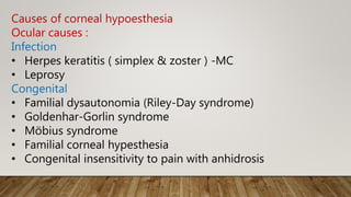
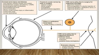
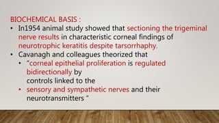
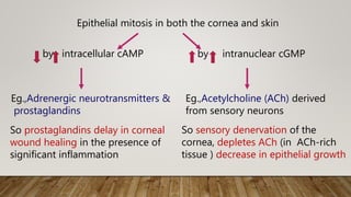
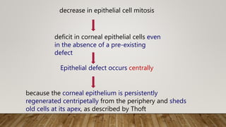
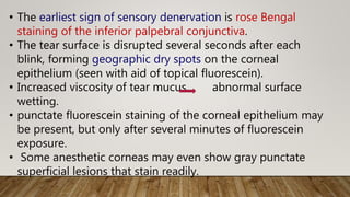
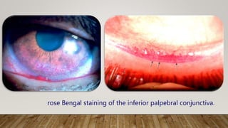
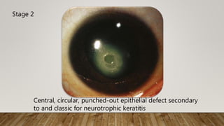
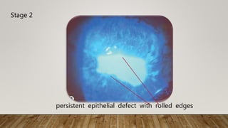
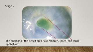
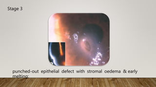
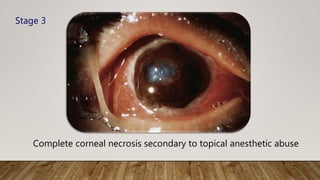
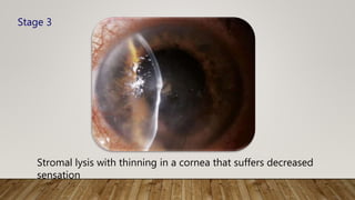
Dr . M. Dinesh
NEUROTROPHIC KERATOPATHY
• Neurotrophic keratitis is a
• Non infectious
• degenerative disease of corneal epithelium
• characterized by impaired healing.
• prevalence < 50/lac individuals
• Absence of corneal sensitivity is the hallmark of the
condition, which ultimately lead to corneal stromal melting
and perforation
• Reduced corneal sensation renders
• the corneal surface prone to occult injury
• decreases reflex tearing
• decrease healing rates of corneal epithelial injuries
• formation of nonhealing epithelial defects that tend to
ulcerate and ultimately perforate (secondary to corneal
sensory denervation )
Causes of corneal hypoesthesia
Ocular causes :
Infection
• Herpes keratitis ( simplex & zoster ) -MC
• Leprosy
Congenital
• Familial dysautonomia (Riley-Day syndrome)
• Goldenhar-Gorlin syndrome
• Möbius syndrome
• Familial corneal hypesthesia
• Congenital insensitivity to pain with anhidrosis
Topical medications
• Anaesthetics (MC cocaine abuse )
• Timolol
• Betaxolol
• Sulfacetamide 30%
• Diclofenac sodium
Corneal dystrophies
• Lattice
• Granular (rare)
Iatrogenic
• Contact lens wear
• Corneal incisions/surgeries
• LASIK
• Trauma to ciliary nerves by laser and surgery (primarily for
retinal conditions)
• Vitrectomy for retinal detachment
• photocoagulation to treat DR
• Routine, single session,
indirect laser for PDR
a/ with development
or worsening of NK
Toxic
• Chemical burns
• Carbon disulfide exposure
• Hydrogen sulfide exposure
Miscellaneous
• Increasing age
• Dark eye color
• Adie's syndrome
• Dihydroxypyrimidine dehydrogenase (DHPD) deficiency
• Any condition causing chronic corneal epithelial injury
or inflammation
Non-ocular causes
Fifth nerve palsy
• neurosurgical procedures or trauma damaging V CN
• Neoplasia (acoustic neuroma)
• Aneurysms
• Facial trauma
• multiple sclerosis
• Stroke
• Congenital hypoplasia of the trigeminal nerve
Systemic causes
• diabetes (NK can be a presenting sign in DM)
• vitamin A deficiency
• drugs (narcoleptics and antipsychotics)
BIOCHEMICAL BASIS :
• In1954 animal study showed that sectioning the trigeminal
nerve results in characteristic corneal findings of
neurotrophic keratitis despite tarsorrhaphy.
• Cavanagh and colleagues theorized that
• “corneal epithelial proliferation is regulated
bidirectionally by
controls linked to the
• sensory and sympathetic nerves and their
neurotransmitters “
Epithelial mitosis in both the cornea and skin
by intracellular cAMP by intranuclear cGMP
Eg.,Adrenergic neurotransmitters &
prostaglandins
Eg.,Acetylcholine (ACh) derived
from sensory neurons
So prostaglandins delay in corneal
wound healing in the presence of
significant inflammation
So sensory denervation of the
cornea, depletes ACh (in ACh-rich
tissue ) decrease in epithelial growth
decrease in epithelial cell mitosis
deficit in corneal epithelial cells even
in the absence of a pre-existing
defect
Epithelial defect occurs centrally
because the corneal epithelium is persistently
regenerated centripetally from the periphery and sheds
old cells at its apex, as described by Thoft
• sensory neurons directly affect the
• development of corneal epithelial cell characteristics (that
inturn)
• maintenance of good epithelial layer integrity
• corneal limbal stem cell deficiency is also seen in eyes with
neurotrophic keratitis
• In animal studies corneal anesthesia is obtained either
chemically or surgically, corneal epithelial effects are modified
by cervical sympathetic denervation.
• These findings support Cavanagh's proposed dual and
antagonistic control system of corneal epithelial regeneration
Animal models on using capsaicin
substance P
show classic changes of neurotrophic keratitis
So substance P act as a direct trophic
molecule on corneal epithelial growth
• Substance P is a neuropeptide present in the cornea
• depleted with sensory denervation,
• depleted with capsaicin specifically.
• Capsaicin is a chilli pepper extract with analgesic properties
,used topically in controlling peripheral nerve pain.
• Substance P also
• stimulate DNA synthesis &
• Stimulate corneal epithelial cell growth.
• calcitonin gene-related peptide (CGRP), which is present in
nearly all substance P-containing neurons in the cornea,
showed synergism with substance P but no trophic effect on
its own.
HISTOPATHOLOGIC FINDINGS
Corneal changes
• Epithelial thickness is decreased
• complete loss of the normal surface desquamating layer
• epithelial cell glycogen depletion
• Surface cells lose all microvilli, leading to impaired tear film
adherence
• Remaining surface epithelial cells show intracellular swelling.
• Abnormal synthesis of basal lamina helps to explain
defective epithelial adhesion
Conjunctival changes
• decreased goblet cell density
• increased length of surface microvilli
• Impression cytology reveals loss of goblet cells as well as their
migration onto the corneal surface
• These corneal and conjunctival epithelial changes are found in
keratitis sicca as well, but they are more severe in
neurotrophic keratitis.
Symptoms
• patients do not commonly complain of ocular surface
symptoms because corneal sensory innervation is impaired
• patients seek medical advice months or years after the
disease has started ,this makes NK challenging,
• blurred vision can be reported due to
• irregular epithelium
• epithelial defects (PED),
• scarring, or
• edema
CLINICAL FINDINGS
• The initial clinical appearance of an anaesthetic cornea
• loss of the usual corneal epithelial sheen
• mild disruptions in the tear surface.
punctate keratitis
epithelial loss
finally stromal ulceration
anaesthetic cornea
(even in the absence of injury)
Due to lack of the afferent limb of the reflex
• Reflex tearing decreases
• Blink rates decreases
• Tear surface abnormalities
• Tear mucus secretions increase
• tear film become more viscous
• Progressive abnormalities of
corneal epithelial microvilli
impair ocular surface
lubrication
irrepairable corneal
injury
• The earliest sign of sensory denervation is rose Bengal
staining of the inferior palpebral conjunctiva.
• The tear surface is disrupted several seconds after each
blink, forming geographic dry spots on the corneal
epithelium (seen with aid of topical fluorescein).
• Increased viscosity of tear mucus abnormal surface
wetting.
• punctate fluorescein staining of the corneal epithelium may
be present, but only after several minutes of fluorescein
exposure.
• Some anesthetic corneas may even show gray punctate
superficial lesions that stain readily.
rose Bengal staining of the inferior palpebral conjunctiva.
• Gaule spots
• Small facets of drying epithelium
• seen in retro illumination.
• compared to dellen
• appear at the limbus in early neurotrophic keratitis.
• constitute Mackie's stage I of neurotrophic keratitis
• This stage may become chronic resulting in
• superficial vascularization,
• stromal scarring,
• epithelial hyperplasia and
• irregularity.
• Rarely, a hyperplastic precorneal membrane will grow
centrally from the limbus to cover intact corneal
epithelium
Clinical stages of neurotrophic keratitis
Mackie classification :
Stage 1 - characterized by mild, nonspecific signs and
symptoms
• Rose Bengal staining of the inf palpebral conjunctivae
• Decreased tear break-up time
• Increased viscosity of tear mucus
• Punctate epithelial staining with fluorescein
• Scattered small facets of dried epithelium (Gaule spots)
Slit-lamp photograph of punctate epithelial erosions
Stage 1
Stage 1
Early central epithelial changes
Stage 2 - involves a nonhealing corneal epithelial defect
• Acute loss of epithelium, usually under the upper lid
• Surrounding rim of loose epithelium
• Stromal edema
• DM folds
• a punched-out oval or circular shape defect– chr of stage
2
• Edges of the defect become smooth and rolled with time
• Aqueous cell and flare
Stage 3
• Corneal ulcer
• Stromal melting
• corneal perforation
Central, circular, punched-out epithelial defect secondary
to and classic for neurotrophic keratitis
Stage 2
Stage 2
persistent epithelial defect with rolled edges
The endings of the deficit area have smooth, rolled, and loose
epithelium
Stage 2
Stage 3
punched-out epithelial defect with stromal oedema & early
melting;
Stage 2
• involves the acute loss of epithelium, often in an area
covered by the upper lid.
• The mechanism of loss is similar to that for recurrent
erosions and is encouraged by the milieu of poor tear
wetting of a rough and abnormal corneal epithelium.
• The area of epithelial loss is usually surrounded by a large
area of loose epithelium.
• Descemet’s Folds develop as the stroma begins to swell.
• Often, aqueous cell and flare are present.
A border of hazy epithelium surrounds a large inferior epithelial
defect, which demonstrates impaired healing secondary to corneal
anesthesia
Stage 2
• Rarely, a sterile hypopyon will form
• This stage is an indication for urgent and appropriate
treatment.
• As this stage persists, the surrounding epithelial cells
become hazy, edematous, and poorly adherent to
Bowman's membrane.
Epithelial defect features in neurotrophic keratitis
• punched-out horizontal, oval, or circular shape (chr of NK )
• The edges of the defect become smooth and rolled as the
defect ages without appreciable epithelial growth.
Neurotrophic ulceration with
hypopyon.
• Stage 3 often ensues
• in the absence of adequate treatment of stages 1 and
2, and
• sometimes despite appropriate treatment.
• Stromal lysis is the hallmark of this stage, sometimes
perforation ensues Inflammation, secondary infection
• The imprudent use of topical corticosteroids promote
stromal lysis and increase the risk for perforation.
Complete corneal necrosis secondary to topical anesthetic abuse
Stage 3
Stromal lysis with thinning in a cornea that suffers decreased
sensation
Stage 3
Clinical evaluation :
• A careful ocular examination supplemented by thorough
medical and
surgical history taking should enable one to determine the
cause for any loss of sensation
• The medical and surgical history may reveal
• previous surgical or traumatic injury to the trigeminal nerve,
• diabetes mellitus
• use of topical medications known to decrease corneal
sensation
• previous ocular laser or surgical procedures
• contact lens use.
• Direct ocular exposure to caustic chemicals or
chronic exposure to carbon disulfide and hydrogen sulfide
must be identified
• The absence of any identifiable cause for corneal
anesthesia, in the setting of severe corneal damage with
advanced stromal lysis, would suggest possible topical
anesthetic abuse, with a history of recent corneal injury.
• The presence of other neurologic deficits may help to
localize neoplasms, injuries, or vascular accidents, which
include the fifth cranial nerve or its brainstem nucleus,
among other local structures.
Acoustic neuromas
• seventh and eighth nerve dysfunction
• Corneal hypesthesia is the second most common finding
(after decreased hearing) in acoustic neuromas
Ocular motility :
• may reveal associated dysfunction of CN III, IV, and VI that
may localize an aneurysm or cavernous sinus pathology.
Pupillary abnormalities
• give clues to status of II cranial nerve
• any defects in the sympathetic innervation of the iris.
• The presence of a relative afferent pupillary defect (RAPD) in
association with corneal hypesthesia would localize the
lesion to the intraconal orbit.
• Pupillary reactions of the Adie's type have also been
associated with alterations in corneal sensation
Evaluation Localise Pathology to
III, IV, VI Ocular motility Aneurism
Cavernous sinus
pathology
VII, VIII Facial N ex. and hearing Acoustic neurinoma
II Aferent pupillary defect Lesion in intraconal
orbit
Sympathetic
inervation
iris
Anisocoria (Eferent pupillary
defect)
Lesion in intraconal
orbit
• Normal eyelid function is critical to the prognosis of NK
Conditons that hastening the progression to stage 3 NK
• Eyelid defects
• Scarred lids
• secondary to removal of periocular infiltrative tumors or
• chemical and thermal burns
• Lagophthalmos (promote corneal epithelial exposure and
drying)
• from cranial nerve VII palsy after resection of an acoustic
neuroma
• proptosis and upper eyelid retraction
• In Thyroid ophthalmopathy
Ocular surface examination
• careful analysis of qualitative and quantitative tear
function.
• Absence of the nasal-lacrimal tearing reflex along with
i/l loss of sensation in the nasal mucosa presents a high-
risk for neurotrophic corneal ulceration and
can even affect the tear production of the unaffected eye.
Corneal examination
• assessment of pattern and degree of corneal sensory deficit
with esthesiometer of Cochet-Bonnet
• It measures corneal sensitivity by the length of nylon line
(b/w 0 & 6 cm in length) required to elicit lid blink or
patient response.
• Patchy hypesthesia indicate- previous HZV keratitis
• Measuring the depth of corneal anesthesia is important,
since lower levels of sensation tend toward more severe
corneal epithelial disease
• Only corneas with readings of 2 cm or less underwent
epithelial sloughing and stromal ulceration in h zoster
keratitis
Biomicroscopic examination of the cornea
• essential to identify corneal conditions that predispose to
corneal sensory deficits.
• dendritic lesion - herpetic disease as the likely cause
• A healing epithelial defect may assume a dendritic shape
but distinguished from herpetic keratitis by the absence of
branching or end bulbs.
• Stromal scarring indicate previous infection.
• Prominence and beading of the corneal nerves may be
subtle signs of lepromatous anterior segment involvement
• Advanced stromal dystrophies
• lattice
• Granular a/with corneal hypoesthesia
Other ocular findings help identify underlying causes of
corneal sensory deficit are
• Iris atrophy with or
without AC inflammation
• Mild AC cell and flare - seen with neurogenic causes of
corneal anesthesia.
• Poor accommodation - signal HZV as the cause for corneal
sensory loss, since the virus may damage ciliary ganglion
motor nerve fibers.
• HZV and HSV
keratouveitis
• leprosy
• Fundus examination –
• optic nerve pallor or swelling on the affected side.
• These findings may localize the lesion to the orbit or retro-
orbital region.
• Background diabetic retinopathy or laser scars from
treatment of retinopathy may also shed light on the cause
of corneal sensory loss.
Indications for immediate and aggressive treatment
• significant fluorescein staining or a frank epithelial defect
• stromal lysis and thinning
• Dense anterior stromal infiltrate indicate infectious keratitis
& cultures should be taken before starting broad-spectrum
topical antibiotic therapy.
Diagnostic Procedures
• Corneal sensitivity should be evaluated
• roughly assessed using a cotton swab or
• quantitatively determined with a corneal aesthesiometer
(e.g. Cochet-Bonnet contact aesthesiometer, CRCERT-
Belmonte non-contact aesthesiometer)
• Corneal staining with fluorescein
• To evaluate corneal/conjunctival integrity, other vital
stains like lissamine green or rose Bengal
• Schirmer test to evaluate tear production, which can be
impaired as a result of reduction in corneal sensitivity.
• Any eye drops should be applied AFTER having tested
corneal sensitivity as they could otherwise alter this
measurement.
• Corneal scrapings and cultures can be performed
to exclude bacterial, viral, fungal or parasitic infections,
which may be associated with reduced corneal sensitivity.
Differential Diagnosis
• The finding of a corneal lesion in absence of ocular
symptoms (due to corneal anesthesia) is highly suspicious
of NK.
• Stage 1 NK should be differentiated from
• dry eye
• topical drug toxicity,
• exposure keratitis,
• contact lens abuse,
• chemical injury and
• limbal stem cells deficiency, which can also be associated
with some degree of NK.
• Herpes infections also reduce corneal sensitivity, pure NK
is sterile.
• Acanthamoeba keratitis often causes intense ocular pain,
but it can also be associated with some degree of corneal
anesthesia.
Management
• should emphasize on prevention of epithelial defects
because of profoundly impaired healing of the epithelium
• depends on the epithelial condition at initial presentation
and the degree of corneal hypesthesia.
• An epithelial defect in the setting of corneal anesthesia is a
serious ocular condition requiring prompt and aggressive
therapy to prevent ulceration and possible perforation
STAGE 1 : punctate keratopathy
• therapeutic goal
• to improve the quality and transparence of epithelium &
• to avoid epithelial breakdown
Treatment includes
1. Use of unpreserved artificial tears, lubricant ointments, to
prevent preservative-induced epithelial toxicity.
2. Therapeutic soft contact lenses at this stage may be
effective but increases the risk for infectious keratitis
3. Eyelid dysfunction must be remedied to prevent exposure
keratopathy and progression
stage 2 : persistent epithelial disease
• therapeutic goal
• to promote PED healing and
• prevent the development of a corneal ulcer.
Treatment includes
1. the use of unpreserved artificial tears, lubricant ointments
2. therapeutic soft contact lenses or patching
3. topical autologous serum application
4. amniotic membrane grafting
5. lateral tarsorrhaphy or botulinum induced ptosis (levator
inj)
Even in the absence of eyelid defects or dysfunction
6. topical Nerve Growth Factor application.
7. Silicone plugs of both puncta can greatly improve tear
function
8. Antibiotic eye drops can be prescribed to prevent
bacterial infections.
• topical tetracycline - speeds healing of epithelial
defects.
8. Topical corticosteroids can be administered to control
inflammation cautiously, as they could induce stromal
melting.
Stage 3 : neurotrophic keratitis
• therapeutic goal
• Promote ulcer healing and
• prevention of corneal perforation
• therapy suggested for stages 1 and 2
• N-acetylcysteine,
• oral tetracycline and medroxyprogesterone can be
prescribed in case of stromal melting.
• When an eye is initially seen at stage 3, preserving the
structural integrity of the globe takes precedence over visual
rehabilitation.
• progression of stromal lysis to perforation can be prevented
by
1. Amniotic membrane transplantation
2. Cyanoacrylate glue
3. Tectonic lamellar keratoplasty,
4. Conjunctival flaps
1.Amniotic membrane transplantation :
• shows additional effectiveness for arresting progressive
melting
• eliminates surrounding stromal infiltrate
• Amniotic membrane in combination with a bandage lens
or stretched across a rigid ring (ProKera) has also
become an effective tool for the management of
nonhealing epithelial defects
2.Cyanoacrylate glue :
• Application of cyanoacrylate glue followed by a bandage
soft contact lens is a noninvasive and effective approach to
address impending perforation
• Small perforation < 2 mm - glue is applied
• Larger defects > 2mm - lamellar or penetrating
keratoplasty.
3. Tectonic lamellar keratoplasty
• For larger defects > 2mm
• lower rejection rate
• more rapid healing,
• preserve most of the endothelium
• Good results have been reported with lamellar grafting
using multilayer amniotic membrane.
so procedure of choice
4. Conjunctival flaps
• despite their effectiveness at arresting stromal lysis and
creating a new epithelial barrier, have a diminished role in
the approach to these ulcerations and perforations because
of the poor cosmetic and visual result.
• In any eye that has suffered ulceration, neurotrophic or
otherwise, a 6-month period of clinical stability is
recommended before considering penetrating keratoplasty
Stromal lysis and thinning with perforation in a corneal
graft performed on a patient with previous herpes
zoster keratitis
• Recent human studies have shown
• substance P and
IGF-1 together,
• R nerve growth factor alone &
• thymosin beta4
• umbilical cord serum and
• autologous serum (both 20% and 50%) eyedrops are also
used
for treatment of neurotrophic ulcers
accelerate epithelial healing
in neurotrophic corneas.
• The therapeutic effect is secondary to
• growth factors,
• fibronectin,
• vitamins, and
• immunoglobulins found in serum
enhanced epithelial
healing
in recalcitrant corneal
ulcers.
• Theoretically the use of topical nonsteroidal agents would
block the influence of prostaglandins locally on corneal
epithelial growth, but their use has not had dramatic
trophic effects on wound healing.
Summary
• A corneal surface lacking adequate sensory innervation is at
risk for
• progressive epitheliopathy
• epithelial sloughing,
• stromal lysis, and
• subsequent corneal perforation in the absence of timely and
appropriate therapy.
• Herpetic infections & damage to V1 are MCC of clinical ds
• Trophic effects of cholinergic sensory nerve fibers on corneal
epithelial cells are lost in any condition that impairs corneal
sensation
• Decrease in epithelial growth rate slows wound healing and
may cause epithelial defects even in the absence of frank
trauma.
• Initial evaluation should
• identify the cause of the corneal sensory loss,
• Identify potential pathology of the central nervous system
along the path of the trigeminal nerve.
• In the absence of an obvious cause, topical anesthetic
abuse should be considered.
• Treatment should be directed toward preventing epithelial
defects and promoting epithelial cell regeneration.
• Preservative-free lubricants,
• autologous serum, and
• tarsorrhaphy
• Autologous serum provides growth factors that research may
eventually provide in pharmacologic form.
mainstays of approach
• cyanoacrylate glue - impending and small perforations and
with lamellar or penetrating keratoplasty - larger defects.
• Prompt identification of corneal anesthesia in the setting of
an epithelial defect will allow appropriate and aggressive
therapy directed toward avoidance of severe stage 3
neurotrophic keratitis.
Topical and systemic tetracyclines can effectively inhibit
MMPs
in animal and human subjects in a mechanism
independent of
their antimicrobial activity.81 High levels of MMPs cause
corneal stromal lysis via collagen degradation and injury
to the
epithelial basement membrane adhesion complexes
resulting in
poor epithelial adherence. MMP-9, a gelatinase in corneal
epithelial cells, has been detected at the edges of
nonhealing
corneal ulcers.82 Oral doxycycline at 50 mg twice a day has
been
demonstrated to inhibit MMP-9, resulting in rapid healing
and
preventing recurrences of recurrent corneal erosions
• Oral doxycycline or
minocycline
• serves to remedy
qualitative tear
dysfunction through its
action in the meibomian
glands
• even aid in preventing
corneal stromal lysis
Specially compounded preservative-free topical steroids,
medroxyprogesterone 1% and methylprednisolone 1%
may
prevent stromal lysis, but must be kept refrigerated to
avoid
contamination. Medroxyprogesterone prevents stromal
melting
by inhibiting local collagenases that degrade the corneal
stroma,
while at the same time exhibiting a mild antiinflammatory
property.86–89 Both doxycycline and corticosteroids can
inhibit
MMP-9. In patients with recurrent corneal erosions
unresponsive
to the conventional therapy, administration of oral
doxycycline
and topical corticosteroids reduces pain and heals
epithelial
defects within 2–10 days.83 Methylprednisolone also
provides
symptomatic relief and resolution of filaments in severe
keratoconjunctivitis associated with Sjögren syndrome.90
Autologous serum drops: These are made from the
patient’s own serum and contain collagenase inhibitors
such as alpha-1, alpha-2 macroglobulins and, in their
undiluted form, were showned to heal trophic herpetic
ulcers in 1973.209 Since then, a number of studies have
reported the usefulness of this approach. Two studies
from
Bonini’s group using murine nerve growth factor (NGF)
(1–10 micrograms of highly purified murine NGF in 50 mL
of physiological saline) to treat human anesthetic
neurotrophic ulcers found that corneal healing began
2–14 days after starting treatment of 10 times daily for
2 days and then six times daily until the ulcers healed. All
patients had complete healing of their corneal ulcers after
10 days to 6 weeks of treatment. Corneal sensitivity
improved in 13/14 eyes, and returned to normal in two
of the 13 eyes. Corneal integrity and sensitivity were
maintained during the follow-up period (range,
3–12 months
THAN Q

Neurotrophic keratopathy

  • 1.
    Dr . M.Dinesh NEUROTROPHIC KERATOPATHY
  • 2.
    • Neurotrophic keratitisis a • Non infectious • degenerative disease of corneal epithelium • characterized by impaired healing. • prevalence < 50/lac individuals • Absence of corneal sensitivity is the hallmark of the condition, which ultimately lead to corneal stromal melting and perforation
  • 3.
    • Reduced cornealsensation renders • the corneal surface prone to occult injury • decreases reflex tearing • decrease healing rates of corneal epithelial injuries • formation of nonhealing epithelial defects that tend to ulcerate and ultimately perforate (secondary to corneal sensory denervation )
  • 4.
    Causes of cornealhypoesthesia Ocular causes : Infection • Herpes keratitis ( simplex & zoster ) -MC • Leprosy Congenital • Familial dysautonomia (Riley-Day syndrome) • Goldenhar-Gorlin syndrome • Möbius syndrome • Familial corneal hypesthesia • Congenital insensitivity to pain with anhidrosis
  • 5.
    Topical medications • Anaesthetics(MC cocaine abuse ) • Timolol • Betaxolol • Sulfacetamide 30% • Diclofenac sodium Corneal dystrophies • Lattice • Granular (rare)
  • 6.
    Iatrogenic • Contact lenswear • Corneal incisions/surgeries • LASIK • Trauma to ciliary nerves by laser and surgery (primarily for retinal conditions) • Vitrectomy for retinal detachment • photocoagulation to treat DR • Routine, single session, indirect laser for PDR a/ with development or worsening of NK
  • 7.
    Toxic • Chemical burns •Carbon disulfide exposure • Hydrogen sulfide exposure Miscellaneous • Increasing age • Dark eye color • Adie's syndrome • Dihydroxypyrimidine dehydrogenase (DHPD) deficiency • Any condition causing chronic corneal epithelial injury or inflammation
  • 8.
    Non-ocular causes Fifth nervepalsy • neurosurgical procedures or trauma damaging V CN • Neoplasia (acoustic neuroma) • Aneurysms • Facial trauma • multiple sclerosis • Stroke • Congenital hypoplasia of the trigeminal nerve Systemic causes • diabetes (NK can be a presenting sign in DM) • vitamin A deficiency • drugs (narcoleptics and antipsychotics)
  • 10.
    BIOCHEMICAL BASIS : •In1954 animal study showed that sectioning the trigeminal nerve results in characteristic corneal findings of neurotrophic keratitis despite tarsorrhaphy. • Cavanagh and colleagues theorized that • “corneal epithelial proliferation is regulated bidirectionally by controls linked to the • sensory and sympathetic nerves and their neurotransmitters “
  • 11.
    Epithelial mitosis inboth the cornea and skin by intracellular cAMP by intranuclear cGMP Eg.,Adrenergic neurotransmitters & prostaglandins Eg.,Acetylcholine (ACh) derived from sensory neurons So prostaglandins delay in corneal wound healing in the presence of significant inflammation So sensory denervation of the cornea, depletes ACh (in ACh-rich tissue ) decrease in epithelial growth
  • 12.
    decrease in epithelialcell mitosis deficit in corneal epithelial cells even in the absence of a pre-existing defect Epithelial defect occurs centrally because the corneal epithelium is persistently regenerated centripetally from the periphery and sheds old cells at its apex, as described by Thoft
  • 13.
    • sensory neuronsdirectly affect the • development of corneal epithelial cell characteristics (that inturn) • maintenance of good epithelial layer integrity • corneal limbal stem cell deficiency is also seen in eyes with neurotrophic keratitis • In animal studies corneal anesthesia is obtained either chemically or surgically, corneal epithelial effects are modified by cervical sympathetic denervation. • These findings support Cavanagh's proposed dual and antagonistic control system of corneal epithelial regeneration
  • 14.
    Animal models onusing capsaicin substance P show classic changes of neurotrophic keratitis So substance P act as a direct trophic molecule on corneal epithelial growth
  • 15.
    • Substance Pis a neuropeptide present in the cornea • depleted with sensory denervation, • depleted with capsaicin specifically. • Capsaicin is a chilli pepper extract with analgesic properties ,used topically in controlling peripheral nerve pain. • Substance P also • stimulate DNA synthesis & • Stimulate corneal epithelial cell growth. • calcitonin gene-related peptide (CGRP), which is present in nearly all substance P-containing neurons in the cornea, showed synergism with substance P but no trophic effect on its own.
  • 16.
    HISTOPATHOLOGIC FINDINGS Corneal changes •Epithelial thickness is decreased • complete loss of the normal surface desquamating layer • epithelial cell glycogen depletion • Surface cells lose all microvilli, leading to impaired tear film adherence • Remaining surface epithelial cells show intracellular swelling. • Abnormal synthesis of basal lamina helps to explain defective epithelial adhesion
  • 17.
    Conjunctival changes • decreasedgoblet cell density • increased length of surface microvilli • Impression cytology reveals loss of goblet cells as well as their migration onto the corneal surface • These corneal and conjunctival epithelial changes are found in keratitis sicca as well, but they are more severe in neurotrophic keratitis.
  • 18.
    Symptoms • patients donot commonly complain of ocular surface symptoms because corneal sensory innervation is impaired • patients seek medical advice months or years after the disease has started ,this makes NK challenging, • blurred vision can be reported due to • irregular epithelium • epithelial defects (PED), • scarring, or • edema
  • 19.
    CLINICAL FINDINGS • Theinitial clinical appearance of an anaesthetic cornea • loss of the usual corneal epithelial sheen • mild disruptions in the tear surface. punctate keratitis epithelial loss finally stromal ulceration anaesthetic cornea (even in the absence of injury)
  • 20.
    Due to lackof the afferent limb of the reflex • Reflex tearing decreases • Blink rates decreases • Tear surface abnormalities • Tear mucus secretions increase • tear film become more viscous • Progressive abnormalities of corneal epithelial microvilli impair ocular surface lubrication irrepairable corneal injury
  • 21.
    • The earliestsign of sensory denervation is rose Bengal staining of the inferior palpebral conjunctiva. • The tear surface is disrupted several seconds after each blink, forming geographic dry spots on the corneal epithelium (seen with aid of topical fluorescein). • Increased viscosity of tear mucus abnormal surface wetting. • punctate fluorescein staining of the corneal epithelium may be present, but only after several minutes of fluorescein exposure. • Some anesthetic corneas may even show gray punctate superficial lesions that stain readily.
  • 22.
    rose Bengal stainingof the inferior palpebral conjunctiva.
  • 23.
    • Gaule spots •Small facets of drying epithelium • seen in retro illumination. • compared to dellen • appear at the limbus in early neurotrophic keratitis. • constitute Mackie's stage I of neurotrophic keratitis
  • 24.
    • This stagemay become chronic resulting in • superficial vascularization, • stromal scarring, • epithelial hyperplasia and • irregularity. • Rarely, a hyperplastic precorneal membrane will grow centrally from the limbus to cover intact corneal epithelium
  • 25.
    Clinical stages ofneurotrophic keratitis Mackie classification : Stage 1 - characterized by mild, nonspecific signs and symptoms • Rose Bengal staining of the inf palpebral conjunctivae • Decreased tear break-up time • Increased viscosity of tear mucus • Punctate epithelial staining with fluorescein • Scattered small facets of dried epithelium (Gaule spots)
  • 26.
    Slit-lamp photograph ofpunctate epithelial erosions Stage 1
  • 27.
    Stage 1 Early centralepithelial changes
  • 28.
    Stage 2 -involves a nonhealing corneal epithelial defect • Acute loss of epithelium, usually under the upper lid • Surrounding rim of loose epithelium • Stromal edema • DM folds • a punched-out oval or circular shape defect– chr of stage 2 • Edges of the defect become smooth and rolled with time • Aqueous cell and flare Stage 3 • Corneal ulcer • Stromal melting • corneal perforation
  • 29.
    Central, circular, punched-outepithelial defect secondary to and classic for neurotrophic keratitis Stage 2
  • 30.
    Stage 2 persistent epithelialdefect with rolled edges
  • 31.
    The endings ofthe deficit area have smooth, rolled, and loose epithelium Stage 2
  • 32.
    Stage 3 punched-out epithelialdefect with stromal oedema & early melting;
  • 33.
    Stage 2 • involvesthe acute loss of epithelium, often in an area covered by the upper lid. • The mechanism of loss is similar to that for recurrent erosions and is encouraged by the milieu of poor tear wetting of a rough and abnormal corneal epithelium. • The area of epithelial loss is usually surrounded by a large area of loose epithelium. • Descemet’s Folds develop as the stroma begins to swell. • Often, aqueous cell and flare are present.
  • 34.
    A border ofhazy epithelium surrounds a large inferior epithelial defect, which demonstrates impaired healing secondary to corneal anesthesia Stage 2
  • 35.
    • Rarely, asterile hypopyon will form • This stage is an indication for urgent and appropriate treatment. • As this stage persists, the surrounding epithelial cells become hazy, edematous, and poorly adherent to Bowman's membrane. Epithelial defect features in neurotrophic keratitis • punched-out horizontal, oval, or circular shape (chr of NK ) • The edges of the defect become smooth and rolled as the defect ages without appreciable epithelial growth.
  • 36.
  • 37.
    • Stage 3often ensues • in the absence of adequate treatment of stages 1 and 2, and • sometimes despite appropriate treatment. • Stromal lysis is the hallmark of this stage, sometimes perforation ensues Inflammation, secondary infection • The imprudent use of topical corticosteroids promote stromal lysis and increase the risk for perforation.
  • 38.
    Complete corneal necrosissecondary to topical anesthetic abuse Stage 3
  • 39.
    Stromal lysis withthinning in a cornea that suffers decreased sensation Stage 3
  • 40.
    Clinical evaluation : •A careful ocular examination supplemented by thorough medical and surgical history taking should enable one to determine the cause for any loss of sensation • The medical and surgical history may reveal • previous surgical or traumatic injury to the trigeminal nerve, • diabetes mellitus • use of topical medications known to decrease corneal sensation • previous ocular laser or surgical procedures • contact lens use.
  • 41.
    • Direct ocularexposure to caustic chemicals or chronic exposure to carbon disulfide and hydrogen sulfide must be identified • The absence of any identifiable cause for corneal anesthesia, in the setting of severe corneal damage with advanced stromal lysis, would suggest possible topical anesthetic abuse, with a history of recent corneal injury.
  • 42.
    • The presenceof other neurologic deficits may help to localize neoplasms, injuries, or vascular accidents, which include the fifth cranial nerve or its brainstem nucleus, among other local structures. Acoustic neuromas • seventh and eighth nerve dysfunction • Corneal hypesthesia is the second most common finding (after decreased hearing) in acoustic neuromas Ocular motility : • may reveal associated dysfunction of CN III, IV, and VI that may localize an aneurysm or cavernous sinus pathology.
  • 43.
    Pupillary abnormalities • giveclues to status of II cranial nerve • any defects in the sympathetic innervation of the iris. • The presence of a relative afferent pupillary defect (RAPD) in association with corneal hypesthesia would localize the lesion to the intraconal orbit. • Pupillary reactions of the Adie's type have also been associated with alterations in corneal sensation
  • 44.
    Evaluation Localise Pathologyto III, IV, VI Ocular motility Aneurism Cavernous sinus pathology VII, VIII Facial N ex. and hearing Acoustic neurinoma II Aferent pupillary defect Lesion in intraconal orbit Sympathetic inervation iris Anisocoria (Eferent pupillary defect) Lesion in intraconal orbit
  • 45.
    • Normal eyelidfunction is critical to the prognosis of NK Conditons that hastening the progression to stage 3 NK • Eyelid defects • Scarred lids • secondary to removal of periocular infiltrative tumors or • chemical and thermal burns • Lagophthalmos (promote corneal epithelial exposure and drying) • from cranial nerve VII palsy after resection of an acoustic neuroma • proptosis and upper eyelid retraction • In Thyroid ophthalmopathy
  • 46.
    Ocular surface examination •careful analysis of qualitative and quantitative tear function. • Absence of the nasal-lacrimal tearing reflex along with i/l loss of sensation in the nasal mucosa presents a high- risk for neurotrophic corneal ulceration and can even affect the tear production of the unaffected eye.
  • 47.
    Corneal examination • assessmentof pattern and degree of corneal sensory deficit with esthesiometer of Cochet-Bonnet • It measures corneal sensitivity by the length of nylon line (b/w 0 & 6 cm in length) required to elicit lid blink or patient response. • Patchy hypesthesia indicate- previous HZV keratitis • Measuring the depth of corneal anesthesia is important, since lower levels of sensation tend toward more severe corneal epithelial disease • Only corneas with readings of 2 cm or less underwent epithelial sloughing and stromal ulceration in h zoster keratitis
  • 48.
    Biomicroscopic examination ofthe cornea • essential to identify corneal conditions that predispose to corneal sensory deficits. • dendritic lesion - herpetic disease as the likely cause • A healing epithelial defect may assume a dendritic shape but distinguished from herpetic keratitis by the absence of branching or end bulbs. • Stromal scarring indicate previous infection. • Prominence and beading of the corneal nerves may be subtle signs of lepromatous anterior segment involvement • Advanced stromal dystrophies • lattice • Granular a/with corneal hypoesthesia
  • 49.
    Other ocular findingshelp identify underlying causes of corneal sensory deficit are • Iris atrophy with or without AC inflammation • Mild AC cell and flare - seen with neurogenic causes of corneal anesthesia. • Poor accommodation - signal HZV as the cause for corneal sensory loss, since the virus may damage ciliary ganglion motor nerve fibers. • HZV and HSV keratouveitis • leprosy
  • 50.
    • Fundus examination– • optic nerve pallor or swelling on the affected side. • These findings may localize the lesion to the orbit or retro- orbital region. • Background diabetic retinopathy or laser scars from treatment of retinopathy may also shed light on the cause of corneal sensory loss.
  • 51.
    Indications for immediateand aggressive treatment • significant fluorescein staining or a frank epithelial defect • stromal lysis and thinning • Dense anterior stromal infiltrate indicate infectious keratitis & cultures should be taken before starting broad-spectrum topical antibiotic therapy.
  • 52.
    Diagnostic Procedures • Cornealsensitivity should be evaluated • roughly assessed using a cotton swab or • quantitatively determined with a corneal aesthesiometer (e.g. Cochet-Bonnet contact aesthesiometer, CRCERT- Belmonte non-contact aesthesiometer) • Corneal staining with fluorescein
  • 53.
    • To evaluatecorneal/conjunctival integrity, other vital stains like lissamine green or rose Bengal • Schirmer test to evaluate tear production, which can be impaired as a result of reduction in corneal sensitivity. • Any eye drops should be applied AFTER having tested corneal sensitivity as they could otherwise alter this measurement. • Corneal scrapings and cultures can be performed to exclude bacterial, viral, fungal or parasitic infections, which may be associated with reduced corneal sensitivity.
  • 54.
    Differential Diagnosis • Thefinding of a corneal lesion in absence of ocular symptoms (due to corneal anesthesia) is highly suspicious of NK. • Stage 1 NK should be differentiated from • dry eye • topical drug toxicity, • exposure keratitis, • contact lens abuse, • chemical injury and • limbal stem cells deficiency, which can also be associated with some degree of NK.
  • 55.
    • Herpes infectionsalso reduce corneal sensitivity, pure NK is sterile. • Acanthamoeba keratitis often causes intense ocular pain, but it can also be associated with some degree of corneal anesthesia.
  • 56.
    Management • should emphasizeon prevention of epithelial defects because of profoundly impaired healing of the epithelium • depends on the epithelial condition at initial presentation and the degree of corneal hypesthesia. • An epithelial defect in the setting of corneal anesthesia is a serious ocular condition requiring prompt and aggressive therapy to prevent ulceration and possible perforation
  • 57.
    STAGE 1 :punctate keratopathy • therapeutic goal • to improve the quality and transparence of epithelium & • to avoid epithelial breakdown Treatment includes 1. Use of unpreserved artificial tears, lubricant ointments, to prevent preservative-induced epithelial toxicity. 2. Therapeutic soft contact lenses at this stage may be effective but increases the risk for infectious keratitis 3. Eyelid dysfunction must be remedied to prevent exposure keratopathy and progression
  • 58.
    stage 2 :persistent epithelial disease • therapeutic goal • to promote PED healing and • prevent the development of a corneal ulcer. Treatment includes 1. the use of unpreserved artificial tears, lubricant ointments 2. therapeutic soft contact lenses or patching 3. topical autologous serum application 4. amniotic membrane grafting 5. lateral tarsorrhaphy or botulinum induced ptosis (levator inj) Even in the absence of eyelid defects or dysfunction 6. topical Nerve Growth Factor application.
  • 59.
    7. Silicone plugsof both puncta can greatly improve tear function 8. Antibiotic eye drops can be prescribed to prevent bacterial infections. • topical tetracycline - speeds healing of epithelial defects. 8. Topical corticosteroids can be administered to control inflammation cautiously, as they could induce stromal melting.
  • 60.
    Stage 3 :neurotrophic keratitis • therapeutic goal • Promote ulcer healing and • prevention of corneal perforation • therapy suggested for stages 1 and 2 • N-acetylcysteine, • oral tetracycline and medroxyprogesterone can be prescribed in case of stromal melting.
  • 61.
    • When aneye is initially seen at stage 3, preserving the structural integrity of the globe takes precedence over visual rehabilitation. • progression of stromal lysis to perforation can be prevented by 1. Amniotic membrane transplantation 2. Cyanoacrylate glue 3. Tectonic lamellar keratoplasty, 4. Conjunctival flaps
  • 62.
    1.Amniotic membrane transplantation: • shows additional effectiveness for arresting progressive melting • eliminates surrounding stromal infiltrate • Amniotic membrane in combination with a bandage lens or stretched across a rigid ring (ProKera) has also become an effective tool for the management of nonhealing epithelial defects
  • 63.
    2.Cyanoacrylate glue : •Application of cyanoacrylate glue followed by a bandage soft contact lens is a noninvasive and effective approach to address impending perforation • Small perforation < 2 mm - glue is applied • Larger defects > 2mm - lamellar or penetrating keratoplasty.
  • 64.
    3. Tectonic lamellarkeratoplasty • For larger defects > 2mm • lower rejection rate • more rapid healing, • preserve most of the endothelium • Good results have been reported with lamellar grafting using multilayer amniotic membrane. so procedure of choice
  • 65.
    4. Conjunctival flaps •despite their effectiveness at arresting stromal lysis and creating a new epithelial barrier, have a diminished role in the approach to these ulcerations and perforations because of the poor cosmetic and visual result. • In any eye that has suffered ulceration, neurotrophic or otherwise, a 6-month period of clinical stability is recommended before considering penetrating keratoplasty
  • 66.
    Stromal lysis andthinning with perforation in a corneal graft performed on a patient with previous herpes zoster keratitis
  • 67.
    • Recent humanstudies have shown • substance P and IGF-1 together, • R nerve growth factor alone & • thymosin beta4 • umbilical cord serum and • autologous serum (both 20% and 50%) eyedrops are also used for treatment of neurotrophic ulcers accelerate epithelial healing in neurotrophic corneas.
  • 68.
    • The therapeuticeffect is secondary to • growth factors, • fibronectin, • vitamins, and • immunoglobulins found in serum enhanced epithelial healing in recalcitrant corneal ulcers.
  • 69.
    • Theoretically theuse of topical nonsteroidal agents would block the influence of prostaglandins locally on corneal epithelial growth, but their use has not had dramatic trophic effects on wound healing.
  • 70.
    Summary • A cornealsurface lacking adequate sensory innervation is at risk for • progressive epitheliopathy • epithelial sloughing, • stromal lysis, and • subsequent corneal perforation in the absence of timely and appropriate therapy. • Herpetic infections & damage to V1 are MCC of clinical ds • Trophic effects of cholinergic sensory nerve fibers on corneal epithelial cells are lost in any condition that impairs corneal sensation
  • 71.
    • Decrease inepithelial growth rate slows wound healing and may cause epithelial defects even in the absence of frank trauma. • Initial evaluation should • identify the cause of the corneal sensory loss, • Identify potential pathology of the central nervous system along the path of the trigeminal nerve. • In the absence of an obvious cause, topical anesthetic abuse should be considered.
  • 72.
    • Treatment shouldbe directed toward preventing epithelial defects and promoting epithelial cell regeneration. • Preservative-free lubricants, • autologous serum, and • tarsorrhaphy • Autologous serum provides growth factors that research may eventually provide in pharmacologic form. mainstays of approach
  • 73.
    • cyanoacrylate glue- impending and small perforations and with lamellar or penetrating keratoplasty - larger defects. • Prompt identification of corneal anesthesia in the setting of an epithelial defect will allow appropriate and aggressive therapy directed toward avoidance of severe stage 3 neurotrophic keratitis.
  • 74.
    Topical and systemictetracyclines can effectively inhibit MMPs in animal and human subjects in a mechanism independent of their antimicrobial activity.81 High levels of MMPs cause corneal stromal lysis via collagen degradation and injury to the epithelial basement membrane adhesion complexes resulting in poor epithelial adherence. MMP-9, a gelatinase in corneal epithelial cells, has been detected at the edges of nonhealing corneal ulcers.82 Oral doxycycline at 50 mg twice a day has been demonstrated to inhibit MMP-9, resulting in rapid healing and preventing recurrences of recurrent corneal erosions • Oral doxycycline or minocycline • serves to remedy qualitative tear dysfunction through its action in the meibomian glands • even aid in preventing corneal stromal lysis
  • 75.
    Specially compounded preservative-freetopical steroids, medroxyprogesterone 1% and methylprednisolone 1% may prevent stromal lysis, but must be kept refrigerated to avoid contamination. Medroxyprogesterone prevents stromal melting by inhibiting local collagenases that degrade the corneal stroma, while at the same time exhibiting a mild antiinflammatory property.86–89 Both doxycycline and corticosteroids can inhibit MMP-9. In patients with recurrent corneal erosions unresponsive to the conventional therapy, administration of oral doxycycline and topical corticosteroids reduces pain and heals epithelial defects within 2–10 days.83 Methylprednisolone also provides symptomatic relief and resolution of filaments in severe keratoconjunctivitis associated with Sjögren syndrome.90
  • 76.
    Autologous serum drops:These are made from the patient’s own serum and contain collagenase inhibitors such as alpha-1, alpha-2 macroglobulins and, in their undiluted form, were showned to heal trophic herpetic ulcers in 1973.209 Since then, a number of studies have reported the usefulness of this approach. Two studies from Bonini’s group using murine nerve growth factor (NGF) (1–10 micrograms of highly purified murine NGF in 50 mL of physiological saline) to treat human anesthetic neurotrophic ulcers found that corneal healing began 2–14 days after starting treatment of 10 times daily for 2 days and then six times daily until the ulcers healed. All patients had complete healing of their corneal ulcers after 10 days to 6 weeks of treatment. Corneal sensitivity improved in 13/14 eyes, and returned to normal in two of the 13 eyes. Corneal integrity and sensitivity were maintained during the follow-up period (range, 3–12 months
  • 77.