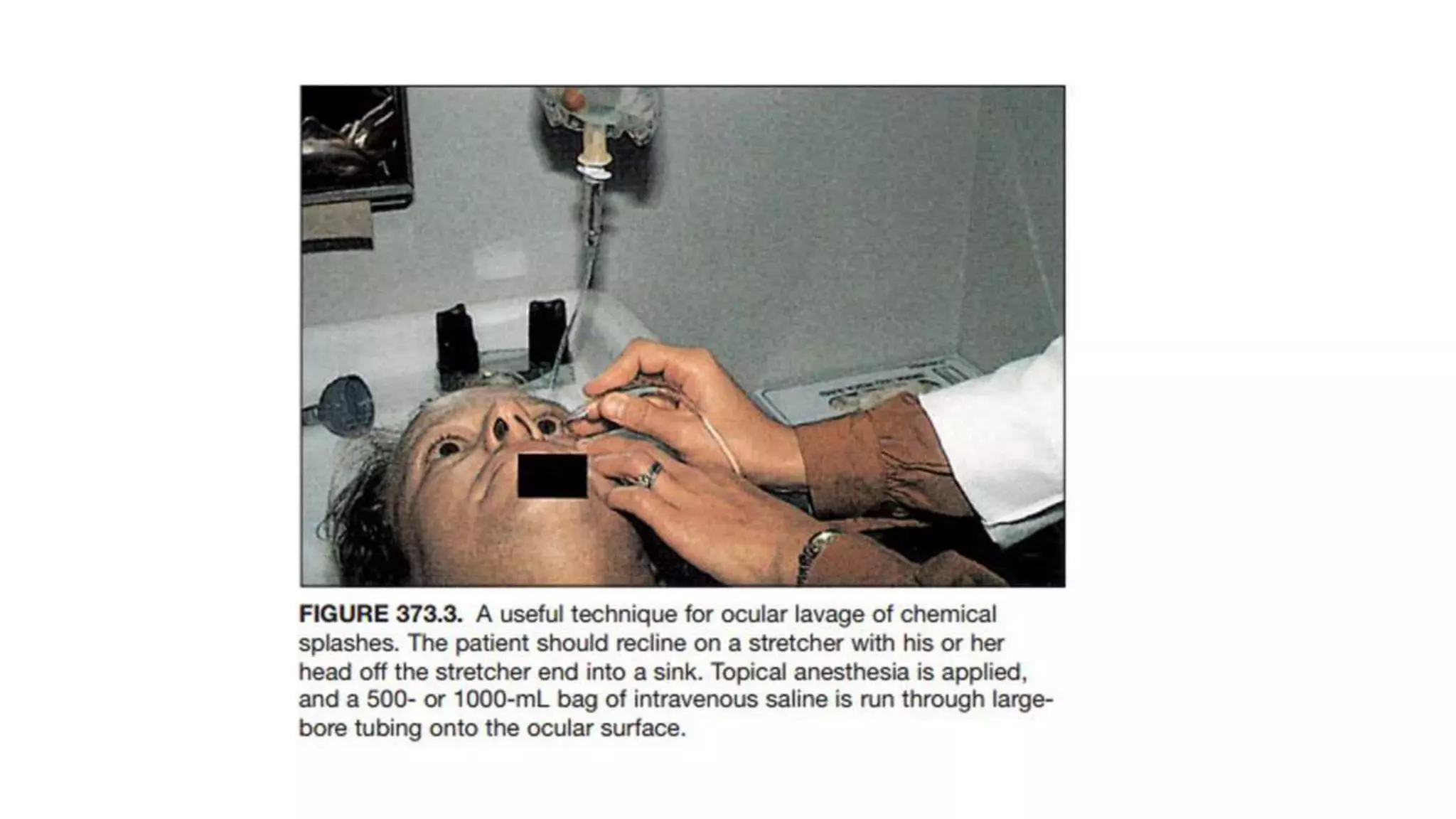
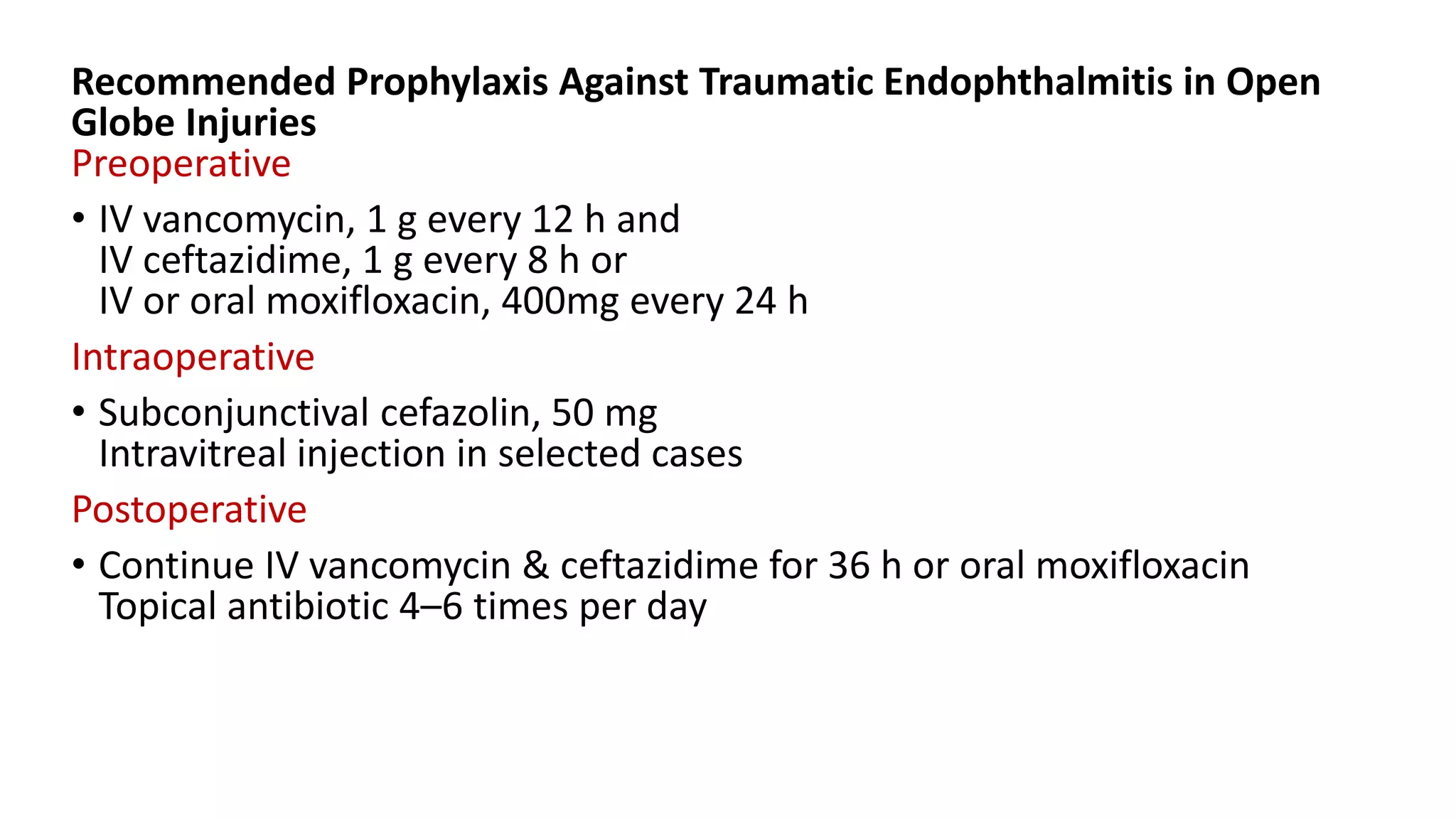
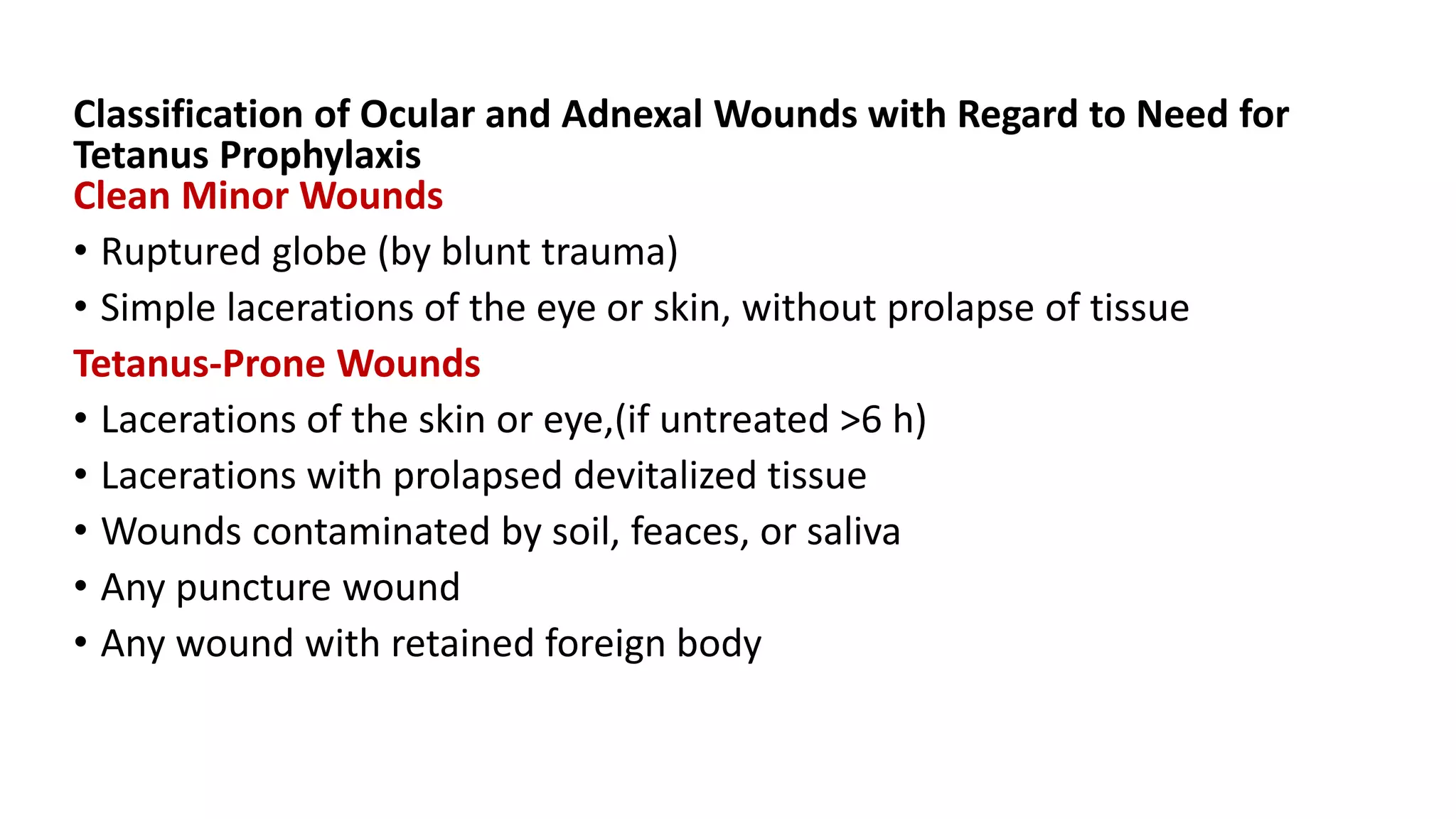
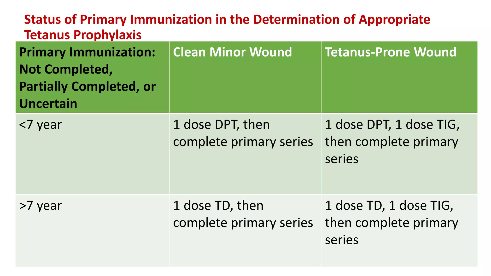
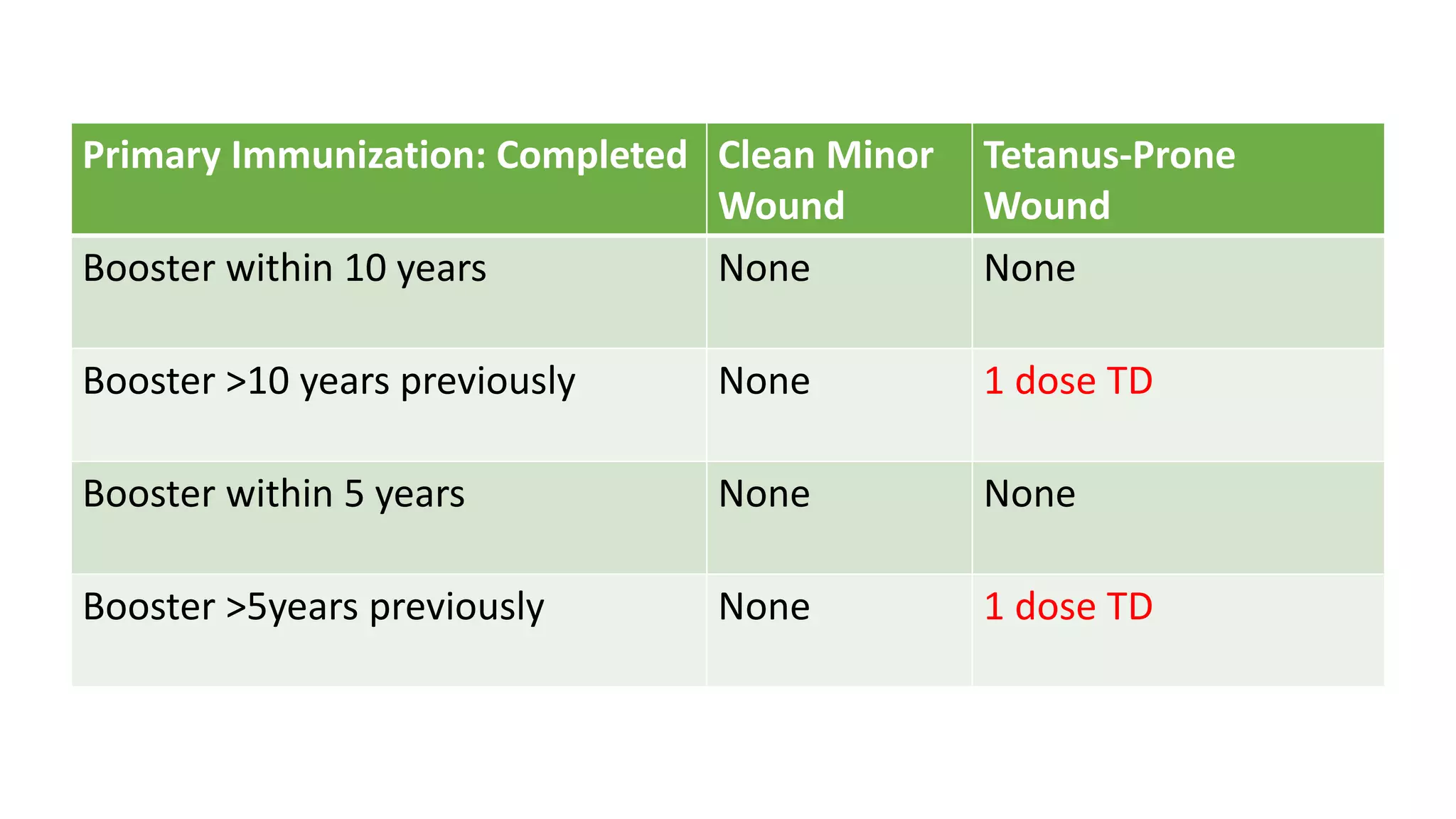
Evaluation and Initial
Management of Patients with
Ocular and Adnexal Trauma
Dr.M.Dinesh
INITIAL EMERGENCY CARE BY NONOPHTHALMIC
SPECIALISTS
ESSENTIAL STEPS IN THE EMERGENCY DEPARTMENT examination of
a patient with ocular trauma include
1. Life-threatening injuries are treated.
General medical and neurosurgical stability and clearance are ensured.
2. The ocular surface is lavaged immediately if there is any history of
chemical ocular injury, especially alkaline substance
3. The past medical history is obtained.
4. The ocular history and the history of the traumatic event are
obtained.
5. An eye examination is performed. If a lacerated or
ruptured eye is seen, use of eyedrops and further examination is
deferred to the ophthalmologist
ESSENTIAL EQUIPMENT FOR THE EXAMINATION OF PATIENTS WITH
OCULAR TRAUMA IN A GENERAL EMERGENCY DEPARTMENT
Vision charts (for near and distance vision)
Pen light
Cobalt blue filter for pen light
Fluorescein strips
Topical anesthetic
Direct ophthalmoscope
Tonopen or Schiøtz tonometer
Eyeshields
Slit-lamp biomicroscope
VISUAL ACUITY :
• Each eye is tested separately.
• A standard Snellen distance chart (at 20 ft)
a near-vision card (at 14 inches with bifocals or reading glasses, is used.
• Alternatively, the ability to see newspaper or magazine
print is documented.
• If none of these maneuvers is possible, it is documented that the patient
can
count fingers,
see hand movements, or
detect light.
• Use of a pinhole device over glasses or over the eye for patients
without glasses ,If vision improves refractive error is the likely cause of
the decrease in vision
• If significant eyelid swelling exists, the eyelid is raised gently
without any pressure being put on the globe.
• Visual loss must not go unexplained.
• Any prior reduction in visual acuity is documented.
EXTERNAL EXAMINATION
• Skin, face, and orbital injuries or deformities are noted.
OCULAR MOTILITY
• The ability of the eyes to move synchronously up, down, right, and left
is assessed.
PUPILS
• Size
• shape
• symmetry
• reaction to light (direct and consensual) and
• presence or lack of an afferent pupillary defect is documented.
Testing for an afferent pupillary defect :
• A strong light source is swung back and forth between the two eyes
(~1 s on each eye).
• The normal reaction is constriction when light shines on each eye.
• An afferent pupillary defect exists if there is dilatation instead of
constriction when the light is swung to the abnormal eye
ANTERIOR SEGMENT
Conjunctiva and Sclera
SCH, swelling (chemosis),
irregularity in the surface,
exposed tissue (may be brown intraocular uveal tissue), and
foreign bodies
Cornea
Lack of clarity or of the light reflex,irregularities,
Fluorescein staining when examined with the cobalt blue light,
Exposed tissue (may be brown iris tissue), and foreign bodies
Anterior Chamber
Depth
presence of blood (hyphema) or white blood cells (hypopyon)
Iris
Irregularities and defects in both the peripheral iris and the pupil
Lens
Position and
degree of cataract are assessed.
POSTERIOR SEGMENT (WITH DIRECT OPHTHALMOSCOPE, UNDILATED
PUPIL)
vitreous - assessed for clarity (of view to posterior pole).
optic disk - assessed for color, margins, and cup:disc ratio.
macula, retina, vs - assessed for presence of blood, foreign bodies,ruptures
IOP (WITH TONOPEN OR SCHIØTZ TONOMETER)
measured only if there is low suspicion for rupture and
never if there is obvious corneal laceration or prolapse of uveal tissue
ADDITIONAL CONSIDERATIONS :
• The ophthalmologist must keep in mind that patients with serious ocular
trauma can present with
near-normal visual acuity and
no obvious anterior or posterior segment findings.
• An examination with the patient’s eyes dilated and with an
indirect ophthalmoscope is often necessary to document the nature of
the eye injury
The following situations require immediate referral to an
ophthalmologist:
1. Decreased vision that did not exist before the injury.
2. Obvious rupture or laceration of the eye.
3. Any internal ocular injury, with or without rupture of the eye.
4. Any question of intraocular foreign body.
5. chemical burns of the cornea, especially with alkaline substances.
6. Lid lacerations that cross the lid margin or penetrate the orbital
septum.
Any lid injury that potentially involves the lacrimal drainage systems
should be referred, including the following:
a. Laceration resulting from an avulsion type history, such as fist, finger.
b. Any skin or lid laceration that points or aims toward medial canthus.
c. Any transmarginal lid laceration involving medial half of eyelid.
d. Fat presenting in any eyelid wound
• If any of the preceding are present, further examination can be deferred
until the patient arrives at an ophthalmology care facility.
• The obvious exception is chemical injuries, which should be lavaged
immediately and continuously during transport.
• Visual acuity should be documented in each eye before transfer from
one hospital to another
• If referral and transport to an ophthalmologist are to be accomplished,
the injured eye should be covered with a metal shield or the end of a
cup that is taped loosely to orbital bones without any pressure being
placed on the eye
• No drops or ointments should be applied to the injured eye.
• If any foreign bodies are protruding from the eye or orbit, they should
not be removed until appropriate evaluation and radiographic imaging
are accomplished.
• If an object is lodged in the orbit and is protruding (e.g., knife, pencil,
stick),object should be stabilized with a Styrofoam cup or a fluffy
bandage to prevent movement during transport.
• Lid lacerations should be gently covered with saline-soaked gauze pads
during transport.
• The injured patient should be transported with the head of the bed
elevated.
• IV line should be accomplished in any patient with a serious ocular
injury.
• If transport to an ophthalmology treatment facility is likely to be
delayed and there is a major ocular injury, broadspectrum antibiotics
should be administered as Prophylaxis of Posttraumatic
Endophthalmitis.
• The patient should not take anything by mouth.
• The injured patient should not receive any narcotics, because these
could delay proper informed consent if operative intervention is
necessary.
• Antiemetics may be important to prevent the increased intrathoracic
and IOP associated with gagging and vomiting.
• The results of anydiagnostic tests performed (blood studies, plain
radiographs, or CT) should accompany the patient on her/his referral.
• If any patient with ocular trauma is managed by the emergency
department physician primarily, a follow-up appointment
with an ophthalmologist is recommended.
• All patients with orbital #s should undergo an ophthalmic examination
with dilatation of the pupils within 1 week of the injury.
EYE CARE BY THE OPHTHALMOLOGIST
IN THE EMERGENCY SETTING
• It is essential that medical and neurosurgical clearance be confirmed.
• A quick review of the referring physician’s examination and diagnostic
test results, conclusions, and recommendations is usually
sufficient.
• Vital signs and physical examination should assure
the ophthalmologist that no grave changes have occurred in the
neurologic or medical status of the transferred patient.
• If a chemical injury has occurred, the globe should be inspected to
ensure an intact status, and then lavage should begin
immediately before a detailed history is obtained
HISTORY
• A detailed description of the traumatic event serves as the focal
point for guiding additional examination and diagnostic
techniques.
• This history should be elicited from the patient and
family members, as well as from any witnesses to the event
• It is important for the physician to determine whether
the history given of the injury matches the severity of the
damage at the examination.
• If the patient describes an injury setting in which fragments are
suspected (explosions, metal-on-metal contact, broken glass or
windshields, broken glass or plastic eyeglasses worn at the time of
injury), the work-up must include a thorough IO examination with the
patient’s pupils dilated and appropriate radiologic studies (e.g., CT or
ultrasound) to rule out a retained fragment.
• Every attempt should be made to determine the composition of a
retained foreign body because composition determines possible ocular
toxicity.
• Inquiry into the possibility of retained wooden foreign bodies should be
initiated because this may require specific diagnostic tests for
detection and specific antibiotic prophylaxis for prevention of
infection.
• If a blunt injury is described, the nature of the offending
object, the force, and the direction of impact are all important
factors in arousing suspicion for posterior scleral rupture.
• Injuries with sharp objects often cause less damage than blunt injuries
because sharp objects enter the eye more easily but cause less
disturbance to the surrounding tissue than do blunt objects.
• High-velocity projectiles are more likely to penetrate the eye,
presumably because the globe has a certain amount of flexibility
and can be pushed off to one side by a slower-moving object.
Lower-velocity missiles are associated with more
damage to the surrounding tissues
• It is important to inquire about
-patient’s preinjury vision
-any history of previous ocular problems
-Any history of previous ocular surgery.
• The most likely points of globe rupture are
-limbus,
-at the insertions of the extraocular muscles,
-in the peripapillary region, and
-at the site of previous intraocular surgery.
• Even nonpenetrating procedures, such as radial keratotomy, create an
eye that is more vulnerable to rupture
This 24-year-old patient was hit in the eye with a fist,
resulting in a ruptured radial keratotomy (RK) wound (6 years
after RK), with extensive iridodialysis and iris prolapse
• The date of the patient’s last tetanus immunization should be
determined.
• Finally, the amount and the time of the patient’s last oral intake should
be ascertained because this dictates the timing of safe anesthesia
and operative intervention if it is necessary
EYE EXAMINATION :
• It is essential that the uninjured eye also be thoroughly
examined, and this examination should be well documented in
the patient’s chart, along with the visual acuity.
• Photography can be invaluable
documentation of the injuries patient before treatment
useful for postoperative comparative purposes and for
medicolegal reasons.
• Alternatively, all lacerations and injuries to the face, head, and neck
should be drawn in the record
VISION :
• Visual acuity must be determined before any additional
examination or intervention.
• Massive swelling of the lids and periorbital area if present,
use of a wire lid speculum,
Desmarres lid retractors, or
bent paper clips to retract the lids.
• If ocular rupture is suspected, care should be taken not to put any
pressure on the globe.
• Each eye must be tested separately, preferably with the patient’s own
spectacles or a pinhole device.
• Patients with ocular injuries present in an intoxicated or semicomatose
state, making determination of visual acuity impossible. The reason for
not assessing visual acuity at that time should be noted, along with a
recommendation for reconsultation when the patient is more alert.
• Determination of visual acuity in all patients with trauma is essential for
prognostic, medicolegal, and comparative purposes during the recovery
period
EXTERNAL EXAMINATION :
• Throughout the examination, the injured eye should be protected from any
external pressure
• Any orbital asymmetry -when the injured side is compared with the non
injured side.
• Differences in globe position between the two sides should be assessed:
Is there displacement of orbital bones?
The anterior–posterior globe position noted – there is any proptosis or
enophthalmos?
Lid position (width of palpebral fissures)
lid and periorbital hemorrhage and swelling
Orbital crepitance (subcutaneous emphysema)
facial hypesthesia should be determined.
OCULAR MOTILITY
• The two eyes are again compared to assess ocular motility.
• Ophthalmologist should determine whether there is diplopia in
any field of gaze.
• If a ruptured globe is suspected, the motility examination should be deferred
because vigorous extraocular muscle contraction can cause extrusion of
intraocular contents.
• Abnormalities in motility may be due to damage to the nerve supplying the
motor innervation, direct muscle trauma, hemorrhage, edema (of the
nerve or muscle, or both), or foreign bodies, or these may be of
central origin (brain trauma).
PUPILS :
• pupil size
• configuration
• symmetry b/w two sides,
• direct and the consensual responses to light -noted
• In comatose patients, the pupillary reactions may provide the only
assessment of visual function
Size :
• dilated pupil (patient with an associated head injury )
-indicate increasing intracranial pressure
-should be evaluated with emergency radiologic imaging and
neurosurgical consultation.
• Dilatation may be due to pressure on the oculomotor nerves at the
tentorial edge
• Pupil dilatation or constriction, with decreased or lacking
direct and consensual responses to light, can be due to injuries
to the pupillary sphincter.
• The traumatic mydriatic pupil is usually oval and dilated in the mid-
position.
Configuration :
• Torn pupillary sphincters and iridodialyses often result in an
irregular pupil.
• Peaking of the pupil occurs with penetrating corneal injuries
complicated by iris incarceration.
• Vitreous prolapse into the anterior chamber can also cause an irregular
pupil.
Symmetry
• Optic nerve injuries (contusion, avulsion, transection) and
extensive retinal injuries (commotio or detachment)- cause afferent
pupillary defect.
• Afferent pupillary defect-defined as a decrease in the
pupillary light response when light is shone in the affected eye
as compared with the unaffected eye (relative afferent pupillary
defect).
• The pupil of the uninjured eye should constrict as much
to consensual light stimulation as to direct light stimulation when a
flashlight is swung across the nose from one eye to the
other
ORBIT
• The spectrum of findings in orbital injuries is large,Injuries that appear to
be trivial on external examination may be extensive
• For a thorough examination,
all blood should be removed with gauze and saline.
Active bleeding should be controlled with tamponade.
• An object that penetrates through the orbit into the brain may leave
only a small entrance wound.
• Orbitocranial injuries have the greatest potential for death
• Lacerations or puncture wounds of the brow, lids, and face should be
carefully inspected and gently probed to determine their direction and
extent.
• The possibility of a retained orbital foreign body should
be kept in mind
• Penetrating objects can enter the anterior cranial fossa through
superior roof of the orbit & may enter
superior orbital fissure
paranasal sinuses, and
cavernous sinus (life-threatening injury to the internal carotid
artery )
Wooden stick enters through
medial canthal area.
CT of this same pt shows extension
of the stick through superior orbital
fissure into cranial cavity.
• Damage to the optic nerve at the orbital apex should be considered if the
patient has visual loss without globe injury
• CT is vital in the assessment of orbital trauma
• The most common orbital #s involve the medial wall & floor.
• A # of the medial orbital wall can occur with
minor force by breaking the paper-thin lamina papyracea, which
separates the orbit nasally from the ethmoidal sinus
Common associated findings include subcutaneous air
significant blood in the nose,
associated injury to the ophthalmic vein
with prominent chemosis,
edema, and ecchymosis
limitation of abduction (with MR incarceration).
Orbital emphysema in a patient with a medial wall #
with dramatic onset after blowing his nose.
Orbital blowout # means #
of the orbital floor,
• The weakest part of the bony
orbit is the posteromedial floor
near the infraorbital groove
( 0.27 mm thick).
Injury with object size larger than TD of globe
sudden increase in intraorbital pressure
# of the orbital floor
herniation of ocular
contents into the
maxillary sinus
enophthalmos
hypoesthesia of the cheek and upper teeth on the side of
injury (damage to the infraorbital nerve at # site)
diplopia & limitation of vertical gaze (caused by
entrapment, edema, or hemorrhage of IR or damage to
the 3rd N to IR).
Evaluation of the orbit is best performed by use of inspection and palpation.
Inspection reveals
-enophthalmos or
-proptosis(s/oretrobulbar hge or edema or subperiosteal hge),
which may be best appreciated by viewing pt from above
-horizontal level of the globes
-distance between canthal tendons and globes,
-alterations in height, width, or inclination of the palpebral
fissures,
-epiphora, and
-flattening of the malar region ( zygomatic fracture )
• Palpation may reveal
-bone discontinuity (step-off) or
-Abnormal mobility,
-pain (at fracture sites),
-depression, or
-Subcutaneous emphysema (in # involving paranasal sinuses)
• Measurements with an exophthalmometer should be documented for the
purpose of establishing a baseline.
• The patient should also be examined for
infraorbital hypoesthesia -- in inferior orbital trauma
jaw mal-occlusion – in mandible–maxillary #
• The eyelids contain loose subcutaneous tissue, and blood
may readily collect in this space.
• Lid ecchymosis can result from
direct trauma or
from dissection from injured parts remote from the lids(eg : # at
the base of the skull that results in periocular ecchymosis (raccoon eyes)
• Orbital roof fractures may be associated with
upper lid ecchymosis and
lateral subconjunctival hemorrhage.
• Lid edema commonly gives the appearance of ptosis
• In extensive lid swelling , it is impossible to differentiate a temporary
mechanical restriction of lid movement (resulting from edema and
blood) from injury to the levator muscle or its nerve.
• Levator function and lid fissure size should be documented for the
purpose of establishing a baseline
• Lid lacerations should be assessed for involvement of the lid
margin, lacrimal drainage system (canaliculus), tarsus, or
levator muscle. If present, meticulous repair -- to minimize deformity
and loss of function
ANTERIOR SEGMENT
• Extensive lid edema and ecchymosis are common in ocular
trauma and create a challenge to the examination of the eye.
• Until rupture or laceration of the globe has been ruled out, no
pressure should be put on the eye.
CORNEA
• Corneal injuries typically cause severe pain(rich in sensory nerve
endings).
• Corneal epithelial defects are detected with the instillation of
fluorescein, a hydrophilic molecule that is repelled by the lipid of the
corneal epithelium, resulting in staining of the underlying stroma if the
epithelium is lacking
• Brilliant green fluorescein uptake in areas lacking epithelium seen under
cobalt blue light .
• Full thickness corneal lacerations often leads to partial or complete
sealing of the laceration(d/t swelling of cut edges of stroma).
• Complete sealing is ensured by the application of 2% fluorescein to the
site of injury. If there is a leak of aqueous from the laceration, the
quenching phenomenon causes the stream of aqueous to fluoresce
bright green (a positive Seidel test)
• Foreign bodies should be searched for in the corneal epithelium, as well
as in the stroma.
• Areas of stromal edema may follow concussive injuries
•
(a) Small full-thickness corneal
wound with a quiet eye and
formed anterior chamber linear
defect at iris at 5 ‘o clock
(b) Same patient, with a positive
Seidel test result after application of
2% fluorescein drops.
CONJUNCTIVA
• The conjunctiva and sclera should likewise be inspected
without and with fluorescein.
• If a conjunctival epithelial defect should be probed at the slit-lamp
examination to ensure that there is scleral involvement.
• The fornices should be examined for foreign bodies if the injury was
associated with shattered glass.
• lids should be everted to rule out foreign bodies after ruling out
ruptured globe.
• SCH is common.
• Bullous SCH should alert possibility of a scleral rupture, especially if any
other predictors of a rupture are present like
-decreased IOP,
-abnormally deep or shallow anterior chamber,
-Vitreous blood.
• Areas of subconjunctival pigmentation indicate a ruptured globe with
exposed uveal tissue.
• Conjunctival chemosis without hemorrhage is common in trauma but is
not as predictive for rupture as is chemosis with associated
hemorrhage
ANTERIOR CHAMBER :
Depth of the AC -- imp predictor of a ruptured globe.
AC shallow -- seen in
-ocular lacerations or
-extensive anterior suprachoroidal hemorrhage.
Localized AC shallowing --
-choroidal hemorrhage or detachment,
-intumescence of lens with rupture of the anterior capsule,
-foreign body
Deep AC – posterior scleral rupture
Contents of AC -- acessed
IRIS
• Iritis -- common in ocular injuries, even when the iris is not primarily
involved.Features include
eye pain,
red eye,
ciliary flush, and
aqueous flare (protein) and cells (WBC/RBC)
• Red blood cells may layer out and form a hyphema.
• Hyphemas are commonly accompanied by significant iris and angle
abnormalities
PUPIL :
• pupillary sphincter may be torn with ocular trauma,
giving the picture of an irregular pupil with a decreased reaction
to light
• The iris root may also be damaged, producing an iridodialysis and a
resultant D-shaped pupil
• Pupils are often peaked when there is iris incarceration in a
corneal wound and may be irregular with vitreous prolapse
into the anterior chamber around broken zonules
• Irregularities in pupil contour can also occur with occult scleral
perforations with peripheral iris prolapse.
Significant iridodialysis resulting in a D-shaped pupil
GONIOSCOPY :
• should not be undertaken in the acute setting unless there is a strong
suspicion that a foreign body is lodged in the angle
• should be performed gently and preferably with a Zeiss four-mirror
gonioscope.
• Blood frequently lines the presenting edge of vitreous when it
traumatically prolapses into the anterior chamber in the acute setting
Clinical (a) and gonioscopic (b) views of an anterior chamber
metallic foreign body
IOP :
• Patients with documented globe rupture or laceration should
forego IOP determination for fear of extrusion of intraocular contents
and infectious contamination.
• Low IOP seen in
-occult ruptured globes
-traumatic iritis.
• Elevated IOP does not rule out a lacerated or ruptured eye ,seen in
-traumatic hyphemas
-injuries from alkaline substances.
• Applanation is preferable, but Tonopen or tactile tensions are also
acceptable.
POSTERIOR SEGMENT
• Indirect ophthalmoscopic examination with pupils dilated.
• Frequently, the ophthalmologist who first examines the patient is the only
one who is able to get a complete fundus examination, as a traumatic cataract
orvitreous hemorrhage may rapidly progress
• Scleral depression should be attempted only if a ruptured or lacerated globe
has been ruled out.
• In patients with associated head injuries, it may be unwise to use mydriatic
agents because neurologic status is often assessed by observation of pupils.
• When the pupils are dilated in any patient, careful documentation in the chart
is essential, with the time, dose, and nature of the dilating agent being noted.
• Systematic evaluation of the vitreous,retina, choroid, macula, optic
nerve, and retinal vasculature is essential.
• The vitreous may be shifted by blunt trauma, thus pulling
away from its firm attachment at the vitreous base.
• This detachment of the vitreous base may cause symptoms of flashes
and floaters and may lead to retinal tears or retinal dialysis.
• Avulsion of the vitreous base is considered pathognomonic
of previous ocular trauma.
• The vitreous may also contain hemorrhage,secondary to rupture of fine
retinal vessels by the compression forces, leading to intraretinal
hemorrhage that may break through the internal limiting membrane into
the vitreous
• Vitreous cells (red and especially white) should be noted, as white blood
cell infiltration may be an early indicator of traumatic endophthalmitis.
• If the history is suggestive of fragments (explosions, metal-on-metal
contact, broken glass or windshields, broken spectacles), there should
be a vigorous search for foreign bodies.
• Foreign bodies lodged in the retina are often associated with
surrounding retinal edema and hemorrhage, confounding precise
localization
Intraocular foreign body surrounded by subretinal
fluid and intraretinal hemorrhage
Commotio retinae (Berlin’s edema)
• common retinal finding in blunt ocular trauma
• occur anteriorly as well as in the macular area
• characterized by a transient opalescent whitening and opacification at
the level of the deep sensory retina.
• Histopathology and OCT show that the primary site of damage
is at the level of the photoreceptor outer segments and retinal
pigment epithelium and thinning of the outer nuclear & plexiform layers.
• Visual loss may result if photoreceptor damage occurs in the fovea.
• Otherwise, commotion retinae gradually resolves, usually without visual
compromise, but often with residual pigmentary disruption.
Extensive posterior pole commotio retinae in a
patient with blunt ocular and orbital trauma.
• Chorioretinitis sclopetaria
• a severe form of blunt trauma, caused by a high-velocity object grazing
the globe but not penetrating it
• a concussive type of injury
• Manifests as
severe choroidal and retinal haemorrhage or
rupture with an intact sclera
Chorioretinitis sclopetaria resulting from a pellet
injury. Note the massive choroidal and retinal hemorrhage
• Retinal tears and holes should be searched for, especially in
the presence of vitreous cells.
• ‘Tobacco dust’ in the vitreous is virtually pathognomonic of a retinal
break.
• The superonasal and inferotemporal quadrants are the most commonly
involved sites for such tears.
• Retinal dialysis results when the retina tears from its attachment at the
ora serrata.
• Careful examination of the macula should be performed to determine
the presence of macular holes
Tobacco dust sign on slit lamp examination
CHOROIDAL RUPTURES (tears in the choroid, Bruch’s membrane,
and retinal pigment epithelium)
• common in blunt ocular trauma.
• usually indirect,occurring in posterior pole away from point of impact.
• may be obscured from view by haemorrhage
• If visible, indirect choroidal ruptures are
crescent shaped,
concentric to the optic disc
• lead to profound visual loss if they occur through the fovea.
• There appears to be a firm chorioretinal attachment surrounding these
ruptures, and they are therefore rarely associated with retinal
detachments
a) Acute choroidal rupture in a
typical location (concentric to the
optic disc) associated with
hemorrhage
(b) Same pt 3 months later. surrounding
pigmentary changes suggesting firm
chorioretinal adhesion.
• The optic nerve should be examined for color, clarity of
margins, and elevation.
• Even in serious optic nerve trauma, the nerve head may acutely appear
normal, but the pupillary examination is not normal.
DIAGNOSTIC TESTS
CT,
U/S
MRI,
OCT all be useful in detecting and clarifying
ocular, orbital, and intracranial lesions
TESTS FOR SPECIFIC SITUATIONS
Orbital Fracture
• CT readily detects orbital fractures and delineates air-fluid
levels in the paranasal sinuses, and with direct or reconstructed
coronal views
• Surgical repair of a fractured orbital floor would be indicated in :
-Significant enophthalmos,
-diplopia in primary gaze or in a functional gaze,
-significant limitation of extraocular movements.
Localization of a Metallic Foreign Body
• For detection nonmetallic foreign bodies and
localization of all foreign bodies
• Ultrasonography can be considered for the detection of intraocular
foreign bodies if the globe is not ruptured
Penetrating Injuries
• Ruling out intracranial involvement by a penetrating object with CT
helical CT is preferred
over conventional CT
Thin overlapping slices
EMERGENCY TREATMENT :
CHEMICAL BURNS :
• Vigorous lavage is the cornerstone in the emergency management of
chemical injuries
• Irrigation is continued until the pH is normalized (N pH of conj.-6.8–7.4)
• No specific chemical antidotes are recommended
• Ammonia injuries most serious because of
high lipid solubility and
consequent rapid intraocular penetration(less than a minute)
• Basic substances saponify fats in cell membranes, leading to cell
disruption and cell death and ready penetration into the eye.
• Acids produce more superficial damage bec acids coagulate and
precipitate proteins in the corneal epithelium and superficial stroma,
thus limiting and localizing the injury.
• The damage is also limited by the buffering effect of the corneal tissue
proteins on the action of the acid
• For all chemical injuries the lids must be everted looked for particulate
matter that may be lodged .
• The cornea and conjunctiva must be examined for retained particulate
matter and for areas of epithelial defects.
• Corneal stromal involvement usually presents as a clouding or
a whitening of stromal tissue but stromal involvement is usually a
secondary phenomenon in all but alkaline injuries.
• Intraocular involvement should likewise be assessed by examining the
status of the lens and the intraocular pressure
• Necrotic tissue should be gently debrided because this tissue causes an
increase in inflammatory mediators that attract neutrophils
• Hourly topical steroid drops are given in the acute stage to assist with
epithelial healing
• Steroids also serve to decrease the progressive neutrophil infiltration
into the corneal stroma, thereby decreasing corneal ulceration.
• Steroids should not be used for more than 10 days if re-epithelialization
has not occurred.
• Vigorous surface lubrication with nonpreserved tear substitutes and a
pressure patch or bandage soft contact lens also aid in epithelial
regeneration, and these aids are often useful acutely
• Topical or oral ascorbate and topical citrate reduce the incidence of
corneal ulceration and promote reepithelialization in eyes with alkali
injuries
• Ascorbate serves as the cofactor in the hydroxylation of proline and
lysine,which is the rate-limiting step in collagen synthesis.
• The intraocular penetration of alkali results in a scorbutic state in the
anterior chamber, which may result in keratocyte ascorbate
depletion and impaired collagen synthesis.
• Citrate serves as a chelator of extracellular calcium that is required for
neutrophil activity.
• Other treatments that may prevent corneal ulceration
in the acute are -oral tetracycline,
-subconjunctival or topical progesterone,
-topical albumin,
-topical fibronectin,
-epidermal growth factor.
• In chronic stages, autologous corneal epithelial stem cell
transplants performed with amniotic membrane grafts have
been shown to promote re-epithelialization in eyes with chronic
epithelial defects
ACUTE RETROBULBAR HEMORRHAGE
• d/t Blunt or lacerating trauma to the orbit
• The hemorrhage itself is controlled by tamponade in the confined retro-
bulbar space, although blood may dissect anteriorly under Tenon’s capsule
and may present as bullous subconjunctival hemorrhage.
• orbital compartment syndrome develops, manifested by
loss of orbital compliance,
proptosis,
ecchymosis,
elevated intraocular pressure,
decreased extraocular motility, and
visual compromise.
• Increased IOP may result in blockage of flow through the central retinal
artery
• Irreversible blindness can occur if the IOP is not quickly lowered.
TREATMENT
Lateral canthotomy and Cantholysis
Lateral canthotomy :
The skin is incised at the lateral canthus to expose the canthal
tendons
Cantholysis :
• The upper and lower limbs of the canthal tendon must be incised with
sharp scissors, releasing the upper and lower eyelids from their lateral
attachments
• allow the orbital diaphragm to move anteriorly. This expands orbital
volume and lowers intraorbital pressure
• Closure is accomplished once swelling has subsided, usually after 2–4
days or later
• if the measures mentioned above are unsuccessful.
aqueous suppressants (β blocker, α agonist, topical or oral CAI).
mannitol (50 mL of 25% sol as a slow IV ) to decrease vitreous vol
Indications for surgical repair and exploration of a potential
open wound of the eyeball include :
• Full-thickness corneal laceration, with a positive Seidel test result.
• Full-thickness corneal laceration, with a negative Seidel but iris
incarceration.
• Full-thickness scleral or corneoscleral lacerations.
• Any clinical suspicion of an intraocular foreign body.
• Suspected posterior scleral rupture.
Any of the following should arouse such suspicion of posterior scleral
rupture :
-bullous subconjunctival hemorrhage,
-abnormally deep anterior chamber,
-hypotony,
-vitreous hemorrhage with inadequate view of the fundus,
-discontinuity in the posterior scleral contour on CT scans,
-intraocular air on CT scans.
• delay in primary repair for more than 24 h is a risk factor for
endophthalmitis
• We attempt to repair lacerated or ruptured eyes as soon as
possible after the work-up has been completed and preferably
within 12 h of injury and no later than 24 h after injury.
PROPHYLAXIS OF POSTTRAUMATIC ENDOPHTHALMITIS
Most Common Organisms Causing Endophthalmitis
• Posttraumatic Endophthalmitis
Staphylococcus epidermidis 25%
Bacillus species 21%
Streptococcus species 11%
Gramnegative organisms, all species 11%
Fungi, all species 11
• Postoperative Endophthalmitis
Staphylococcus epidermidis 38%
Streptococcus species 28%
Staphylococcus aureus 24%
• Bleb-Associated Endophthalmitis
Streptococcus species 50%
Haemophilus influenzae 25%
Recommended Prophylaxis Against Traumatic Endophthalmitis in Open
Globe Injuries
Preoperative
• IV vancomycin, 1 g every 12 h and
IV ceftazidime, 1 g every 8 h or
IV or oral moxifloxacin, 400mg every 24 h
Intraoperative
• Subconjunctival cefazolin, 50 mg
Intravitreal injection in selected cases
Postoperative
• Continue IV vancomycin & ceftazidime for 36 h or oral moxifloxacin
Topical antibiotic 4–6 times per day
Classification of Ocular and Adnexal Wounds with Regard to Need for
Tetanus Prophylaxis
Clean Minor Wounds
• Ruptured globe (by blunt trauma)
• Simple lacerations of the eye or skin, without prolapse of tissue
Tetanus-Prone Wounds
• Lacerations of the skin or eye,(if untreated >6 h)
• Lacerations with prolapsed devitalized tissue
• Wounds contaminated by soil, feaces, or saliva
• Any puncture wound
• Any wound with retained foreign body
Status of Primary Immunization in the Determination of Appropriate
Tetanus Prophylaxis
Primary Immunization:
Not Completed,
Partially Completed, or
Uncertain
Clean Minor Wound Tetanus-Prone Wound
<7 year 1 dose DPT, then
complete primary series
1 dose DPT, 1 dose TIG,
then complete primary
series
>7 year 1 dose TD, then
complete primary series
1 dose TD, 1 dose TIG,
then complete primary
series
Primary Immunization: Completed Clean Minor
Wound
Tetanus-Prone
Wound
Booster within 10 years None None
Booster >10 years previously None 1 dose TD
Booster within 5 years None None
Booster >5years previously None 1 dose TD
THAN Q
• Use a bright torch which can be focussed to give a narrow, even beam of light. Perform the test in a
semi-darkened room. If the room is too dark it will be difficult to observe the pupil responses,
particularly in heavily pigmented eyes.
• Ask the patient to look at a distant object, and to keep looking at it. Use a Snellen chart, or a picture.
This is to prevent the near-pupil response (a constriction in pupil size when moving focus from a
distant to a near object). While performing the test, take care not to get in the way of the fixation
target.
• Move the whole torch deliberately from side to side so that the beam of light is directed directly
into each eye. Do not swing the beam from side to side around a central axis (e.g. by holding it in
front of the person's nose) as this can also stimulate the near response.
• Keep the light source at the same distance from each eye to ensure that the light stimulus is equally
bright in both.
• Keep the beam of light steadily on the first eye for at least 3 seconds. This allows the pupil size to
stabilise. Note whether the pupil of the eye being illuminated reacts briskly and constricts fully to
the light. Also note what happens to the pupil of the other eye: does it also constrict briskly?
• Move the light quickly to shine in the other eye. Again, hold the light steady for 3 seconds. Note
whether the pupil being illuminated stays the same size, or whether it gets bigger. Note also what
happens to the other eye.
• As there is a lot to look at, repeat the test, observing what happens to the pupils of both eyes when
one and then the other eye is illuminated.

Evaluation and initial management of patients with ocular and adnexal trauma

  • 1.
    Evaluation and Initial Managementof Patients with Ocular and Adnexal Trauma Dr.M.Dinesh
  • 2.
    INITIAL EMERGENCY CAREBY NONOPHTHALMIC SPECIALISTS ESSENTIAL STEPS IN THE EMERGENCY DEPARTMENT examination of a patient with ocular trauma include 1. Life-threatening injuries are treated. General medical and neurosurgical stability and clearance are ensured. 2. The ocular surface is lavaged immediately if there is any history of chemical ocular injury, especially alkaline substance
  • 3.
    3. The pastmedical history is obtained. 4. The ocular history and the history of the traumatic event are obtained. 5. An eye examination is performed. If a lacerated or ruptured eye is seen, use of eyedrops and further examination is deferred to the ophthalmologist
  • 4.
    ESSENTIAL EQUIPMENT FORTHE EXAMINATION OF PATIENTS WITH OCULAR TRAUMA IN A GENERAL EMERGENCY DEPARTMENT Vision charts (for near and distance vision) Pen light Cobalt blue filter for pen light Fluorescein strips Topical anesthetic Direct ophthalmoscope Tonopen or Schiøtz tonometer Eyeshields Slit-lamp biomicroscope
  • 5.
    VISUAL ACUITY : •Each eye is tested separately. • A standard Snellen distance chart (at 20 ft) a near-vision card (at 14 inches with bifocals or reading glasses, is used. • Alternatively, the ability to see newspaper or magazine print is documented. • If none of these maneuvers is possible, it is documented that the patient can count fingers, see hand movements, or detect light.
  • 6.
    • Use ofa pinhole device over glasses or over the eye for patients without glasses ,If vision improves refractive error is the likely cause of the decrease in vision • If significant eyelid swelling exists, the eyelid is raised gently without any pressure being put on the globe. • Visual loss must not go unexplained. • Any prior reduction in visual acuity is documented.
  • 7.
    EXTERNAL EXAMINATION • Skin,face, and orbital injuries or deformities are noted. OCULAR MOTILITY • The ability of the eyes to move synchronously up, down, right, and left is assessed. PUPILS • Size • shape • symmetry • reaction to light (direct and consensual) and • presence or lack of an afferent pupillary defect is documented.
  • 8.
    Testing for anafferent pupillary defect : • A strong light source is swung back and forth between the two eyes (~1 s on each eye). • The normal reaction is constriction when light shines on each eye. • An afferent pupillary defect exists if there is dilatation instead of constriction when the light is swung to the abnormal eye
  • 10.
    ANTERIOR SEGMENT Conjunctiva andSclera SCH, swelling (chemosis), irregularity in the surface, exposed tissue (may be brown intraocular uveal tissue), and foreign bodies
  • 11.
    Cornea Lack of clarityor of the light reflex,irregularities, Fluorescein staining when examined with the cobalt blue light, Exposed tissue (may be brown iris tissue), and foreign bodies Anterior Chamber Depth presence of blood (hyphema) or white blood cells (hypopyon) Iris Irregularities and defects in both the peripheral iris and the pupil Lens Position and degree of cataract are assessed.
  • 12.
    POSTERIOR SEGMENT (WITHDIRECT OPHTHALMOSCOPE, UNDILATED PUPIL) vitreous - assessed for clarity (of view to posterior pole). optic disk - assessed for color, margins, and cup:disc ratio. macula, retina, vs - assessed for presence of blood, foreign bodies,ruptures IOP (WITH TONOPEN OR SCHIØTZ TONOMETER) measured only if there is low suspicion for rupture and never if there is obvious corneal laceration or prolapse of uveal tissue
  • 13.
    ADDITIONAL CONSIDERATIONS : •The ophthalmologist must keep in mind that patients with serious ocular trauma can present with near-normal visual acuity and no obvious anterior or posterior segment findings. • An examination with the patient’s eyes dilated and with an indirect ophthalmoscope is often necessary to document the nature of the eye injury
  • 14.
    The following situationsrequire immediate referral to an ophthalmologist: 1. Decreased vision that did not exist before the injury. 2. Obvious rupture or laceration of the eye. 3. Any internal ocular injury, with or without rupture of the eye. 4. Any question of intraocular foreign body. 5. chemical burns of the cornea, especially with alkaline substances. 6. Lid lacerations that cross the lid margin or penetrate the orbital septum.
  • 15.
    Any lid injurythat potentially involves the lacrimal drainage systems should be referred, including the following: a. Laceration resulting from an avulsion type history, such as fist, finger. b. Any skin or lid laceration that points or aims toward medial canthus. c. Any transmarginal lid laceration involving medial half of eyelid. d. Fat presenting in any eyelid wound
  • 16.
    • If anyof the preceding are present, further examination can be deferred until the patient arrives at an ophthalmology care facility. • The obvious exception is chemical injuries, which should be lavaged immediately and continuously during transport. • Visual acuity should be documented in each eye before transfer from one hospital to another
  • 18.
    • If referraland transport to an ophthalmologist are to be accomplished, the injured eye should be covered with a metal shield or the end of a cup that is taped loosely to orbital bones without any pressure being placed on the eye • No drops or ointments should be applied to the injured eye. • If any foreign bodies are protruding from the eye or orbit, they should not be removed until appropriate evaluation and radiographic imaging are accomplished. • If an object is lodged in the orbit and is protruding (e.g., knife, pencil, stick),object should be stabilized with a Styrofoam cup or a fluffy bandage to prevent movement during transport. • Lid lacerations should be gently covered with saline-soaked gauze pads during transport.
  • 20.
    • The injuredpatient should be transported with the head of the bed elevated. • IV line should be accomplished in any patient with a serious ocular injury. • If transport to an ophthalmology treatment facility is likely to be delayed and there is a major ocular injury, broadspectrum antibiotics should be administered as Prophylaxis of Posttraumatic Endophthalmitis. • The patient should not take anything by mouth. • The injured patient should not receive any narcotics, because these could delay proper informed consent if operative intervention is necessary.
  • 21.
    • Antiemetics maybe important to prevent the increased intrathoracic and IOP associated with gagging and vomiting. • The results of anydiagnostic tests performed (blood studies, plain radiographs, or CT) should accompany the patient on her/his referral. • If any patient with ocular trauma is managed by the emergency department physician primarily, a follow-up appointment with an ophthalmologist is recommended. • All patients with orbital #s should undergo an ophthalmic examination with dilatation of the pupils within 1 week of the injury.
  • 22.
    EYE CARE BYTHE OPHTHALMOLOGIST IN THE EMERGENCY SETTING • It is essential that medical and neurosurgical clearance be confirmed. • A quick review of the referring physician’s examination and diagnostic test results, conclusions, and recommendations is usually sufficient. • Vital signs and physical examination should assure the ophthalmologist that no grave changes have occurred in the neurologic or medical status of the transferred patient. • If a chemical injury has occurred, the globe should be inspected to ensure an intact status, and then lavage should begin immediately before a detailed history is obtained
  • 23.
    HISTORY • A detaileddescription of the traumatic event serves as the focal point for guiding additional examination and diagnostic techniques. • This history should be elicited from the patient and family members, as well as from any witnesses to the event • It is important for the physician to determine whether the history given of the injury matches the severity of the damage at the examination.
  • 24.
    • If thepatient describes an injury setting in which fragments are suspected (explosions, metal-on-metal contact, broken glass or windshields, broken glass or plastic eyeglasses worn at the time of injury), the work-up must include a thorough IO examination with the patient’s pupils dilated and appropriate radiologic studies (e.g., CT or ultrasound) to rule out a retained fragment. • Every attempt should be made to determine the composition of a retained foreign body because composition determines possible ocular toxicity. • Inquiry into the possibility of retained wooden foreign bodies should be initiated because this may require specific diagnostic tests for detection and specific antibiotic prophylaxis for prevention of infection.
  • 25.
    • If ablunt injury is described, the nature of the offending object, the force, and the direction of impact are all important factors in arousing suspicion for posterior scleral rupture. • Injuries with sharp objects often cause less damage than blunt injuries because sharp objects enter the eye more easily but cause less disturbance to the surrounding tissue than do blunt objects. • High-velocity projectiles are more likely to penetrate the eye, presumably because the globe has a certain amount of flexibility and can be pushed off to one side by a slower-moving object. Lower-velocity missiles are associated with more damage to the surrounding tissues
  • 26.
    • It isimportant to inquire about -patient’s preinjury vision -any history of previous ocular problems -Any history of previous ocular surgery. • The most likely points of globe rupture are -limbus, -at the insertions of the extraocular muscles, -in the peripapillary region, and -at the site of previous intraocular surgery. • Even nonpenetrating procedures, such as radial keratotomy, create an eye that is more vulnerable to rupture
  • 27.
    This 24-year-old patientwas hit in the eye with a fist, resulting in a ruptured radial keratotomy (RK) wound (6 years after RK), with extensive iridodialysis and iris prolapse
  • 28.
    • The dateof the patient’s last tetanus immunization should be determined. • Finally, the amount and the time of the patient’s last oral intake should be ascertained because this dictates the timing of safe anesthesia and operative intervention if it is necessary
  • 29.
    EYE EXAMINATION : •It is essential that the uninjured eye also be thoroughly examined, and this examination should be well documented in the patient’s chart, along with the visual acuity. • Photography can be invaluable documentation of the injuries patient before treatment useful for postoperative comparative purposes and for medicolegal reasons. • Alternatively, all lacerations and injuries to the face, head, and neck should be drawn in the record
  • 30.
    VISION : • Visualacuity must be determined before any additional examination or intervention. • Massive swelling of the lids and periorbital area if present, use of a wire lid speculum, Desmarres lid retractors, or bent paper clips to retract the lids. • If ocular rupture is suspected, care should be taken not to put any pressure on the globe. • Each eye must be tested separately, preferably with the patient’s own spectacles or a pinhole device.
  • 31.
    • Patients withocular injuries present in an intoxicated or semicomatose state, making determination of visual acuity impossible. The reason for not assessing visual acuity at that time should be noted, along with a recommendation for reconsultation when the patient is more alert. • Determination of visual acuity in all patients with trauma is essential for prognostic, medicolegal, and comparative purposes during the recovery period
  • 32.
    EXTERNAL EXAMINATION : •Throughout the examination, the injured eye should be protected from any external pressure • Any orbital asymmetry -when the injured side is compared with the non injured side. • Differences in globe position between the two sides should be assessed: Is there displacement of orbital bones? The anterior–posterior globe position noted – there is any proptosis or enophthalmos? Lid position (width of palpebral fissures) lid and periorbital hemorrhage and swelling Orbital crepitance (subcutaneous emphysema) facial hypesthesia should be determined.
  • 33.
    OCULAR MOTILITY • Thetwo eyes are again compared to assess ocular motility. • Ophthalmologist should determine whether there is diplopia in any field of gaze. • If a ruptured globe is suspected, the motility examination should be deferred because vigorous extraocular muscle contraction can cause extrusion of intraocular contents. • Abnormalities in motility may be due to damage to the nerve supplying the motor innervation, direct muscle trauma, hemorrhage, edema (of the nerve or muscle, or both), or foreign bodies, or these may be of central origin (brain trauma).
  • 34.
    PUPILS : • pupilsize • configuration • symmetry b/w two sides, • direct and the consensual responses to light -noted • In comatose patients, the pupillary reactions may provide the only assessment of visual function
  • 35.
    Size : • dilatedpupil (patient with an associated head injury ) -indicate increasing intracranial pressure -should be evaluated with emergency radiologic imaging and neurosurgical consultation. • Dilatation may be due to pressure on the oculomotor nerves at the tentorial edge • Pupil dilatation or constriction, with decreased or lacking direct and consensual responses to light, can be due to injuries to the pupillary sphincter. • The traumatic mydriatic pupil is usually oval and dilated in the mid- position.
  • 36.
    Configuration : • Tornpupillary sphincters and iridodialyses often result in an irregular pupil. • Peaking of the pupil occurs with penetrating corneal injuries complicated by iris incarceration. • Vitreous prolapse into the anterior chamber can also cause an irregular pupil.
  • 37.
    Symmetry • Optic nerveinjuries (contusion, avulsion, transection) and extensive retinal injuries (commotio or detachment)- cause afferent pupillary defect. • Afferent pupillary defect-defined as a decrease in the pupillary light response when light is shone in the affected eye as compared with the unaffected eye (relative afferent pupillary defect). • The pupil of the uninjured eye should constrict as much to consensual light stimulation as to direct light stimulation when a flashlight is swung across the nose from one eye to the other
  • 38.
    ORBIT • The spectrumof findings in orbital injuries is large,Injuries that appear to be trivial on external examination may be extensive • For a thorough examination, all blood should be removed with gauze and saline. Active bleeding should be controlled with tamponade. • An object that penetrates through the orbit into the brain may leave only a small entrance wound. • Orbitocranial injuries have the greatest potential for death
  • 39.
    • Lacerations orpuncture wounds of the brow, lids, and face should be carefully inspected and gently probed to determine their direction and extent. • The possibility of a retained orbital foreign body should be kept in mind • Penetrating objects can enter the anterior cranial fossa through superior roof of the orbit & may enter superior orbital fissure paranasal sinuses, and cavernous sinus (life-threatening injury to the internal carotid artery )
  • 40.
    Wooden stick entersthrough medial canthal area. CT of this same pt shows extension of the stick through superior orbital fissure into cranial cavity.
  • 41.
    • Damage tothe optic nerve at the orbital apex should be considered if the patient has visual loss without globe injury • CT is vital in the assessment of orbital trauma • The most common orbital #s involve the medial wall & floor. • A # of the medial orbital wall can occur with minor force by breaking the paper-thin lamina papyracea, which separates the orbit nasally from the ethmoidal sinus Common associated findings include subcutaneous air significant blood in the nose, associated injury to the ophthalmic vein with prominent chemosis, edema, and ecchymosis limitation of abduction (with MR incarceration).
  • 42.
    Orbital emphysema ina patient with a medial wall # with dramatic onset after blowing his nose.
  • 43.
    Orbital blowout #means # of the orbital floor, • The weakest part of the bony orbit is the posteromedial floor near the infraorbital groove ( 0.27 mm thick).
  • 44.
    Injury with objectsize larger than TD of globe sudden increase in intraorbital pressure # of the orbital floor herniation of ocular contents into the maxillary sinus enophthalmos hypoesthesia of the cheek and upper teeth on the side of injury (damage to the infraorbital nerve at # site) diplopia & limitation of vertical gaze (caused by entrapment, edema, or hemorrhage of IR or damage to the 3rd N to IR).
  • 45.
    Evaluation of theorbit is best performed by use of inspection and palpation. Inspection reveals -enophthalmos or -proptosis(s/oretrobulbar hge or edema or subperiosteal hge), which may be best appreciated by viewing pt from above -horizontal level of the globes -distance between canthal tendons and globes, -alterations in height, width, or inclination of the palpebral fissures, -epiphora, and -flattening of the malar region ( zygomatic fracture )
  • 46.
    • Palpation mayreveal -bone discontinuity (step-off) or -Abnormal mobility, -pain (at fracture sites), -depression, or -Subcutaneous emphysema (in # involving paranasal sinuses) • Measurements with an exophthalmometer should be documented for the purpose of establishing a baseline. • The patient should also be examined for infraorbital hypoesthesia -- in inferior orbital trauma jaw mal-occlusion – in mandible–maxillary #
  • 47.
    • The eyelidscontain loose subcutaneous tissue, and blood may readily collect in this space. • Lid ecchymosis can result from direct trauma or from dissection from injured parts remote from the lids(eg : # at the base of the skull that results in periocular ecchymosis (raccoon eyes) • Orbital roof fractures may be associated with upper lid ecchymosis and lateral subconjunctival hemorrhage.
  • 48.
    • Lid edemacommonly gives the appearance of ptosis • In extensive lid swelling , it is impossible to differentiate a temporary mechanical restriction of lid movement (resulting from edema and blood) from injury to the levator muscle or its nerve. • Levator function and lid fissure size should be documented for the purpose of establishing a baseline • Lid lacerations should be assessed for involvement of the lid margin, lacrimal drainage system (canaliculus), tarsus, or levator muscle. If present, meticulous repair -- to minimize deformity and loss of function
  • 49.
    ANTERIOR SEGMENT • Extensivelid edema and ecchymosis are common in ocular trauma and create a challenge to the examination of the eye. • Until rupture or laceration of the globe has been ruled out, no pressure should be put on the eye. CORNEA • Corneal injuries typically cause severe pain(rich in sensory nerve endings). • Corneal epithelial defects are detected with the instillation of fluorescein, a hydrophilic molecule that is repelled by the lipid of the corneal epithelium, resulting in staining of the underlying stroma if the epithelium is lacking
  • 50.
    • Brilliant greenfluorescein uptake in areas lacking epithelium seen under cobalt blue light . • Full thickness corneal lacerations often leads to partial or complete sealing of the laceration(d/t swelling of cut edges of stroma). • Complete sealing is ensured by the application of 2% fluorescein to the site of injury. If there is a leak of aqueous from the laceration, the quenching phenomenon causes the stream of aqueous to fluoresce bright green (a positive Seidel test) • Foreign bodies should be searched for in the corneal epithelium, as well as in the stroma. • Areas of stromal edema may follow concussive injuries
  • 51.
    • (a) Small full-thicknesscorneal wound with a quiet eye and formed anterior chamber linear defect at iris at 5 ‘o clock (b) Same patient, with a positive Seidel test result after application of 2% fluorescein drops.
  • 52.
    CONJUNCTIVA • The conjunctivaand sclera should likewise be inspected without and with fluorescein. • If a conjunctival epithelial defect should be probed at the slit-lamp examination to ensure that there is scleral involvement. • The fornices should be examined for foreign bodies if the injury was associated with shattered glass. • lids should be everted to rule out foreign bodies after ruling out ruptured globe.
  • 53.
    • SCH iscommon. • Bullous SCH should alert possibility of a scleral rupture, especially if any other predictors of a rupture are present like -decreased IOP, -abnormally deep or shallow anterior chamber, -Vitreous blood. • Areas of subconjunctival pigmentation indicate a ruptured globe with exposed uveal tissue. • Conjunctival chemosis without hemorrhage is common in trauma but is not as predictive for rupture as is chemosis with associated hemorrhage
  • 54.
    ANTERIOR CHAMBER : Depthof the AC -- imp predictor of a ruptured globe. AC shallow -- seen in -ocular lacerations or -extensive anterior suprachoroidal hemorrhage. Localized AC shallowing -- -choroidal hemorrhage or detachment, -intumescence of lens with rupture of the anterior capsule, -foreign body Deep AC – posterior scleral rupture Contents of AC -- acessed
  • 55.
    IRIS • Iritis --common in ocular injuries, even when the iris is not primarily involved.Features include eye pain, red eye, ciliary flush, and aqueous flare (protein) and cells (WBC/RBC) • Red blood cells may layer out and form a hyphema. • Hyphemas are commonly accompanied by significant iris and angle abnormalities
  • 56.
    PUPIL : • pupillarysphincter may be torn with ocular trauma, giving the picture of an irregular pupil with a decreased reaction to light • The iris root may also be damaged, producing an iridodialysis and a resultant D-shaped pupil • Pupils are often peaked when there is iris incarceration in a corneal wound and may be irregular with vitreous prolapse into the anterior chamber around broken zonules • Irregularities in pupil contour can also occur with occult scleral perforations with peripheral iris prolapse.
  • 57.
  • 58.
    GONIOSCOPY : • shouldnot be undertaken in the acute setting unless there is a strong suspicion that a foreign body is lodged in the angle • should be performed gently and preferably with a Zeiss four-mirror gonioscope. • Blood frequently lines the presenting edge of vitreous when it traumatically prolapses into the anterior chamber in the acute setting
  • 59.
    Clinical (a) andgonioscopic (b) views of an anterior chamber metallic foreign body
  • 60.
    IOP : • Patientswith documented globe rupture or laceration should forego IOP determination for fear of extrusion of intraocular contents and infectious contamination. • Low IOP seen in -occult ruptured globes -traumatic iritis. • Elevated IOP does not rule out a lacerated or ruptured eye ,seen in -traumatic hyphemas -injuries from alkaline substances. • Applanation is preferable, but Tonopen or tactile tensions are also acceptable.
  • 61.
    POSTERIOR SEGMENT • Indirectophthalmoscopic examination with pupils dilated. • Frequently, the ophthalmologist who first examines the patient is the only one who is able to get a complete fundus examination, as a traumatic cataract orvitreous hemorrhage may rapidly progress • Scleral depression should be attempted only if a ruptured or lacerated globe has been ruled out. • In patients with associated head injuries, it may be unwise to use mydriatic agents because neurologic status is often assessed by observation of pupils. • When the pupils are dilated in any patient, careful documentation in the chart is essential, with the time, dose, and nature of the dilating agent being noted.
  • 62.
    • Systematic evaluationof the vitreous,retina, choroid, macula, optic nerve, and retinal vasculature is essential. • The vitreous may be shifted by blunt trauma, thus pulling away from its firm attachment at the vitreous base. • This detachment of the vitreous base may cause symptoms of flashes and floaters and may lead to retinal tears or retinal dialysis. • Avulsion of the vitreous base is considered pathognomonic of previous ocular trauma. • The vitreous may also contain hemorrhage,secondary to rupture of fine retinal vessels by the compression forces, leading to intraretinal hemorrhage that may break through the internal limiting membrane into the vitreous
  • 63.
    • Vitreous cells(red and especially white) should be noted, as white blood cell infiltration may be an early indicator of traumatic endophthalmitis. • If the history is suggestive of fragments (explosions, metal-on-metal contact, broken glass or windshields, broken spectacles), there should be a vigorous search for foreign bodies. • Foreign bodies lodged in the retina are often associated with surrounding retinal edema and hemorrhage, confounding precise localization
  • 64.
    Intraocular foreign bodysurrounded by subretinal fluid and intraretinal hemorrhage
  • 65.
    Commotio retinae (Berlin’sedema) • common retinal finding in blunt ocular trauma • occur anteriorly as well as in the macular area • characterized by a transient opalescent whitening and opacification at the level of the deep sensory retina. • Histopathology and OCT show that the primary site of damage is at the level of the photoreceptor outer segments and retinal pigment epithelium and thinning of the outer nuclear & plexiform layers. • Visual loss may result if photoreceptor damage occurs in the fovea. • Otherwise, commotion retinae gradually resolves, usually without visual compromise, but often with residual pigmentary disruption.
  • 66.
    Extensive posterior polecommotio retinae in a patient with blunt ocular and orbital trauma.
  • 67.
    • Chorioretinitis sclopetaria •a severe form of blunt trauma, caused by a high-velocity object grazing the globe but not penetrating it • a concussive type of injury • Manifests as severe choroidal and retinal haemorrhage or rupture with an intact sclera
  • 68.
    Chorioretinitis sclopetaria resultingfrom a pellet injury. Note the massive choroidal and retinal hemorrhage
  • 69.
    • Retinal tearsand holes should be searched for, especially in the presence of vitreous cells. • ‘Tobacco dust’ in the vitreous is virtually pathognomonic of a retinal break. • The superonasal and inferotemporal quadrants are the most commonly involved sites for such tears. • Retinal dialysis results when the retina tears from its attachment at the ora serrata. • Careful examination of the macula should be performed to determine the presence of macular holes
  • 70.
    Tobacco dust signon slit lamp examination
  • 71.
    CHOROIDAL RUPTURES (tearsin the choroid, Bruch’s membrane, and retinal pigment epithelium) • common in blunt ocular trauma. • usually indirect,occurring in posterior pole away from point of impact. • may be obscured from view by haemorrhage • If visible, indirect choroidal ruptures are crescent shaped, concentric to the optic disc • lead to profound visual loss if they occur through the fovea. • There appears to be a firm chorioretinal attachment surrounding these ruptures, and they are therefore rarely associated with retinal detachments
  • 72.
    a) Acute choroidalrupture in a typical location (concentric to the optic disc) associated with hemorrhage (b) Same pt 3 months later. surrounding pigmentary changes suggesting firm chorioretinal adhesion.
  • 73.
    • The opticnerve should be examined for color, clarity of margins, and elevation. • Even in serious optic nerve trauma, the nerve head may acutely appear normal, but the pupillary examination is not normal.
  • 74.
    DIAGNOSTIC TESTS CT, U/S MRI, OCT allbe useful in detecting and clarifying ocular, orbital, and intracranial lesions
  • 75.
    TESTS FOR SPECIFICSITUATIONS Orbital Fracture • CT readily detects orbital fractures and delineates air-fluid levels in the paranasal sinuses, and with direct or reconstructed coronal views • Surgical repair of a fractured orbital floor would be indicated in : -Significant enophthalmos, -diplopia in primary gaze or in a functional gaze, -significant limitation of extraocular movements.
  • 76.
    Localization of aMetallic Foreign Body • For detection nonmetallic foreign bodies and localization of all foreign bodies • Ultrasonography can be considered for the detection of intraocular foreign bodies if the globe is not ruptured Penetrating Injuries • Ruling out intracranial involvement by a penetrating object with CT helical CT is preferred over conventional CT Thin overlapping slices
  • 77.
    EMERGENCY TREATMENT : CHEMICALBURNS : • Vigorous lavage is the cornerstone in the emergency management of chemical injuries • Irrigation is continued until the pH is normalized (N pH of conj.-6.8–7.4) • No specific chemical antidotes are recommended • Ammonia injuries most serious because of high lipid solubility and consequent rapid intraocular penetration(less than a minute)
  • 78.
    • Basic substancessaponify fats in cell membranes, leading to cell disruption and cell death and ready penetration into the eye. • Acids produce more superficial damage bec acids coagulate and precipitate proteins in the corneal epithelium and superficial stroma, thus limiting and localizing the injury. • The damage is also limited by the buffering effect of the corneal tissue proteins on the action of the acid
  • 79.
    • For allchemical injuries the lids must be everted looked for particulate matter that may be lodged . • The cornea and conjunctiva must be examined for retained particulate matter and for areas of epithelial defects. • Corneal stromal involvement usually presents as a clouding or a whitening of stromal tissue but stromal involvement is usually a secondary phenomenon in all but alkaline injuries. • Intraocular involvement should likewise be assessed by examining the status of the lens and the intraocular pressure
  • 81.
    • Necrotic tissueshould be gently debrided because this tissue causes an increase in inflammatory mediators that attract neutrophils • Hourly topical steroid drops are given in the acute stage to assist with epithelial healing • Steroids also serve to decrease the progressive neutrophil infiltration into the corneal stroma, thereby decreasing corneal ulceration. • Steroids should not be used for more than 10 days if re-epithelialization has not occurred. • Vigorous surface lubrication with nonpreserved tear substitutes and a pressure patch or bandage soft contact lens also aid in epithelial regeneration, and these aids are often useful acutely
  • 82.
    • Topical ororal ascorbate and topical citrate reduce the incidence of corneal ulceration and promote reepithelialization in eyes with alkali injuries • Ascorbate serves as the cofactor in the hydroxylation of proline and lysine,which is the rate-limiting step in collagen synthesis. • The intraocular penetration of alkali results in a scorbutic state in the anterior chamber, which may result in keratocyte ascorbate depletion and impaired collagen synthesis. • Citrate serves as a chelator of extracellular calcium that is required for neutrophil activity.
  • 83.
    • Other treatmentsthat may prevent corneal ulceration in the acute are -oral tetracycline, -subconjunctival or topical progesterone, -topical albumin, -topical fibronectin, -epidermal growth factor. • In chronic stages, autologous corneal epithelial stem cell transplants performed with amniotic membrane grafts have been shown to promote re-epithelialization in eyes with chronic epithelial defects
  • 84.
    ACUTE RETROBULBAR HEMORRHAGE •d/t Blunt or lacerating trauma to the orbit • The hemorrhage itself is controlled by tamponade in the confined retro- bulbar space, although blood may dissect anteriorly under Tenon’s capsule and may present as bullous subconjunctival hemorrhage. • orbital compartment syndrome develops, manifested by loss of orbital compliance, proptosis, ecchymosis, elevated intraocular pressure, decreased extraocular motility, and visual compromise.
  • 85.
    • Increased IOPmay result in blockage of flow through the central retinal artery • Irreversible blindness can occur if the IOP is not quickly lowered. TREATMENT Lateral canthotomy and Cantholysis Lateral canthotomy : The skin is incised at the lateral canthus to expose the canthal tendons
  • 87.
    Cantholysis : • Theupper and lower limbs of the canthal tendon must be incised with sharp scissors, releasing the upper and lower eyelids from their lateral attachments • allow the orbital diaphragm to move anteriorly. This expands orbital volume and lowers intraorbital pressure • Closure is accomplished once swelling has subsided, usually after 2–4 days or later • if the measures mentioned above are unsuccessful. aqueous suppressants (β blocker, α agonist, topical or oral CAI). mannitol (50 mL of 25% sol as a slow IV ) to decrease vitreous vol
  • 89.
    Indications for surgicalrepair and exploration of a potential open wound of the eyeball include : • Full-thickness corneal laceration, with a positive Seidel test result. • Full-thickness corneal laceration, with a negative Seidel but iris incarceration. • Full-thickness scleral or corneoscleral lacerations. • Any clinical suspicion of an intraocular foreign body. • Suspected posterior scleral rupture.
  • 90.
    Any of thefollowing should arouse such suspicion of posterior scleral rupture : -bullous subconjunctival hemorrhage, -abnormally deep anterior chamber, -hypotony, -vitreous hemorrhage with inadequate view of the fundus, -discontinuity in the posterior scleral contour on CT scans, -intraocular air on CT scans.
  • 91.
    • delay inprimary repair for more than 24 h is a risk factor for endophthalmitis • We attempt to repair lacerated or ruptured eyes as soon as possible after the work-up has been completed and preferably within 12 h of injury and no later than 24 h after injury.
  • 92.
    PROPHYLAXIS OF POSTTRAUMATICENDOPHTHALMITIS Most Common Organisms Causing Endophthalmitis • Posttraumatic Endophthalmitis Staphylococcus epidermidis 25% Bacillus species 21% Streptococcus species 11% Gramnegative organisms, all species 11% Fungi, all species 11
  • 93.
    • Postoperative Endophthalmitis Staphylococcusepidermidis 38% Streptococcus species 28% Staphylococcus aureus 24% • Bleb-Associated Endophthalmitis Streptococcus species 50% Haemophilus influenzae 25%
  • 94.
    Recommended Prophylaxis AgainstTraumatic Endophthalmitis in Open Globe Injuries Preoperative • IV vancomycin, 1 g every 12 h and IV ceftazidime, 1 g every 8 h or IV or oral moxifloxacin, 400mg every 24 h Intraoperative • Subconjunctival cefazolin, 50 mg Intravitreal injection in selected cases Postoperative • Continue IV vancomycin & ceftazidime for 36 h or oral moxifloxacin Topical antibiotic 4–6 times per day
  • 95.
    Classification of Ocularand Adnexal Wounds with Regard to Need for Tetanus Prophylaxis Clean Minor Wounds • Ruptured globe (by blunt trauma) • Simple lacerations of the eye or skin, without prolapse of tissue Tetanus-Prone Wounds • Lacerations of the skin or eye,(if untreated >6 h) • Lacerations with prolapsed devitalized tissue • Wounds contaminated by soil, feaces, or saliva • Any puncture wound • Any wound with retained foreign body
  • 96.
    Status of PrimaryImmunization in the Determination of Appropriate Tetanus Prophylaxis Primary Immunization: Not Completed, Partially Completed, or Uncertain Clean Minor Wound Tetanus-Prone Wound <7 year 1 dose DPT, then complete primary series 1 dose DPT, 1 dose TIG, then complete primary series >7 year 1 dose TD, then complete primary series 1 dose TD, 1 dose TIG, then complete primary series
  • 97.
    Primary Immunization: CompletedClean Minor Wound Tetanus-Prone Wound Booster within 10 years None None Booster >10 years previously None 1 dose TD Booster within 5 years None None Booster >5years previously None 1 dose TD
  • 98.
  • 100.
    • Use abright torch which can be focussed to give a narrow, even beam of light. Perform the test in a semi-darkened room. If the room is too dark it will be difficult to observe the pupil responses, particularly in heavily pigmented eyes. • Ask the patient to look at a distant object, and to keep looking at it. Use a Snellen chart, or a picture. This is to prevent the near-pupil response (a constriction in pupil size when moving focus from a distant to a near object). While performing the test, take care not to get in the way of the fixation target. • Move the whole torch deliberately from side to side so that the beam of light is directed directly into each eye. Do not swing the beam from side to side around a central axis (e.g. by holding it in front of the person's nose) as this can also stimulate the near response. • Keep the light source at the same distance from each eye to ensure that the light stimulus is equally bright in both. • Keep the beam of light steadily on the first eye for at least 3 seconds. This allows the pupil size to stabilise. Note whether the pupil of the eye being illuminated reacts briskly and constricts fully to the light. Also note what happens to the pupil of the other eye: does it also constrict briskly? • Move the light quickly to shine in the other eye. Again, hold the light steady for 3 seconds. Note whether the pupil being illuminated stays the same size, or whether it gets bigger. Note also what happens to the other eye. • As there is a lot to look at, repeat the test, observing what happens to the pupils of both eyes when one and then the other eye is illuminated.