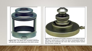
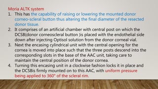
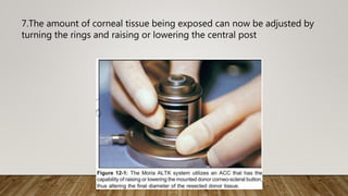
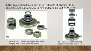
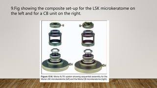
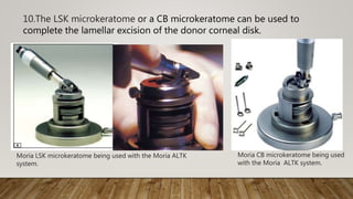
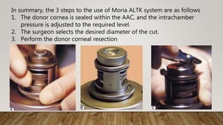
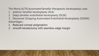
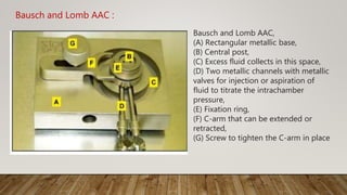
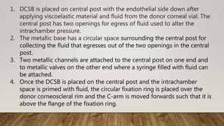
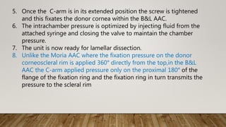
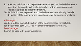
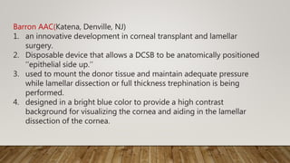
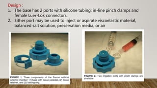
SlideShare a Scribd company logo
Artificial anterior
chamber
DR.M.DINESH
AAC(Artificial anterior chambers)
1. first described by Ward and Nesburn in 1976.
2. utilized for
1. manual lamellar dissection,
2. automated lamellar dissection of the donor cornea using a
microkeratome and
3. Surgical objective is to obtain
1. a lamellar corneal disk of desired thickness &diameter,
2. an even surface with an uniplanar cut to augment
1. the donor-host corneal interface,
2. ease of operation,
3. avoidance of disk perforation
Types of AAC
1. Reusable AAC
1. Moria AAC (manual), and Moria ALTK system (automated
with microkeratome)
2. Bausch and Lomb AAC (manual).
2. Disposable AAC
1. Barron Disposable AAC
2. Coronet AAC
Moria AAC
1. utilizes the Evolution3 console
2. fully compatible with all Moria microkeratomes including the LSK,
M2, and the CB units.
3. It runs on wall current and has a battery back-up for uninterrupted
use.
4. It has two high pumps that provide a quick and stable vacuum for
the
procedure.
5. The Moria unit for lamellar surgical procedures is called the ALTK
system(Automated lamellar therapeutic keratoplasty)
Moria AAC for ALK
1. The Hanna trephine seats well on the Moria AAC and allows for
stable
partial thickness trephination of the donor cornea from the
epithelial side. This is followed by manual lamellar dissection of
the donor corneal tissue
2. Unlike the Moria ALTK system, with the Moria AAC there is no
control of the amount of corneal tissue that is exposed within the
ring.
3. Hence the diameter of tissue resection is determined by the
diameter of the blade in the Hanna trephine.
Moria ALTK system
1. This has the capability of raising or lowering the mounted donor
corneo-scleral button thus altering the final diameter of the resected
donor tissue.
2. It comprises of an artificial chamber with central post on which the
DCSB(donor corneoscleral button )is placed with the endothelial side
down after injecting Optisol solution from the donor corneal vial.
3. Next the encasing cylindrical unit with the central opening for the
cornea is moved into place such that the three posts descend into the
corresponding slots in the base of the AAC unit, taking care to
maintain the central position of the donor cornea.
4. Turning this encasing unit in a clockwise fashion locks it in place and
the DCSBis firmly mounted on to this AAC, with uniform pressure
being applied to 360° of the scleral rim.
5. The intrachamber pressure can be titrated by injecting additional
fluid from the attached syringe into the chamber, and locking the
valve if the pressure is low, or alternatively aspirate fluid from the
chamber if the intrachamber pressure is too high. The pressure
can be checked using a Barraquer tonometer
6. Depending on whether a LSK(LASIK) or CB(Corrazio-Barraquer)
microkeratome is used with this unit, the appropriate ring is
chosen and screwed on to the unit.
7.The amount of corneal tissue being exposed can now be adjusted by
turning the rings and raising or lowering the central post
8.The applanation lenses provide an estimate of diameter to be
resected, ranging from 6 to 11 mm and for a CB unit on the right.
(A) Moria ALTK system with applanating lenses
ranging from 6.0 to 9.0 mm in diameter.
(B) Moria ALTK system showing
sequential assembly prior to use
9.Fig showing the composite set-up for the LSK microkeratome on
the left and for a CB unit on the right.
10.The LSK microkeratome or a CB microkeratome can be used to
complete the lamellar excision of the donor corneal disk.
Moria LSK microkeratome being used with the Moria ALTK
system.
Moria CB microkeratome being used
with the Moria ALTK system.
In summary, the 3 steps to the use of Moria ALTK system are as follows
1. The donor cornea is sealed within the AAC, and the intrachamber
pressure is adjusted to the required level.
2. The surgeon selects the desired diameter of the cut.
3. Perform the donor corneal resection
The Moria ALTK(Automated lamellar therapeutic keratoplasty) uses
1. anterior lamellar keratoplasty (ALK)
2. Deep lamellar endothelial keratoplasty (DLEK)
3. Descemet Stripping Automated Endothelial Keratoplasty (DSAEK)
Advantages :
1. Reduced corneal astigmatism
2. smooth keratectomy with seamless-edge margin
Bausch and Lomb AAC :
Bausch and Lomb AAC,
(A) Rectangular metallic base,
(B) Central post,
(C) Excess fluid collects in this space,
(D) Two metallic channels with metallic
valves for injection or aspiration of
fluid to titrate the intrachamber
pressure,
(E) Fixation ring,
(F) C-arm that can be extended or
retracted,
(G) Screw to tighten the C-arm in place
1. DCSB is placed on central post with the endothelial side down after
applying viscoelastic material and fluid from the donor corneal vial. The
central post has two openings for egress of fluid used to alter the
intrachamber pressure.
2. The metallic base has a circular space surrounding the central post for
collecting the fluid that egresses out of the two openings in the central
post.
3. Two metallic channels are attached to the central post on one end and
to metallic valves on the other end where a syringe filled with fluid can
be attached.
4. Once the DCSB is placed on the central post and the intrachamber
space is primed with fluid, the circular fixation ring is placed over the
donor corneoscleral rim and the C-arm is moved forwards such that it is
above the flange of the fixation ring.
5. Once the C-arm is in its extended position the screw is tightened
and this fixates the donor cornea within the B&L AAC.
6. The intrachamber pressure is optimized by injecting fluid from the
attached syringe and closing the valve to maintain the chamber
pressure.
7. The unit is now ready for lamellar dissection.
8. Unlike the Moria AAC where the fixation pressure on the donor
corneoscleral rim is applied 360° directly from the top,in the B&L
AAC the C-arm applied pressure only on the proximal 180° of the
flange of the fixation ring and the fixation ring in turn transmits the
pressure to the scleral rim
9. A Barron radial vacuum trephine (Katena, Inc.) of the desired diameter is
placed on the moistened, epithelial surface of the donor cornea and
suction is applied to fixate the trephine.
10. Partial thickness trephination to desired corneal depth is f/by lamellar
dissection of the donor cornea to obtain a lamellar donor corneal disk
Advantages :
can be used for manual dissection of the donor lamellar corneal disk.
can be used for both DLEK and for anterior lamellar keratoplasty.
Disadvantages :
Cannot be used with a microkeratome.
Barron AAC(Katena, Denville, NJ)
1. an innovative development in corneal transplant and lamellar
surgery.
2. Disposable device that allows a DCSB to be anatomically positioned
‘‘epithelial side up.’’
3. used to mount the donor tissue and maintain adequate pressure
while lamellar dissection or full thickness trephination is being
performed.
4. designed in a bright blue color to provide a high contrast
background for visualizing the cornea and aiding in the lamellar
dissection of the cornea.
Design :
1. The base has 2 ports with silicone tubing: in-line pinch clamps and
female Luer-Lok connectors.
2. Either port may be used to inject or aspirate viscoelastic material,
balanced salt solution, preservation media, or air
3.The design of the chamber allows the
surgeon to firmly hold the 14- to 18-mm
DCSB on a bed of viscoelastic material,
pressurize it, and cut it from the epithelial
side.
4.The chamber is designed with a peripheral angle so that the surgeon
can still use a regular set of lamellar dissection instruments with the
disposable
chamber
DCSB is placed onto the base
the tissue retainer is carefully placed over the
unit with the guideposts aligned;
tissue retainer is advanced to the bottom of the
base.
AAC is then secured by placing the locking ring
over the unit and turning it until the locked position
is reached.
Main steps
step-by-step description of preparing the DCSB using Barron AAC
1.Instruments req
1. Disassemble the components of Barron AAC
2. Have available the lamellar dissection instruments,
3. donor trephine punch,
4. guarded trifaceted diamond knife
5. gentian violet marking pen,
6. corneal preservation media
7. balanced salt solution
8. cohesive viscoelastic material,
9. 25-gauge needle,
10. Merocel sponges
2. Prepare 2 syringes and fill them with desired fluid or air medium (ie,
Optisol-GS solution and balanced salt solution).
3. Connect the syringes to the 2 ports attached to the base of the
artificial anterior chamber. Keep the pinch locks in the open position.
4. Purge the first port with balanced salt solution and snap the pinch lock
shut. Remove the excess solution from the base of the chamber.
5. Using Optisol-GS solution, fill the second port until the fluid covers the
base of the chamber. Leave the pinch lock in the open position
6. Apply viscoelastic material to the endothelial side of corneoscleral
button.
7. Place the DCSB on the base of the AAC, avoiding any air pockets to
develop under the graft once the DCSB is on the base.
8. Place the tissue retainer over the DCSB , aligning the guideposts;
then, advance the tissue retainer to the bottom of the chamber
base.
9. Place the locking ring over the tissue retainer. While holding the
tissue retainer with 1 hand, use the other hand to rotate the locking
ring into the closed position. The DCSB is now securely in place.
10. Infuse the Optisol-GS solution through the irrigation
port and shut the pinch lock once the desired pressure is achieved.
11.Proceed with the lamellar dissection as follows:
• Scrape off the epithelium of donor graft. Mark the temporal side
and the horizontal direction on donor cornea.
• Prepare the surface markings on cornea as reference.
• Make a peripheral incision using the trifaceted diamond knife set at
a depth of 0.4 mm.
• Proceed with typical lamellar dissection
12. Release some pressure within the AAC by unlocking the pinch lock on
the Optisol-GS solution irrigation port.
13. Place a Merocel sponge soaked in Optisol-GS solution on the corneal
surface and proceed with the preparation of the procedures for the
recipient
14. To remove the corneoscleral button, release the locking ring with 1
hand while holding onto the tissue retainer with the other hand.
15. Grasp the wings of the tissue retainer and gently remove the retainer
from chamber base.
16. Take the retainer ring off while keeping the cornea on the chamber
base by gently using a needle to retain the donor tissue on the AAC.
17. While the surgeon is separating the cornea from the chamber, the
assistant should maintain pressure through the Optisol-GS solution
irrigation port to prevent tissue collapse or buckling.
18. Mount the donor cornea (epithelial side down) onto a punch block
using the previously made central marking on the surface of the
cornea as a guide. Use a donor trephine to punch out the donor
disc. Dry the periphery using a Merocel sponge. Ensure that the
posterior lamellar disc can be separated from the anterior corneal
layers.
19. Fold the disc (endothelial side in) with an approximate 60/40 ratio.
Make sure that a very thin layer of viscoelastic material protects the
endothelial layer. The disc may now be inserted into the anterior
chamber.
advantages
1. diminish donor-host mismatch with full-thickness PK
2. reduced cost,
3. less time consuming and cumbersome
disadvantage
1. its design does not allow for the use of the chamber in conjunction
with a microkeratome.
2. it may take some experience to get AACustomed to the removal of
the dissected donor cornea from the AAC
complication
1. inability to create a seal between the base of the chamber, the
donor cornea, and the tissue retainer ring.
Coronet AAC
1. Supplied with 2 tissue retaining heads (small 14.5-16mm, large 16-
18mm)
2. Single chamber design can be adapted to fit graft tissue between
14.5mm and 18.0mm
3. Provides improved approximation b/w the donor button and patient
cornea
4. Designed to allow required intraocular pressure to be recreated
5. Constructed to provide a leak free chamber
6. Graticule cross hairs for improved precision
7. Suitable for use with both manual trephines and femto-second lasers
8. Weighted and broad base gives a stable platform
9. Supplied sterile, single use only, one per box, 5 year shelf life
Tissue retaining heads
Prerequisite :
The DCSB should have a minimal scleral skirt of 3.6 mm for successful
use in an AAC for lamellar or penetrating keratoplasty
(epithelial approach)
New non contact nonmechanical trephination using
1. femtosecond laser
2. excimer laser
3. Er:YAG laser
Advantages
1. it is performed from the epithelial side in both donor & recipient;
2. it avoids mechanical distortion during trephination by creating
smooth and congruent cut edges
3. eliminates a major factor for postkeratoplasty astigmatism
Caution
1. right exposure parameters, such as repetition rate and rotation
speed
to reduce the thermal effects of the laser
THAN Q
Ref:
1. Corneal Endothelial Transplant by Thomas John MD USA
2. Use of a Disposable AAC for Trans-Epithelial Trephination and
Endothelial Keratoplasty 2007 Lippincott Williams & Wilkins,
The Moria automated lamellar therapeutic keratoplasty system
(Moria/Microtek Inc, Doylestown, PA) includes
1. a reusable stainless steel AACused to support the corneoscleral rim.
2. designed with a gearless track on the base plate to AACept the use
of a microkeratome head.
3. The grooved base plate enables the microkeratome to pass across
the cornea maintaining the same plane and direction.
4. The Moria chamber offers the surgeon a reusable chamber and a
fast method of preparing donor tissue of any diameter
The Bausch & Lomb AACis also available to surgeons (Bausch & Lomb,
St Louis, MO)

More Related Content

What's hot

Endothelial keratoplasty
Endothelial keratoplastyEndothelial keratoplasty
Endothelial keratoplasty
ShreyaBhargava10
 
Complications of trabeculectomy
Complications of trabeculectomyComplications of trabeculectomy
Complications of trabeculectomy
Sumeet Agrawal
 
Bandage Contact Lens
Bandage Contact LensBandage Contact Lens
Bandage Contact Lens
Loknath Goswami
 
Trabeculectomy
TrabeculectomyTrabeculectomy
Trabeculectomy
Sadhwini Harish
 
Pseudoexfoliation syndrome
Pseudoexfoliation syndromePseudoexfoliation syndrome
Pseudoexfoliation syndrome
Gloria George
 
DME Management Guidelines
DME Management GuidelinesDME Management Guidelines
DME Management Guidelines
Reyad Yossif
 
Vitreous substitutes
Vitreous substitutesVitreous substitutes
Vitreous substitutes
SSSIHMS-PG
 
Accommodating IOLs
Accommodating IOLsAccommodating IOLs
Accommodating IOLs
Visionary Ophthamology
 
Surgical strategies for small pupils - Malyugin Ring
Surgical strategies for small pupils - Malyugin RingSurgical strategies for small pupils - Malyugin Ring
Surgical strategies for small pupils - Malyugin Ring
MicroSurgical Technology
 
Femtosecond laser assisted cataract surgery
Femtosecond laser assisted cataract surgeryFemtosecond laser assisted cataract surgery
Femtosecond laser assisted cataract surgery
VIMSAROPHTHALMOLOGYD
 
Iridocorneal endothelial syndrome
Iridocorneal endothelial syndromeIridocorneal endothelial syndrome
Iridocorneal endothelial syndrome
SSSIHMS-PG
 
Corneal collagen cross linking
Corneal collagen cross linkingCorneal collagen cross linking
Corneal collagen cross linkingPaavan Kalra
 
MIVS
MIVSMIVS
Choroidal neovascular membranes (CNVM)
Choroidal neovascular membranes (CNVM)Choroidal neovascular membranes (CNVM)
Choroidal neovascular membranes (CNVM)
Md Riyaj Ali
 
Minimally invasive glaucoma surgery
Minimally invasive glaucoma surgery Minimally invasive glaucoma surgery
Minimally invasive glaucoma surgery
aditisingh77985
 
Pellucid marginal degeneration
Pellucid marginal degenerationPellucid marginal degeneration
Pellucid marginal degeneration
Pushpraj Singh
 
Corneal degenerations
Corneal degenerationsCorneal degenerations
Corneal degenerations
drkvasantha
 
Complications of cataract surgery
Complications of cataract surgeryComplications of cataract surgery
Complications of cataract surgery
Dr Laltanpuia Chhangte
 
Peripheral fundus & its disorders
Peripheral fundus & its disordersPeripheral fundus & its disorders
Peripheral fundus & its disorders
Rohit Rao
 
Femtosecond laser assistedcataractsurgery
Femtosecond laser assistedcataractsurgeryFemtosecond laser assistedcataractsurgery
Femtosecond laser assistedcataractsurgerysolinskyeyecare
 

What's hot (20)

Endothelial keratoplasty
Endothelial keratoplastyEndothelial keratoplasty
Endothelial keratoplasty
 
Complications of trabeculectomy
Complications of trabeculectomyComplications of trabeculectomy
Complications of trabeculectomy
 
Bandage Contact Lens
Bandage Contact LensBandage Contact Lens
Bandage Contact Lens
 
Trabeculectomy
TrabeculectomyTrabeculectomy
Trabeculectomy
 
Pseudoexfoliation syndrome
Pseudoexfoliation syndromePseudoexfoliation syndrome
Pseudoexfoliation syndrome
 
DME Management Guidelines
DME Management GuidelinesDME Management Guidelines
DME Management Guidelines
 
Vitreous substitutes
Vitreous substitutesVitreous substitutes
Vitreous substitutes
 
Accommodating IOLs
Accommodating IOLsAccommodating IOLs
Accommodating IOLs
 
Surgical strategies for small pupils - Malyugin Ring
Surgical strategies for small pupils - Malyugin RingSurgical strategies for small pupils - Malyugin Ring
Surgical strategies for small pupils - Malyugin Ring
 
Femtosecond laser assisted cataract surgery
Femtosecond laser assisted cataract surgeryFemtosecond laser assisted cataract surgery
Femtosecond laser assisted cataract surgery
 
Iridocorneal endothelial syndrome
Iridocorneal endothelial syndromeIridocorneal endothelial syndrome
Iridocorneal endothelial syndrome
 
Corneal collagen cross linking
Corneal collagen cross linkingCorneal collagen cross linking
Corneal collagen cross linking
 
MIVS
MIVSMIVS
MIVS
 
Choroidal neovascular membranes (CNVM)
Choroidal neovascular membranes (CNVM)Choroidal neovascular membranes (CNVM)
Choroidal neovascular membranes (CNVM)
 
Minimally invasive glaucoma surgery
Minimally invasive glaucoma surgery Minimally invasive glaucoma surgery
Minimally invasive glaucoma surgery
 
Pellucid marginal degeneration
Pellucid marginal degenerationPellucid marginal degeneration
Pellucid marginal degeneration
 
Corneal degenerations
Corneal degenerationsCorneal degenerations
Corneal degenerations
 
Complications of cataract surgery
Complications of cataract surgeryComplications of cataract surgery
Complications of cataract surgery
 
Peripheral fundus & its disorders
Peripheral fundus & its disordersPeripheral fundus & its disorders
Peripheral fundus & its disorders
 
Femtosecond laser assistedcataractsurgery
Femtosecond laser assistedcataractsurgeryFemtosecond laser assistedcataractsurgery
Femtosecond laser assistedcataractsurgery
 

Similar to Artificial anterior chamber

Lt0540 a ac graftrope
Lt0540 a ac graftropeLt0540 a ac graftrope
Lt0540 a ac graftropedrnaula
 
Manual small incision cataract surgery
Manual small incision cataract surgeryManual small incision cataract surgery
Manual small incision cataract surgery
medusae1
 
Recent advances in dcr
Recent advances in dcrRecent advances in dcr
Recent advances in dcr
Dinesh Madduri
 
PHACO STEP.pptx
PHACO STEP.pptxPHACO STEP.pptx
PHACO STEP.pptx
dianlistyanti1
 
Dr Prakash Bam MSICS.pptx
Dr Prakash Bam MSICS.pptxDr Prakash Bam MSICS.pptx
Dr Prakash Bam MSICS.pptx
PrakashBam
 
Phacoemulsification part 3
Phacoemulsification part 3Phacoemulsification part 3
Phacoemulsification part 3
Priyanka Raj
 
Obturation of root canal systems
Obturation of root canal systemsObturation of root canal systems
Obturation of root canal systems
Mohammed Sa'ad
 
Lec 7 - Anas Mahdee.pdf.................
Lec 7 - Anas Mahdee.pdf.................Lec 7 - Anas Mahdee.pdf.................
Lec 7 - Anas Mahdee.pdf.................
AboAmjed1
 
faizal s ppt op.pptx
faizal s ppt op.pptxfaizal s ppt op.pptx
faizal s ppt op.pptx
44faizalm
 
Glaucoma drainage devices[1]
Glaucoma drainage devices[1]Glaucoma drainage devices[1]
Glaucoma drainage devices[1]
paresh nichlani
 
Glaucoma drainage devices
Glaucoma drainage devicesGlaucoma drainage devices
Glaucoma drainage devices
vaishusmail
 
Cataract surgery revisited
Cataract surgery revisitedCataract surgery revisited
Cataract surgery revisited
DINESH and SONALEE
 
Corrocoat Composite Nozzle Insert
Corrocoat Composite Nozzle InsertCorrocoat Composite Nozzle Insert
Corrocoat Composite Nozzle Insert
3M
 
Immobilization in radiotherapy
Immobilization  in  radiotherapyImmobilization  in  radiotherapy
Immobilization in radiotherapy
Kiran Ramakrishna
 
The “Bony Bankart Bridge” Procedure Shoulder Instability | Shoudler Surgery |...
The “Bony Bankart Bridge” Procedure Shoulder Instability | Shoudler Surgery |...The “Bony Bankart Bridge” Procedure Shoulder Instability | Shoudler Surgery |...
The “Bony Bankart Bridge” Procedure Shoulder Instability | Shoudler Surgery |...
Peter Millett MD
 
mgmtofpcr-171026135845iooojjjjjikjjjjjkkkk
mgmtofpcr-171026135845iooojjjjjikjjjjjkkkkmgmtofpcr-171026135845iooojjjjjikjjjjjkkkk
mgmtofpcr-171026135845iooojjjjjikjjjjjkkkk
preetiagarwal53
 
Mgmt of pcr
Mgmt of pcrMgmt of pcr
Mgmt of pcr
Poonam Shrestha
 
Intracapsular Cataract extraction
Intracapsular Cataract extraction Intracapsular Cataract extraction
Intracapsular Cataract extraction
JESLIN JOSE
 
Gdd
Gdd Gdd
Gdd
Nilay P
 
Dental implant surgery
Dental implant surgeryDental implant surgery
Dental implant surgery
s farrokhi
 

Similar to Artificial anterior chamber (20)

Lt0540 a ac graftrope
Lt0540 a ac graftropeLt0540 a ac graftrope
Lt0540 a ac graftrope
 
Manual small incision cataract surgery
Manual small incision cataract surgeryManual small incision cataract surgery
Manual small incision cataract surgery
 
Recent advances in dcr
Recent advances in dcrRecent advances in dcr
Recent advances in dcr
 
PHACO STEP.pptx
PHACO STEP.pptxPHACO STEP.pptx
PHACO STEP.pptx
 
Dr Prakash Bam MSICS.pptx
Dr Prakash Bam MSICS.pptxDr Prakash Bam MSICS.pptx
Dr Prakash Bam MSICS.pptx
 
Phacoemulsification part 3
Phacoemulsification part 3Phacoemulsification part 3
Phacoemulsification part 3
 
Obturation of root canal systems
Obturation of root canal systemsObturation of root canal systems
Obturation of root canal systems
 
Lec 7 - Anas Mahdee.pdf.................
Lec 7 - Anas Mahdee.pdf.................Lec 7 - Anas Mahdee.pdf.................
Lec 7 - Anas Mahdee.pdf.................
 
faizal s ppt op.pptx
faizal s ppt op.pptxfaizal s ppt op.pptx
faizal s ppt op.pptx
 
Glaucoma drainage devices[1]
Glaucoma drainage devices[1]Glaucoma drainage devices[1]
Glaucoma drainage devices[1]
 
Glaucoma drainage devices
Glaucoma drainage devicesGlaucoma drainage devices
Glaucoma drainage devices
 
Cataract surgery revisited
Cataract surgery revisitedCataract surgery revisited
Cataract surgery revisited
 
Corrocoat Composite Nozzle Insert
Corrocoat Composite Nozzle InsertCorrocoat Composite Nozzle Insert
Corrocoat Composite Nozzle Insert
 
Immobilization in radiotherapy
Immobilization  in  radiotherapyImmobilization  in  radiotherapy
Immobilization in radiotherapy
 
The “Bony Bankart Bridge” Procedure Shoulder Instability | Shoudler Surgery |...
The “Bony Bankart Bridge” Procedure Shoulder Instability | Shoudler Surgery |...The “Bony Bankart Bridge” Procedure Shoulder Instability | Shoudler Surgery |...
The “Bony Bankart Bridge” Procedure Shoulder Instability | Shoudler Surgery |...
 
mgmtofpcr-171026135845iooojjjjjikjjjjjkkkk
mgmtofpcr-171026135845iooojjjjjikjjjjjkkkkmgmtofpcr-171026135845iooojjjjjikjjjjjkkkk
mgmtofpcr-171026135845iooojjjjjikjjjjjkkkk
 
Mgmt of pcr
Mgmt of pcrMgmt of pcr
Mgmt of pcr
 
Intracapsular Cataract extraction
Intracapsular Cataract extraction Intracapsular Cataract extraction
Intracapsular Cataract extraction
 
Gdd
Gdd Gdd
Gdd
 
Dental implant surgery
Dental implant surgeryDental implant surgery
Dental implant surgery
 

More from Dinesh Madduri

Anterior segment OCT & UBM
Anterior segment OCT & UBMAnterior segment OCT & UBM
Anterior segment OCT & UBM
Dinesh Madduri
 
corneo scleral wound healing in sics
corneo scleral wound healing in sics corneo scleral wound healing in sics
corneo scleral wound healing in sics
Dinesh Madduri
 
ocular Toxoplasmosis
ocular Toxoplasmosisocular Toxoplasmosis
ocular Toxoplasmosis
Dinesh Madduri
 
Sics steps
Sics stepsSics steps
Sics steps
Dinesh Madduri
 
Oct in post seg disorders
Oct in post seg disordersOct in post seg disorders
Oct in post seg disorders
Dinesh Madduri
 
Slitlampexamination dinesh
Slitlampexamination dineshSlitlampexamination dinesh
Slitlampexamination dinesh
Dinesh Madduri
 
Neurotrophic keratopathy
Neurotrophic keratopathyNeurotrophic keratopathy
Neurotrophic keratopathy
Dinesh Madduri
 
Keratoconus f dinesh
Keratoconus f dineshKeratoconus f dinesh
Keratoconus f dinesh
Dinesh Madduri
 
Confocal microscopy dinesh
Confocal microscopy dineshConfocal microscopy dinesh
Confocal microscopy dinesh
Dinesh Madduri
 
Evaluation and initial management of patients with ocular and adnexal trauma
Evaluation and initial management of patients with ocular and adnexal traumaEvaluation and initial management of patients with ocular and adnexal trauma
Evaluation and initial management of patients with ocular and adnexal trauma
Dinesh Madduri
 
Cmv retinitis
Cmv retinitisCmv retinitis
Cmv retinitis
Dinesh Madduri
 
Biologicals in uveitis
Biologicals in uveitisBiologicals in uveitis
Biologicals in uveitis
Dinesh Madduri
 
Bacterial keratitis
Bacterial keratitisBacterial keratitis
Bacterial keratitis
Dinesh Madduri
 

More from Dinesh Madduri (14)

Anterior segment OCT & UBM
Anterior segment OCT & UBMAnterior segment OCT & UBM
Anterior segment OCT & UBM
 
corneo scleral wound healing in sics
corneo scleral wound healing in sics corneo scleral wound healing in sics
corneo scleral wound healing in sics
 
ocular Toxoplasmosis
ocular Toxoplasmosisocular Toxoplasmosis
ocular Toxoplasmosis
 
Sics steps
Sics stepsSics steps
Sics steps
 
Oct in post seg disorders
Oct in post seg disordersOct in post seg disorders
Oct in post seg disorders
 
Slitlampexamination dinesh
Slitlampexamination dineshSlitlampexamination dinesh
Slitlampexamination dinesh
 
Neurotrophic keratopathy
Neurotrophic keratopathyNeurotrophic keratopathy
Neurotrophic keratopathy
 
Keratoconus f dinesh
Keratoconus f dineshKeratoconus f dinesh
Keratoconus f dinesh
 
Confocal microscopy dinesh
Confocal microscopy dineshConfocal microscopy dinesh
Confocal microscopy dinesh
 
Evaluation and initial management of patients with ocular and adnexal trauma
Evaluation and initial management of patients with ocular and adnexal traumaEvaluation and initial management of patients with ocular and adnexal trauma
Evaluation and initial management of patients with ocular and adnexal trauma
 
Cmv retinitis
Cmv retinitisCmv retinitis
Cmv retinitis
 
Biologicals in uveitis
Biologicals in uveitisBiologicals in uveitis
Biologicals in uveitis
 
Botox
BotoxBotox
Botox
 
Bacterial keratitis
Bacterial keratitisBacterial keratitis
Bacterial keratitis
 

Recently uploaded

TESDA TM1 REVIEWER FOR NATIONAL ASSESSMENT WRITTEN AND ORAL QUESTIONS WITH A...
TESDA TM1 REVIEWER  FOR NATIONAL ASSESSMENT WRITTEN AND ORAL QUESTIONS WITH A...TESDA TM1 REVIEWER  FOR NATIONAL ASSESSMENT WRITTEN AND ORAL QUESTIONS WITH A...
TESDA TM1 REVIEWER FOR NATIONAL ASSESSMENT WRITTEN AND ORAL QUESTIONS WITH A...
EugeneSaldivar
 
Introduction to Quality Improvement Essentials
Introduction to Quality Improvement EssentialsIntroduction to Quality Improvement Essentials
Introduction to Quality Improvement Essentials
Excellence Foundation for South Sudan
 
Synthetic Fiber Construction in lab .pptx
Synthetic Fiber Construction in lab .pptxSynthetic Fiber Construction in lab .pptx
Synthetic Fiber Construction in lab .pptx
Pavel ( NSTU)
 
2024.06.01 Introducing a competency framework for languag learning materials ...
2024.06.01 Introducing a competency framework for languag learning materials ...2024.06.01 Introducing a competency framework for languag learning materials ...
2024.06.01 Introducing a competency framework for languag learning materials ...
Sandy Millin
 
Students, digital devices and success - Andreas Schleicher - 27 May 2024..pptx
Students, digital devices and success - Andreas Schleicher - 27 May 2024..pptxStudents, digital devices and success - Andreas Schleicher - 27 May 2024..pptx
Students, digital devices and success - Andreas Schleicher - 27 May 2024..pptx
EduSkills OECD
 
How to Split Bills in the Odoo 17 POS Module
How to Split Bills in the Odoo 17 POS ModuleHow to Split Bills in the Odoo 17 POS Module
How to Split Bills in the Odoo 17 POS Module
Celine George
 
Phrasal Verbs.XXXXXXXXXXXXXXXXXXXXXXXXXX
Phrasal Verbs.XXXXXXXXXXXXXXXXXXXXXXXXXXPhrasal Verbs.XXXXXXXXXXXXXXXXXXXXXXXXXX
Phrasal Verbs.XXXXXXXXXXXXXXXXXXXXXXXXXX
MIRIAMSALINAS13
 
Ethnobotany and Ethnopharmacology ......
Ethnobotany and Ethnopharmacology ......Ethnobotany and Ethnopharmacology ......
Ethnobotany and Ethnopharmacology ......
Ashokrao Mane college of Pharmacy Peth-Vadgaon
 
The Challenger.pdf DNHS Official Publication
The Challenger.pdf DNHS Official PublicationThe Challenger.pdf DNHS Official Publication
The Challenger.pdf DNHS Official Publication
Delapenabediema
 
ESC Beyond Borders _From EU to You_ InfoPack general.pdf
ESC Beyond Borders _From EU to You_ InfoPack general.pdfESC Beyond Borders _From EU to You_ InfoPack general.pdf
ESC Beyond Borders _From EU to You_ InfoPack general.pdf
Fundacja Rozwoju Społeczeństwa Przedsiębiorczego
 
The approach at University of Liverpool.pptx
The approach at University of Liverpool.pptxThe approach at University of Liverpool.pptx
The approach at University of Liverpool.pptx
Jisc
 
Additional Benefits for Employee Website.pdf
Additional Benefits for Employee Website.pdfAdditional Benefits for Employee Website.pdf
Additional Benefits for Employee Website.pdf
joachimlavalley1
 
Cambridge International AS A Level Biology Coursebook - EBook (MaryFosbery J...
Cambridge International AS  A Level Biology Coursebook - EBook (MaryFosbery J...Cambridge International AS  A Level Biology Coursebook - EBook (MaryFosbery J...
Cambridge International AS A Level Biology Coursebook - EBook (MaryFosbery J...
AzmatAli747758
 
How to Make a Field invisible in Odoo 17
How to Make a Field invisible in Odoo 17How to Make a Field invisible in Odoo 17
How to Make a Field invisible in Odoo 17
Celine George
 
Sectors of the Indian Economy - Class 10 Study Notes pdf
Sectors of the Indian Economy - Class 10 Study Notes pdfSectors of the Indian Economy - Class 10 Study Notes pdf
Sectors of the Indian Economy - Class 10 Study Notes pdf
Vivekanand Anglo Vedic Academy
 
MARUTI SUZUKI- A Successful Joint Venture in India.pptx
MARUTI SUZUKI- A Successful Joint Venture in India.pptxMARUTI SUZUKI- A Successful Joint Venture in India.pptx
MARUTI SUZUKI- A Successful Joint Venture in India.pptx
bennyroshan06
 
Home assignment II on Spectroscopy 2024 Answers.pdf
Home assignment II on Spectroscopy 2024 Answers.pdfHome assignment II on Spectroscopy 2024 Answers.pdf
Home assignment II on Spectroscopy 2024 Answers.pdf
Tamralipta Mahavidyalaya
 
Language Across the Curriculm LAC B.Ed.
Language Across the  Curriculm LAC B.Ed.Language Across the  Curriculm LAC B.Ed.
Language Across the Curriculm LAC B.Ed.
Atul Kumar Singh
 
1.4 modern child centered education - mahatma gandhi-2.pptx
1.4 modern child centered education - mahatma gandhi-2.pptx1.4 modern child centered education - mahatma gandhi-2.pptx
1.4 modern child centered education - mahatma gandhi-2.pptx
JosvitaDsouza2
 
The Roman Empire A Historical Colossus.pdf
The Roman Empire A Historical Colossus.pdfThe Roman Empire A Historical Colossus.pdf
The Roman Empire A Historical Colossus.pdf
kaushalkr1407
 

Recently uploaded (20)

TESDA TM1 REVIEWER FOR NATIONAL ASSESSMENT WRITTEN AND ORAL QUESTIONS WITH A...
TESDA TM1 REVIEWER  FOR NATIONAL ASSESSMENT WRITTEN AND ORAL QUESTIONS WITH A...TESDA TM1 REVIEWER  FOR NATIONAL ASSESSMENT WRITTEN AND ORAL QUESTIONS WITH A...
TESDA TM1 REVIEWER FOR NATIONAL ASSESSMENT WRITTEN AND ORAL QUESTIONS WITH A...
 
Introduction to Quality Improvement Essentials
Introduction to Quality Improvement EssentialsIntroduction to Quality Improvement Essentials
Introduction to Quality Improvement Essentials
 
Synthetic Fiber Construction in lab .pptx
Synthetic Fiber Construction in lab .pptxSynthetic Fiber Construction in lab .pptx
Synthetic Fiber Construction in lab .pptx
 
2024.06.01 Introducing a competency framework for languag learning materials ...
2024.06.01 Introducing a competency framework for languag learning materials ...2024.06.01 Introducing a competency framework for languag learning materials ...
2024.06.01 Introducing a competency framework for languag learning materials ...
 
Students, digital devices and success - Andreas Schleicher - 27 May 2024..pptx
Students, digital devices and success - Andreas Schleicher - 27 May 2024..pptxStudents, digital devices and success - Andreas Schleicher - 27 May 2024..pptx
Students, digital devices and success - Andreas Schleicher - 27 May 2024..pptx
 
How to Split Bills in the Odoo 17 POS Module
How to Split Bills in the Odoo 17 POS ModuleHow to Split Bills in the Odoo 17 POS Module
How to Split Bills in the Odoo 17 POS Module
 
Phrasal Verbs.XXXXXXXXXXXXXXXXXXXXXXXXXX
Phrasal Verbs.XXXXXXXXXXXXXXXXXXXXXXXXXXPhrasal Verbs.XXXXXXXXXXXXXXXXXXXXXXXXXX
Phrasal Verbs.XXXXXXXXXXXXXXXXXXXXXXXXXX
 
Ethnobotany and Ethnopharmacology ......
Ethnobotany and Ethnopharmacology ......Ethnobotany and Ethnopharmacology ......
Ethnobotany and Ethnopharmacology ......
 
The Challenger.pdf DNHS Official Publication
The Challenger.pdf DNHS Official PublicationThe Challenger.pdf DNHS Official Publication
The Challenger.pdf DNHS Official Publication
 
ESC Beyond Borders _From EU to You_ InfoPack general.pdf
ESC Beyond Borders _From EU to You_ InfoPack general.pdfESC Beyond Borders _From EU to You_ InfoPack general.pdf
ESC Beyond Borders _From EU to You_ InfoPack general.pdf
 
The approach at University of Liverpool.pptx
The approach at University of Liverpool.pptxThe approach at University of Liverpool.pptx
The approach at University of Liverpool.pptx
 
Additional Benefits for Employee Website.pdf
Additional Benefits for Employee Website.pdfAdditional Benefits for Employee Website.pdf
Additional Benefits for Employee Website.pdf
 
Cambridge International AS A Level Biology Coursebook - EBook (MaryFosbery J...
Cambridge International AS  A Level Biology Coursebook - EBook (MaryFosbery J...Cambridge International AS  A Level Biology Coursebook - EBook (MaryFosbery J...
Cambridge International AS A Level Biology Coursebook - EBook (MaryFosbery J...
 
How to Make a Field invisible in Odoo 17
How to Make a Field invisible in Odoo 17How to Make a Field invisible in Odoo 17
How to Make a Field invisible in Odoo 17
 
Sectors of the Indian Economy - Class 10 Study Notes pdf
Sectors of the Indian Economy - Class 10 Study Notes pdfSectors of the Indian Economy - Class 10 Study Notes pdf
Sectors of the Indian Economy - Class 10 Study Notes pdf
 
MARUTI SUZUKI- A Successful Joint Venture in India.pptx
MARUTI SUZUKI- A Successful Joint Venture in India.pptxMARUTI SUZUKI- A Successful Joint Venture in India.pptx
MARUTI SUZUKI- A Successful Joint Venture in India.pptx
 
Home assignment II on Spectroscopy 2024 Answers.pdf
Home assignment II on Spectroscopy 2024 Answers.pdfHome assignment II on Spectroscopy 2024 Answers.pdf
Home assignment II on Spectroscopy 2024 Answers.pdf
 
Language Across the Curriculm LAC B.Ed.
Language Across the  Curriculm LAC B.Ed.Language Across the  Curriculm LAC B.Ed.
Language Across the Curriculm LAC B.Ed.
 
1.4 modern child centered education - mahatma gandhi-2.pptx
1.4 modern child centered education - mahatma gandhi-2.pptx1.4 modern child centered education - mahatma gandhi-2.pptx
1.4 modern child centered education - mahatma gandhi-2.pptx
 
The Roman Empire A Historical Colossus.pdf
The Roman Empire A Historical Colossus.pdfThe Roman Empire A Historical Colossus.pdf
The Roman Empire A Historical Colossus.pdf
 

Artificial anterior chamber

  • 2. AAC(Artificial anterior chambers) 1. first described by Ward and Nesburn in 1976. 2. utilized for 1. manual lamellar dissection, 2. automated lamellar dissection of the donor cornea using a microkeratome and 3. Surgical objective is to obtain 1. a lamellar corneal disk of desired thickness &diameter, 2. an even surface with an uniplanar cut to augment 1. the donor-host corneal interface, 2. ease of operation, 3. avoidance of disk perforation
  • 3. Types of AAC 1. Reusable AAC 1. Moria AAC (manual), and Moria ALTK system (automated with microkeratome) 2. Bausch and Lomb AAC (manual). 2. Disposable AAC 1. Barron Disposable AAC 2. Coronet AAC
  • 4. Moria AAC 1. utilizes the Evolution3 console 2. fully compatible with all Moria microkeratomes including the LSK, M2, and the CB units. 3. It runs on wall current and has a battery back-up for uninterrupted use. 4. It has two high pumps that provide a quick and stable vacuum for the procedure. 5. The Moria unit for lamellar surgical procedures is called the ALTK system(Automated lamellar therapeutic keratoplasty)
  • 5. Moria AAC for ALK 1. The Hanna trephine seats well on the Moria AAC and allows for stable partial thickness trephination of the donor cornea from the epithelial side. This is followed by manual lamellar dissection of the donor corneal tissue 2. Unlike the Moria ALTK system, with the Moria AAC there is no control of the amount of corneal tissue that is exposed within the ring. 3. Hence the diameter of tissue resection is determined by the diameter of the blade in the Hanna trephine.
  • 6.
  • 7. Moria ALTK system 1. This has the capability of raising or lowering the mounted donor corneo-scleral button thus altering the final diameter of the resected donor tissue. 2. It comprises of an artificial chamber with central post on which the DCSB(donor corneoscleral button )is placed with the endothelial side down after injecting Optisol solution from the donor corneal vial. 3. Next the encasing cylindrical unit with the central opening for the cornea is moved into place such that the three posts descend into the corresponding slots in the base of the AAC unit, taking care to maintain the central position of the donor cornea. 4. Turning this encasing unit in a clockwise fashion locks it in place and the DCSBis firmly mounted on to this AAC, with uniform pressure being applied to 360° of the scleral rim.
  • 8.
  • 9. 5. The intrachamber pressure can be titrated by injecting additional fluid from the attached syringe into the chamber, and locking the valve if the pressure is low, or alternatively aspirate fluid from the chamber if the intrachamber pressure is too high. The pressure can be checked using a Barraquer tonometer 6. Depending on whether a LSK(LASIK) or CB(Corrazio-Barraquer) microkeratome is used with this unit, the appropriate ring is chosen and screwed on to the unit.
  • 10. 7.The amount of corneal tissue being exposed can now be adjusted by turning the rings and raising or lowering the central post
  • 11. 8.The applanation lenses provide an estimate of diameter to be resected, ranging from 6 to 11 mm and for a CB unit on the right. (A) Moria ALTK system with applanating lenses ranging from 6.0 to 9.0 mm in diameter. (B) Moria ALTK system showing sequential assembly prior to use
  • 12. 9.Fig showing the composite set-up for the LSK microkeratome on the left and for a CB unit on the right.
  • 13. 10.The LSK microkeratome or a CB microkeratome can be used to complete the lamellar excision of the donor corneal disk. Moria LSK microkeratome being used with the Moria ALTK system. Moria CB microkeratome being used with the Moria ALTK system.
  • 14. In summary, the 3 steps to the use of Moria ALTK system are as follows 1. The donor cornea is sealed within the AAC, and the intrachamber pressure is adjusted to the required level. 2. The surgeon selects the desired diameter of the cut. 3. Perform the donor corneal resection
  • 15. The Moria ALTK(Automated lamellar therapeutic keratoplasty) uses 1. anterior lamellar keratoplasty (ALK) 2. Deep lamellar endothelial keratoplasty (DLEK) 3. Descemet Stripping Automated Endothelial Keratoplasty (DSAEK) Advantages : 1. Reduced corneal astigmatism 2. smooth keratectomy with seamless-edge margin
  • 16. Bausch and Lomb AAC : Bausch and Lomb AAC, (A) Rectangular metallic base, (B) Central post, (C) Excess fluid collects in this space, (D) Two metallic channels with metallic valves for injection or aspiration of fluid to titrate the intrachamber pressure, (E) Fixation ring, (F) C-arm that can be extended or retracted, (G) Screw to tighten the C-arm in place
  • 17. 1. DCSB is placed on central post with the endothelial side down after applying viscoelastic material and fluid from the donor corneal vial. The central post has two openings for egress of fluid used to alter the intrachamber pressure. 2. The metallic base has a circular space surrounding the central post for collecting the fluid that egresses out of the two openings in the central post. 3. Two metallic channels are attached to the central post on one end and to metallic valves on the other end where a syringe filled with fluid can be attached. 4. Once the DCSB is placed on the central post and the intrachamber space is primed with fluid, the circular fixation ring is placed over the donor corneoscleral rim and the C-arm is moved forwards such that it is above the flange of the fixation ring.
  • 18. 5. Once the C-arm is in its extended position the screw is tightened and this fixates the donor cornea within the B&L AAC. 6. The intrachamber pressure is optimized by injecting fluid from the attached syringe and closing the valve to maintain the chamber pressure. 7. The unit is now ready for lamellar dissection. 8. Unlike the Moria AAC where the fixation pressure on the donor corneoscleral rim is applied 360° directly from the top,in the B&L AAC the C-arm applied pressure only on the proximal 180° of the flange of the fixation ring and the fixation ring in turn transmits the pressure to the scleral rim
  • 19. 9. A Barron radial vacuum trephine (Katena, Inc.) of the desired diameter is placed on the moistened, epithelial surface of the donor cornea and suction is applied to fixate the trephine. 10. Partial thickness trephination to desired corneal depth is f/by lamellar dissection of the donor cornea to obtain a lamellar donor corneal disk Advantages : can be used for manual dissection of the donor lamellar corneal disk. can be used for both DLEK and for anterior lamellar keratoplasty. Disadvantages : Cannot be used with a microkeratome.
  • 20. Barron AAC(Katena, Denville, NJ) 1. an innovative development in corneal transplant and lamellar surgery. 2. Disposable device that allows a DCSB to be anatomically positioned ‘‘epithelial side up.’’ 3. used to mount the donor tissue and maintain adequate pressure while lamellar dissection or full thickness trephination is being performed. 4. designed in a bright blue color to provide a high contrast background for visualizing the cornea and aiding in the lamellar dissection of the cornea.
  • 21. Design : 1. The base has 2 ports with silicone tubing: in-line pinch clamps and female Luer-Lok connectors. 2. Either port may be used to inject or aspirate viscoelastic material, balanced salt solution, preservation media, or air
  • 22. 3.The design of the chamber allows the surgeon to firmly hold the 14- to 18-mm DCSB on a bed of viscoelastic material, pressurize it, and cut it from the epithelial side.
  • 23. 4.The chamber is designed with a peripheral angle so that the surgeon can still use a regular set of lamellar dissection instruments with the disposable chamber
  • 24. DCSB is placed onto the base the tissue retainer is carefully placed over the unit with the guideposts aligned; tissue retainer is advanced to the bottom of the base. AAC is then secured by placing the locking ring over the unit and turning it until the locked position is reached. Main steps
  • 25. step-by-step description of preparing the DCSB using Barron AAC 1.Instruments req 1. Disassemble the components of Barron AAC 2. Have available the lamellar dissection instruments, 3. donor trephine punch, 4. guarded trifaceted diamond knife 5. gentian violet marking pen, 6. corneal preservation media 7. balanced salt solution 8. cohesive viscoelastic material, 9. 25-gauge needle, 10. Merocel sponges
  • 26. 2. Prepare 2 syringes and fill them with desired fluid or air medium (ie, Optisol-GS solution and balanced salt solution). 3. Connect the syringes to the 2 ports attached to the base of the artificial anterior chamber. Keep the pinch locks in the open position. 4. Purge the first port with balanced salt solution and snap the pinch lock shut. Remove the excess solution from the base of the chamber. 5. Using Optisol-GS solution, fill the second port until the fluid covers the base of the chamber. Leave the pinch lock in the open position
  • 27. 6. Apply viscoelastic material to the endothelial side of corneoscleral button. 7. Place the DCSB on the base of the AAC, avoiding any air pockets to develop under the graft once the DCSB is on the base. 8. Place the tissue retainer over the DCSB , aligning the guideposts; then, advance the tissue retainer to the bottom of the chamber base. 9. Place the locking ring over the tissue retainer. While holding the tissue retainer with 1 hand, use the other hand to rotate the locking ring into the closed position. The DCSB is now securely in place. 10. Infuse the Optisol-GS solution through the irrigation port and shut the pinch lock once the desired pressure is achieved.
  • 28. 11.Proceed with the lamellar dissection as follows: • Scrape off the epithelium of donor graft. Mark the temporal side and the horizontal direction on donor cornea. • Prepare the surface markings on cornea as reference. • Make a peripheral incision using the trifaceted diamond knife set at a depth of 0.4 mm. • Proceed with typical lamellar dissection
  • 29. 12. Release some pressure within the AAC by unlocking the pinch lock on the Optisol-GS solution irrigation port. 13. Place a Merocel sponge soaked in Optisol-GS solution on the corneal surface and proceed with the preparation of the procedures for the recipient 14. To remove the corneoscleral button, release the locking ring with 1 hand while holding onto the tissue retainer with the other hand. 15. Grasp the wings of the tissue retainer and gently remove the retainer from chamber base. 16. Take the retainer ring off while keeping the cornea on the chamber base by gently using a needle to retain the donor tissue on the AAC. 17. While the surgeon is separating the cornea from the chamber, the assistant should maintain pressure through the Optisol-GS solution irrigation port to prevent tissue collapse or buckling.
  • 30. 18. Mount the donor cornea (epithelial side down) onto a punch block using the previously made central marking on the surface of the cornea as a guide. Use a donor trephine to punch out the donor disc. Dry the periphery using a Merocel sponge. Ensure that the posterior lamellar disc can be separated from the anterior corneal layers. 19. Fold the disc (endothelial side in) with an approximate 60/40 ratio. Make sure that a very thin layer of viscoelastic material protects the endothelial layer. The disc may now be inserted into the anterior chamber.
  • 31. advantages 1. diminish donor-host mismatch with full-thickness PK 2. reduced cost, 3. less time consuming and cumbersome disadvantage 1. its design does not allow for the use of the chamber in conjunction with a microkeratome. 2. it may take some experience to get AACustomed to the removal of the dissected donor cornea from the AAC complication 1. inability to create a seal between the base of the chamber, the donor cornea, and the tissue retainer ring.
  • 32. Coronet AAC 1. Supplied with 2 tissue retaining heads (small 14.5-16mm, large 16- 18mm) 2. Single chamber design can be adapted to fit graft tissue between 14.5mm and 18.0mm 3. Provides improved approximation b/w the donor button and patient cornea 4. Designed to allow required intraocular pressure to be recreated 5. Constructed to provide a leak free chamber 6. Graticule cross hairs for improved precision 7. Suitable for use with both manual trephines and femto-second lasers 8. Weighted and broad base gives a stable platform 9. Supplied sterile, single use only, one per box, 5 year shelf life
  • 34. Prerequisite : The DCSB should have a minimal scleral skirt of 3.6 mm for successful use in an AAC for lamellar or penetrating keratoplasty (epithelial approach)
  • 35. New non contact nonmechanical trephination using 1. femtosecond laser 2. excimer laser 3. Er:YAG laser Advantages 1. it is performed from the epithelial side in both donor & recipient; 2. it avoids mechanical distortion during trephination by creating smooth and congruent cut edges 3. eliminates a major factor for postkeratoplasty astigmatism Caution 1. right exposure parameters, such as repetition rate and rotation speed to reduce the thermal effects of the laser
  • 36. THAN Q Ref: 1. Corneal Endothelial Transplant by Thomas John MD USA 2. Use of a Disposable AAC for Trans-Epithelial Trephination and Endothelial Keratoplasty 2007 Lippincott Williams & Wilkins,
  • 37.
  • 38. The Moria automated lamellar therapeutic keratoplasty system (Moria/Microtek Inc, Doylestown, PA) includes 1. a reusable stainless steel AACused to support the corneoscleral rim. 2. designed with a gearless track on the base plate to AACept the use of a microkeratome head. 3. The grooved base plate enables the microkeratome to pass across the cornea maintaining the same plane and direction. 4. The Moria chamber offers the surgeon a reusable chamber and a fast method of preparing donor tissue of any diameter The Bausch & Lomb AACis also available to surgeons (Bausch & Lomb, St Louis, MO)