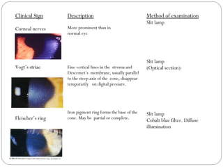
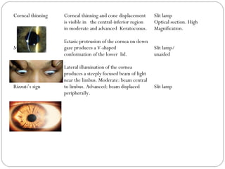
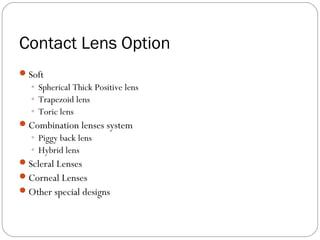
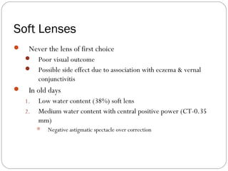
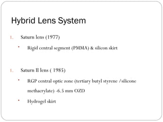
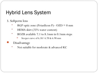
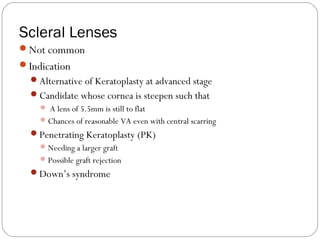
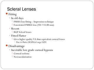
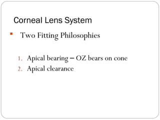
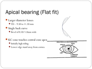
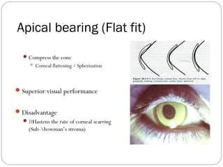
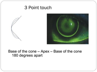
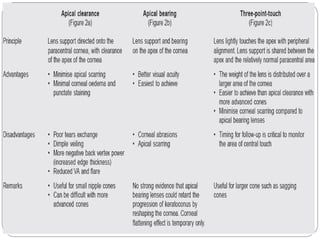
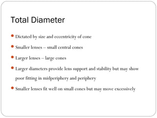
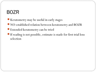
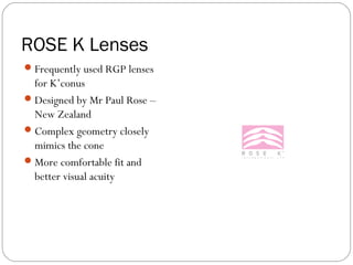
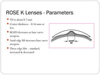
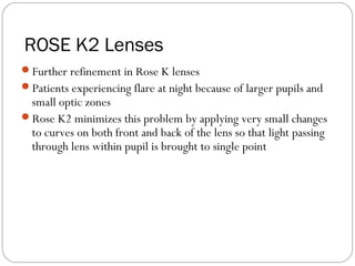
Keratoconic Contact Lenses
Rabindra Adhikary
ravinems@iom.edu.np
Introduction
Definition
◦ Bilateral, non inflammatory
condition
◦ Localized manifestation of mild
connective tissue disorder
◦ Causing corneal thinning and
ectasia
 following a weakening of the
corneal collagen
This disease produces
irregular astigmatism
myopia (from anterior corneal displacement)
central scarring
IntroductionIntroduction
Signs
Scissors reflex (swirling
retinoscopy reflex)
Distorted/ irregular Keratometer
mires with steep readings
Prominent corneal nerves
Symptoms
Frequently changing spectacle Rx
and axis of astigmatism
Ghosting/ monocular diplopia
Glare at night
Haloes around lights
Blurred/ distorted vision
Clinical Sign
Corneal nerves
Vogt’s striae
Fleischer’s ring
Description
More prominent than in
normal eye
Fine vertical lines in the stroma and
Descemet’s membrane, usually parallel
to the steep axis of the cone, disappear
temporarily on digital pressure.
Iron pigment ring forms the base of the
cone. May be partial or complete.
Method of examination
Slit lamp
Slit lamp
(Optical section)
Slit lamp
Cobalt blue filter. Diffuse
illumination
Corneal thinning
Munson’s sign
Rizzuti’s sign
Corneal thinning and cone displacement
is visible in the central-inferior region
in moderate and advanced Keratoconus.
Ectasic protrusion of the cornea on down
gaze produces a V-shaped
conformation of the lower lid.
Lateral illumination of the cornea
produces a steeply focused beam of light
near the limbus. Moderate: beam central
to limbus. Advanced: beam displaced
peripherally.
Slit lamp
Optical section. High
Magnification.
Slit lamp/
unaided
Slit lamp
Association with ocular & systemic conditions
◦ Atopic dermatitis
◦ Vernal catarrh,
◦ Down's syndrome,
◦ Retinitis pigmentosa,
◦ Infantile tapetoretinal degeneration,
◦ Marfan's syndrome,
◦ Aniridia, and blue sclera
◦ Ehlers –Danlos syndrome
◦ Osteogenesis Imperfecta
◦ Mitral Valve prolapse
Management
Spectacle
Contact lenses
Surgical
◦ Keratoplasty
◦ Epikeratoplasty
◦ Penetrating Keratoplasty
Contact Lens Option
Counterproductive
Spectacle vision is reasonable
Good unaided VA in one eye
Indicated
Enthusiastic & motivated patient
Significant Visual gain
Contact Lens Option
Soft
◦ Spherical Thick Positive lens
◦ Trapezoid lens
◦ Toric lens
Combination lenses system
◦ Piggy back lens
◦ Hybrid lens
Scleral Lenses
Corneal Lenses
Other special designs
Soft Lenses
 Never the lens of first choice
 Poor visual outcome
 Possible side effect due to association with eczema & vernal
conjunctivitis
 In old days
1. Low water content (38%) soft lens
2. Medium water content with central positive power (CT-0.35
mm)
 Negative astigmatic spectacle over correction
Soft lenses
HEMA Trapezoid Lenses
◦ Multiple fenestrated
◦ Fit like optic zone of fenestrated scleral lenses
◦ Central thickness -0.6 mm
 Good vision with out astigmatic overcorrection
Correctly fit lens
◦ Produce small peripheral circulating bubbles
Disadvantage
◦ Difficult to manufacture
◦ Lens flexure
Toric lenses
Less role
Chances of localized oedema due to lens thickness difference
Combined Lens System
 Combination of RGP & Soft lens
◦ Visual result of RGP
◦ Comfort of Soft lenses
 Two Possibilities
1. Piggy back lenses
– Two separate lenses
1. Hybrid lenses
 A single lens fabricated from two materials
Piggy Back Lens SystemPiggy Back Lens System
Helpful when RGP lens is
intolerable due to staining
and patient reluctant to
surgery
Soft lens provides comfort
RGP lens provides visual
correction
Disadvantages
◦ RGP rides low with little or no movement
◦ Localized hypoxia & Neovascularization
◦ Difficult to handle/maintain two types of lenses
Hybrid Lens System
1. Saturn lens (1977)
• Rigid central segment (PMMA) & silicon skirt
1. Saturn II lens ( 1985)
• RGP central optic zone (tertiary butyl styrene /silicone
methacrylate) -6.5 mm OZD
• Hydrogel skirt
Hybrid Lens System
3. Softperm lens
• RGP optic zone (Petasilicon P)– OZD = 8 mm
• HEMA skirt (25% water content)
• BOZR available 7.1 to 8.1mm in 0.1mm steps
 Steeper curve of 6.50/ 6.70 & 6.90 mm
 Disadvantage
• Not suitable for moderate & advanced KC
Scleral Lenses
Not common
Indication
Alternative of Keratoplasty at advanced stage
Candidate whose cornea is steepen such that
 A lens of 5.5mm is still to flat
Chances of reasonable VA even with central scarring
Penetrating Keratoplasty (PK)
Needing a larger graft
Possible graft rejection
Down’s syndrome
Scleral Lenses
Fitting
◦ In old days
 PMMA lens fitting – Impression technique
 Fenestrated PMMA lens (TD >14.00 mm)
◦ Recent
 RGP Scleral lenses
◦ Fitted Flatter
 Gives higher quality VA than equivalent corneal lenses
◦ Due to flatter BOZR & Large OZD
Disadvantage
◦ Inevitable low grade corneal hypoxia
 Corneal oedema
 Neovascularization
Corneal Lens System
 Two Fitting Philosophies
1. Apical bearing – OZ bears on cone
2. Apical clearance
Apical bearing (Flat fit)
Larger diameter lenses
TD – 9.50 to 11.50 mm
Single back curve
Bevel of 0.50/1.0mm wide
KC cone touches central cone apex
Initially high riding
Lower edge stand away from cornea
Apical bearing (Flat fit)
Compress the cone
 Corneal flattening / Spherization
Superior visual performance
Disadvantage
??Hastens the rate of corneal scarring
(Sub-bowman’s stroma)
Apical clearance
Small diameter & thin lenses (USA)
TD of 6.00 mm to 8.00 mm
BOZR – 5.00mm to 7.5 mm
With Two flatter peripheral curves
• Advantage
– Less role on corneal scarring
– Well tolerated by atopic eye disease
• Disadvantage
– Optical
• Flare/monocular diplopia
– OZD is only 4 mm
3 Point touch
Also known as ‘divided support’
Most weight of the lens is on
almost normal peripheral cornea
Central cornea is supported by
slight touch
Bearing is not heavy to cause
abrasion & scarring
Things to avoid
Peripheral fit too tight causing
sealing off the tear exchange
behind optic zone
Excessive movement that
causes discomfort and corneal
scarring
3 Point touch
Base of the cone – Apex – Base of the cone
180 degrees apart
Total Diameter
Dictated by size and eccentricity of cone
Smaller lenses – small central cones
Larger lenses – large cones
Larger diameters provide lens support and stability but may show
poor fitting in midperiphery and periphery
Smaller lenses fit well on small cones but may move excessively
BOZR
Keratometry may be useful in early stages
NO established relation between keratometry and BOZR
Extended keratometry can be tried
If reading is not possible, estimate is made for first trial lens
selection
Optic Zone Diameter
Usually smaller OZD preferred to conform to the aspheric
cornea ( between 6 to 7.2 mm)
More advanced the cone, smaller the OZD
Intermediate & Peripheral Curves
Intermediate curve is about 2 mm flatter than the base curve ( BOZR)
Peripheral curve (PC) is about 2 mm flatter than the intermediate
curve
Steep PC – inadequate tear reservoir, restricted lens movement, lens
adherence
Flat PC – lid irritation, excessive lens movement
Material
Good wettability and dimensional stability preferred over high
Dk
Materials with high Dk are generally more flexible, so low Dk
materials may be preferred
Special design lenses
Soper lens system
Given by Soper and Jarrett
 Based on sagittal depth
Mild (7.5-mm diameter, 6.0-mm optic zone diameter)
Moderate (8.5-mm diameter, 7.0-mm optic zone diameter)
Advanced (9.5-mm diameter, 8.0-mm optic zone diameter)
Soper lens system
The initial trial lens is selected on the basis of degree of
advancement of the cone.
more advanced the cone larger the diameter of lens is
recommended
For smaller and more centrally located the apex smaller
diameter lens.
McGuire lens system
Modification of the Soper lens design
Nipple (8.1 mm diameter, 5.5 mm optic zone)
Oval (8.6 mm diameter, 6 mm optic zone)
Globus (9.1 mm diameter, 6.5 mm optic zone)
Four peripheral curves
Three inner curves are each 0.3 mm wide and the peripheral
curve is 0.4 mm wide.
NiCone lens
Available from Lancaster Contact Lens Co, Lancaster
Three base curves and one constant peripheral curve of 12.25
mm.
Designated by the numbers 1 to 3
The Number 1 cone set is for patients with Keratometry
readings between 40 and 52 D
Number 2 set covers from 53 to 65 D
Number 3 set is for readings >65 D.
NiCone lens
The second base curve is a 0.3-mm "transition zone" between
the central base curve and the "third base curve," which rests on
normal peripheral cornea.
ROSE K Lenses
Frequently used RGP lenses
for K’conus
Designed by Mr Paul Rose –
New Zealand
Complex geometry closely
mimics the cone
More comfortable fit and
better visual acuity
ROSE K Lenses - Parameters
TD is about 8.7 mm
Center thickness – 0.16 mm or
less
BOZD decreases as base curve
steepens
Axial edge lift increases base curve
steepens
Three edge lifts – standard,
increased & decreased
ROSE K Lenses - Parameters
Front surface lenticulations used in some bases
BOZR – 4.75 to 8.00 mm
OD – 7.90 to 10.20 mm
Material – Boston ES
Design – Spherical back surface, full back surface toric or a toric
periphery only
ROSE K2 Lenses
Further refinement in Rose K lenses
Patients experiencing flare at night because of larger pupils and
small optic zones
Rose K2 minimizes this problem by applying very small changes
to curves on both front and back of the lens so that light passing
through lens within pupil is brought to single point
REFERENCES
Contact lenses
A.J. Phillips
IACLE modules
Class notes
Internet
Thank you!!

Contact lens in keratoconus

  • 1.
    Keratoconic Contact Lenses RabindraAdhikary ravinems@iom.edu.np
  • 2.
    Introduction Definition ◦ Bilateral, noninflammatory condition ◦ Localized manifestation of mild connective tissue disorder ◦ Causing corneal thinning and ectasia  following a weakening of the corneal collagen
  • 3.
    This disease produces irregularastigmatism myopia (from anterior corneal displacement) central scarring IntroductionIntroduction
  • 4.
    Signs Scissors reflex (swirling retinoscopyreflex) Distorted/ irregular Keratometer mires with steep readings Prominent corneal nerves Symptoms Frequently changing spectacle Rx and axis of astigmatism Ghosting/ monocular diplopia Glare at night Haloes around lights Blurred/ distorted vision
  • 5.
    Clinical Sign Corneal nerves Vogt’sstriae Fleischer’s ring Description More prominent than in normal eye Fine vertical lines in the stroma and Descemet’s membrane, usually parallel to the steep axis of the cone, disappear temporarily on digital pressure. Iron pigment ring forms the base of the cone. May be partial or complete. Method of examination Slit lamp Slit lamp (Optical section) Slit lamp Cobalt blue filter. Diffuse illumination
  • 6.
    Corneal thinning Munson’s sign Rizzuti’ssign Corneal thinning and cone displacement is visible in the central-inferior region in moderate and advanced Keratoconus. Ectasic protrusion of the cornea on down gaze produces a V-shaped conformation of the lower lid. Lateral illumination of the cornea produces a steeply focused beam of light near the limbus. Moderate: beam central to limbus. Advanced: beam displaced peripherally. Slit lamp Optical section. High Magnification. Slit lamp/ unaided Slit lamp
  • 8.
    Association with ocular& systemic conditions ◦ Atopic dermatitis ◦ Vernal catarrh, ◦ Down's syndrome, ◦ Retinitis pigmentosa, ◦ Infantile tapetoretinal degeneration, ◦ Marfan's syndrome, ◦ Aniridia, and blue sclera ◦ Ehlers –Danlos syndrome ◦ Osteogenesis Imperfecta ◦ Mitral Valve prolapse
  • 9.
  • 10.
    Contact Lens Option Counterproductive Spectaclevision is reasonable Good unaided VA in one eye Indicated Enthusiastic & motivated patient Significant Visual gain
  • 11.
    Contact Lens Option Soft ◦Spherical Thick Positive lens ◦ Trapezoid lens ◦ Toric lens Combination lenses system ◦ Piggy back lens ◦ Hybrid lens Scleral Lenses Corneal Lenses Other special designs
  • 12.
    Soft Lenses  Neverthe lens of first choice  Poor visual outcome  Possible side effect due to association with eczema & vernal conjunctivitis  In old days 1. Low water content (38%) soft lens 2. Medium water content with central positive power (CT-0.35 mm)  Negative astigmatic spectacle over correction
  • 13.
    Soft lenses HEMA TrapezoidLenses ◦ Multiple fenestrated ◦ Fit like optic zone of fenestrated scleral lenses ◦ Central thickness -0.6 mm  Good vision with out astigmatic overcorrection Correctly fit lens ◦ Produce small peripheral circulating bubbles Disadvantage ◦ Difficult to manufacture ◦ Lens flexure
  • 14.
    Toric lenses Less role Chancesof localized oedema due to lens thickness difference
  • 15.
    Combined Lens System Combination of RGP & Soft lens ◦ Visual result of RGP ◦ Comfort of Soft lenses  Two Possibilities 1. Piggy back lenses – Two separate lenses 1. Hybrid lenses  A single lens fabricated from two materials
  • 16.
    Piggy Back LensSystemPiggy Back Lens System Helpful when RGP lens is intolerable due to staining and patient reluctant to surgery Soft lens provides comfort RGP lens provides visual correction
  • 17.
    Disadvantages ◦ RGP rideslow with little or no movement ◦ Localized hypoxia & Neovascularization ◦ Difficult to handle/maintain two types of lenses
  • 18.
    Hybrid Lens System 1.Saturn lens (1977) • Rigid central segment (PMMA) & silicon skirt 1. Saturn II lens ( 1985) • RGP central optic zone (tertiary butyl styrene /silicone methacrylate) -6.5 mm OZD • Hydrogel skirt
  • 19.
    Hybrid Lens System 3.Softperm lens • RGP optic zone (Petasilicon P)– OZD = 8 mm • HEMA skirt (25% water content) • BOZR available 7.1 to 8.1mm in 0.1mm steps  Steeper curve of 6.50/ 6.70 & 6.90 mm  Disadvantage • Not suitable for moderate & advanced KC
  • 20.
    Scleral Lenses Not common Indication Alternativeof Keratoplasty at advanced stage Candidate whose cornea is steepen such that  A lens of 5.5mm is still to flat Chances of reasonable VA even with central scarring Penetrating Keratoplasty (PK) Needing a larger graft Possible graft rejection Down’s syndrome
  • 21.
    Scleral Lenses Fitting ◦ Inold days  PMMA lens fitting – Impression technique  Fenestrated PMMA lens (TD >14.00 mm) ◦ Recent  RGP Scleral lenses ◦ Fitted Flatter  Gives higher quality VA than equivalent corneal lenses ◦ Due to flatter BOZR & Large OZD Disadvantage ◦ Inevitable low grade corneal hypoxia  Corneal oedema  Neovascularization
  • 22.
    Corneal Lens System Two Fitting Philosophies 1. Apical bearing – OZ bears on cone 2. Apical clearance
  • 23.
    Apical bearing (Flatfit) Larger diameter lenses TD – 9.50 to 11.50 mm Single back curve Bevel of 0.50/1.0mm wide KC cone touches central cone apex Initially high riding Lower edge stand away from cornea
  • 24.
    Apical bearing (Flatfit) Compress the cone  Corneal flattening / Spherization Superior visual performance Disadvantage ??Hastens the rate of corneal scarring (Sub-bowman’s stroma)
  • 25.
    Apical clearance Small diameter& thin lenses (USA) TD of 6.00 mm to 8.00 mm BOZR – 5.00mm to 7.5 mm With Two flatter peripheral curves
  • 26.
    • Advantage – Lessrole on corneal scarring – Well tolerated by atopic eye disease • Disadvantage – Optical • Flare/monocular diplopia – OZD is only 4 mm
  • 27.
    3 Point touch Alsoknown as ‘divided support’ Most weight of the lens is on almost normal peripheral cornea Central cornea is supported by slight touch Bearing is not heavy to cause abrasion & scarring
  • 28.
    Things to avoid Peripheralfit too tight causing sealing off the tear exchange behind optic zone Excessive movement that causes discomfort and corneal scarring
  • 29.
    3 Point touch Baseof the cone – Apex – Base of the cone 180 degrees apart
  • 31.
    Total Diameter Dictated bysize and eccentricity of cone Smaller lenses – small central cones Larger lenses – large cones Larger diameters provide lens support and stability but may show poor fitting in midperiphery and periphery Smaller lenses fit well on small cones but may move excessively
  • 32.
    BOZR Keratometry may beuseful in early stages NO established relation between keratometry and BOZR Extended keratometry can be tried If reading is not possible, estimate is made for first trial lens selection
  • 33.
    Optic Zone Diameter Usuallysmaller OZD preferred to conform to the aspheric cornea ( between 6 to 7.2 mm) More advanced the cone, smaller the OZD
  • 34.
    Intermediate & PeripheralCurves Intermediate curve is about 2 mm flatter than the base curve ( BOZR) Peripheral curve (PC) is about 2 mm flatter than the intermediate curve Steep PC – inadequate tear reservoir, restricted lens movement, lens adherence Flat PC – lid irritation, excessive lens movement
  • 35.
    Material Good wettability anddimensional stability preferred over high Dk Materials with high Dk are generally more flexible, so low Dk materials may be preferred
  • 36.
  • 37.
    Soper lens system Givenby Soper and Jarrett  Based on sagittal depth Mild (7.5-mm diameter, 6.0-mm optic zone diameter) Moderate (8.5-mm diameter, 7.0-mm optic zone diameter) Advanced (9.5-mm diameter, 8.0-mm optic zone diameter)
  • 38.
    Soper lens system Theinitial trial lens is selected on the basis of degree of advancement of the cone. more advanced the cone larger the diameter of lens is recommended For smaller and more centrally located the apex smaller diameter lens.
  • 39.
    McGuire lens system Modificationof the Soper lens design Nipple (8.1 mm diameter, 5.5 mm optic zone) Oval (8.6 mm diameter, 6 mm optic zone) Globus (9.1 mm diameter, 6.5 mm optic zone) Four peripheral curves Three inner curves are each 0.3 mm wide and the peripheral curve is 0.4 mm wide.
  • 40.
    NiCone lens Available fromLancaster Contact Lens Co, Lancaster Three base curves and one constant peripheral curve of 12.25 mm. Designated by the numbers 1 to 3 The Number 1 cone set is for patients with Keratometry readings between 40 and 52 D Number 2 set covers from 53 to 65 D Number 3 set is for readings >65 D.
  • 41.
    NiCone lens The secondbase curve is a 0.3-mm "transition zone" between the central base curve and the "third base curve," which rests on normal peripheral cornea.
  • 42.
    ROSE K Lenses Frequentlyused RGP lenses for K’conus Designed by Mr Paul Rose – New Zealand Complex geometry closely mimics the cone More comfortable fit and better visual acuity
  • 43.
    ROSE K Lenses- Parameters TD is about 8.7 mm Center thickness – 0.16 mm or less BOZD decreases as base curve steepens Axial edge lift increases base curve steepens Three edge lifts – standard, increased & decreased
  • 44.
    ROSE K Lenses- Parameters Front surface lenticulations used in some bases BOZR – 4.75 to 8.00 mm OD – 7.90 to 10.20 mm Material – Boston ES Design – Spherical back surface, full back surface toric or a toric periphery only
  • 45.
    ROSE K2 Lenses Furtherrefinement in Rose K lenses Patients experiencing flare at night because of larger pupils and small optic zones Rose K2 minimizes this problem by applying very small changes to curves on both front and back of the lens so that light passing through lens within pupil is brought to single point
  • 46.
  • 47.