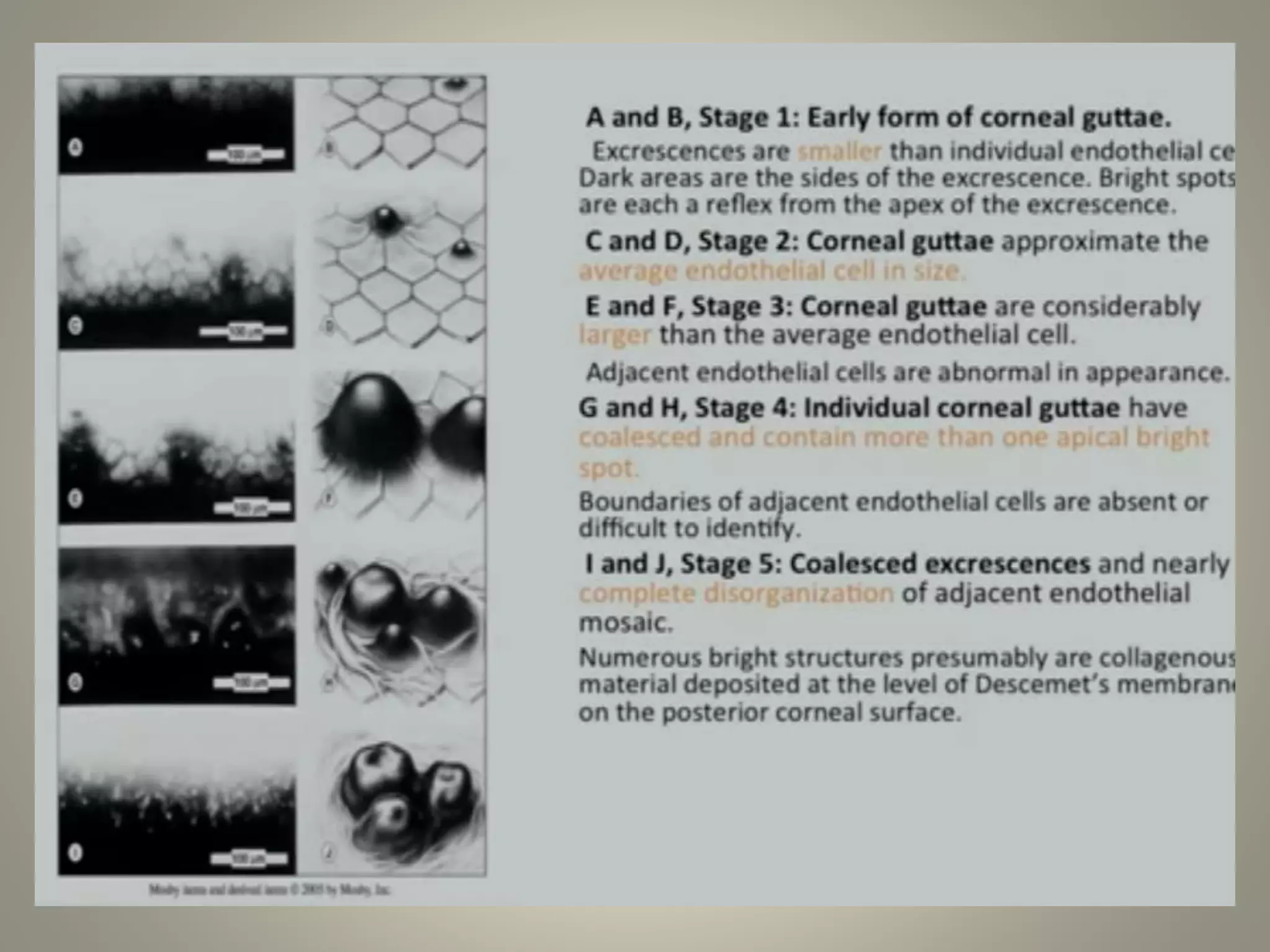
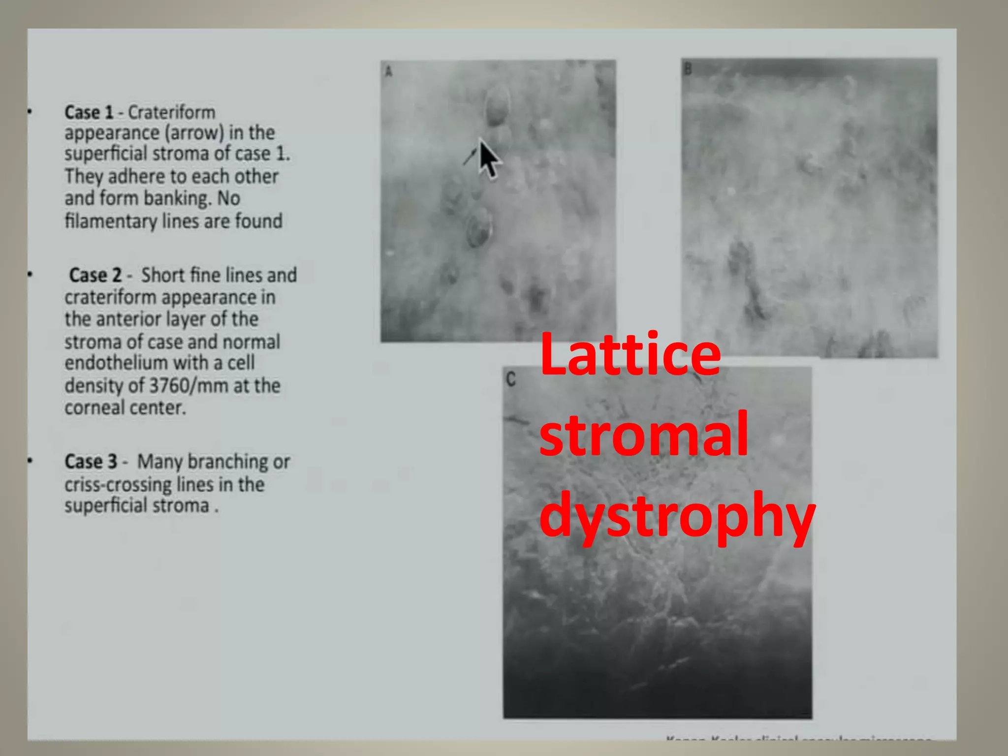
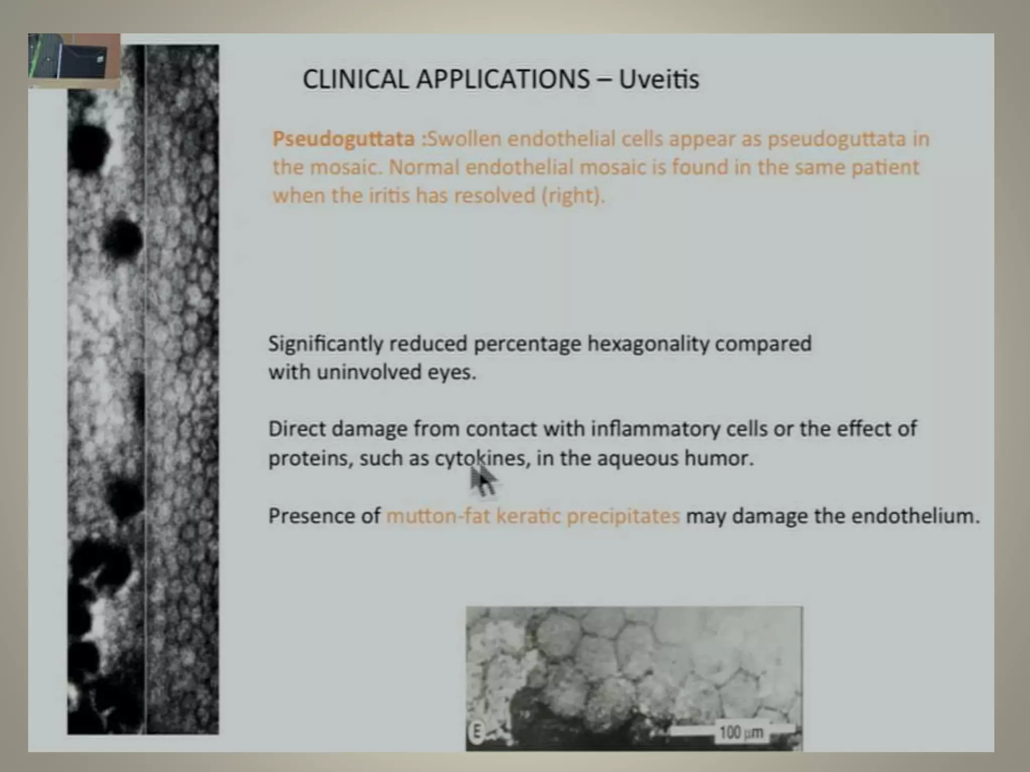
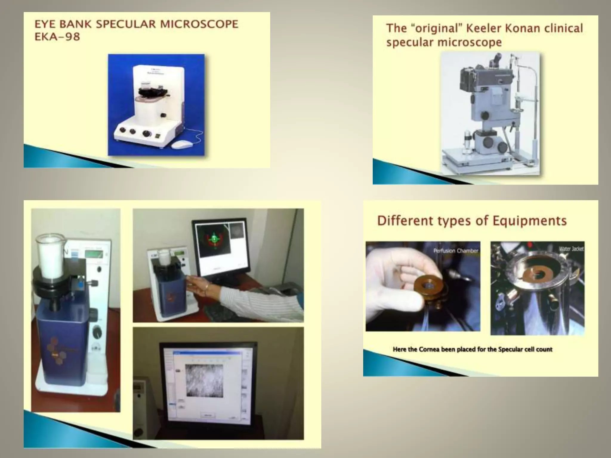
Specular microscopy
Dr.Ruchi sood
• Introduction
• Principle
• Types
• Procedure and analysis
• Clinical applications
• Limitation
Principle
Types
• Good resolution and magnification
• Patients discomfort, infections, artifactsContact
• Comfort, less infections/trauma, broader field of view
• less resolution and magnification
Non-
contact
• 10-15 times more resolution and field of view
• Improves optics so less reflections hindranceWide-field
Procedure
• Explain the procedure
• Patient should be seated comfortably for a good scan.
• NON Contact- blink
• Contact- topical anesthesia coupling fluid used
• Internal fixation target is used to keep eye straight
Image analysis
Clinical applications
 To study the normal endothelium morphology.
 To diagnose and study pathological changes in corneal
endothelial diseases.
 Eye banking- assessment of donor cornea status.
 Pre operative endothelial health – surgery decision making.
 Research and monitoring of epithelium in below
Aging
DM
CONTACT LENS
WEARER
POST SURGERY
GLAUCOMAS
UVEITIS
TRAUMA
COMPARISION OF
DIFFERENT
SURGICAL TECH.
• Corneal endothelial disorders
Fuch’s endothelial dystrophy
Posterior polymorphous dystrophy
Iridocorneal endothelial disorder
Lattice corneal dystrophy
DM tear
Fuch’s endothelial corneal
dystrophy
Ice syndrome
Lattice
stromal
dystrophy
DIABETICS
Surgery Endothelial cell loss
PHOCOEMULSIFICATION 5-8% in 6 months
10.5% at 1 year
SICS 4.21% at 6 weeks
PKP 11%+-20% at 6 months
20% at 1 year
DALK 14.2%+-11.7% at 1 month
8.6% at 1 year
DSAEK 34%-+22% at 6 months
Same at 1 year
DMEK 35% at 6 months
38% at 1year
PPV 9%+-14.6% at 3 months
TRABECULAECTOMY 7%
summary
• Explain the procedure to the patient
• Image the region 3x at the same sitting
• Use same analysis method during follow-up
• To obtain maximum efficacy count 75-100 cells or more
Specular microscopy

Specular microscopy

  • 1.
  • 2.
    • Introduction • Principle •Types • Procedure and analysis • Clinical applications • Limitation
  • 4.
  • 8.
    Types • Good resolutionand magnification • Patients discomfort, infections, artifactsContact • Comfort, less infections/trauma, broader field of view • less resolution and magnification Non- contact • 10-15 times more resolution and field of view • Improves optics so less reflections hindranceWide-field
  • 9.
    Procedure • Explain theprocedure • Patient should be seated comfortably for a good scan. • NON Contact- blink • Contact- topical anesthesia coupling fluid used • Internal fixation target is used to keep eye straight
  • 12.
  • 22.
  • 23.
     To studythe normal endothelium morphology.  To diagnose and study pathological changes in corneal endothelial diseases.  Eye banking- assessment of donor cornea status.  Pre operative endothelial health – surgery decision making.  Research and monitoring of epithelium in below Aging DM CONTACT LENS WEARER POST SURGERY GLAUCOMAS UVEITIS TRAUMA COMPARISION OF DIFFERENT SURGICAL TECH.
  • 24.
    • Corneal endothelialdisorders Fuch’s endothelial dystrophy Posterior polymorphous dystrophy Iridocorneal endothelial disorder Lattice corneal dystrophy DM tear
  • 26.
  • 30.
  • 31.
  • 33.
  • 39.
    Surgery Endothelial cellloss PHOCOEMULSIFICATION 5-8% in 6 months 10.5% at 1 year SICS 4.21% at 6 weeks PKP 11%+-20% at 6 months 20% at 1 year DALK 14.2%+-11.7% at 1 month 8.6% at 1 year DSAEK 34%-+22% at 6 months Same at 1 year DMEK 35% at 6 months 38% at 1year PPV 9%+-14.6% at 3 months TRABECULAECTOMY 7%
  • 40.
    summary • Explain theprocedure to the patient • Image the region 3x at the same sitting • Use same analysis method during follow-up • To obtain maximum efficacy count 75-100 cells or more