Download as PDF, PPTX
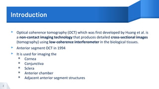
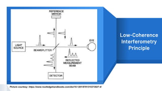
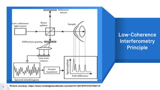
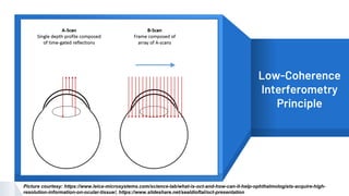
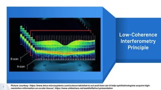
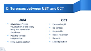
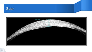
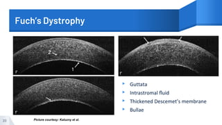
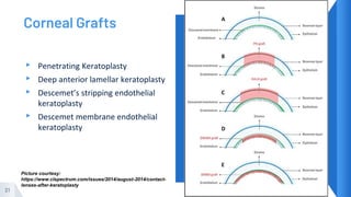
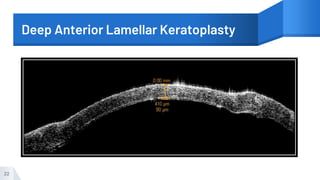
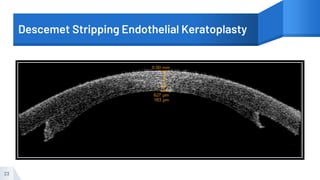
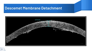
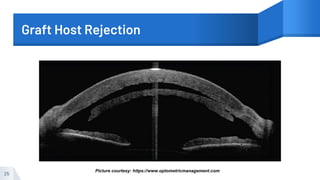
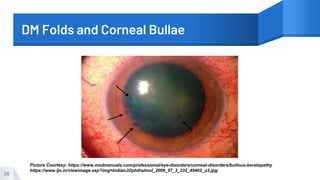
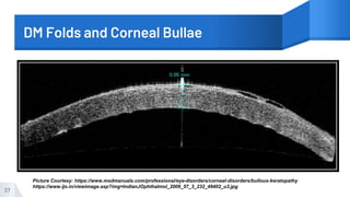
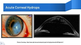
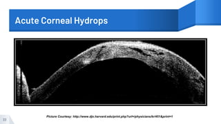
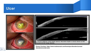
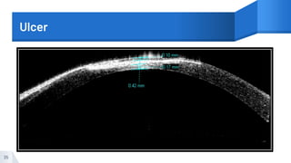
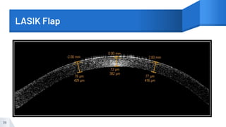
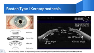
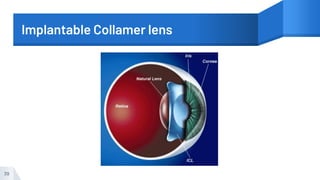
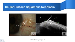
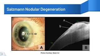
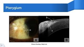
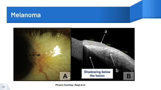
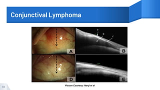
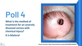
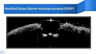
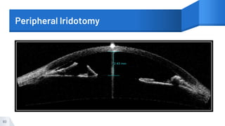
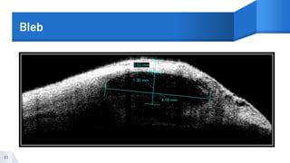
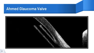


This document discusses the application of anterior segment optical coherence tomography (AS-OCT) in diagnosing various ocular conditions. It provides an overview of AS-OCT imaging principles and compares it to ultrasound biomicroscopy. The document then examines the use of AS-OCT in diagnosing and monitoring conditions of the cornea, conjunctiva, and anterior chamber angle/glaucoma. Examples of pathologies that can be identified include corneal scars, Fuchs' dystrophy, graft rejection, angle closure, and bleb assessment after glaucoma surgery.


