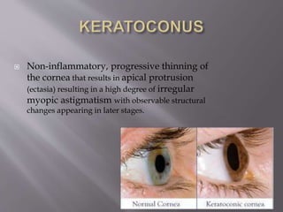
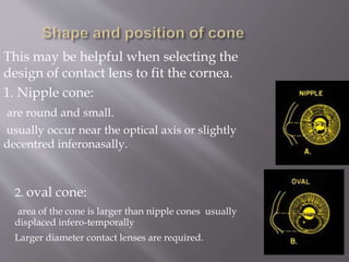
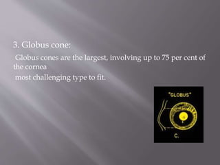
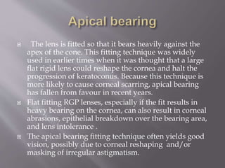
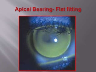
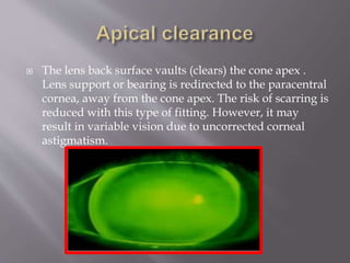
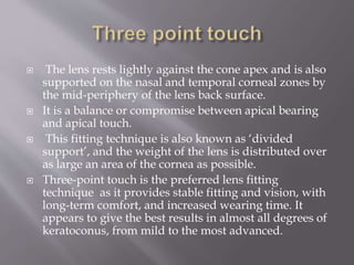
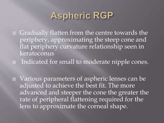
KAMAL THAKUR
3RD YEAR BSC OPTO
NETHRADHAMA SCHOOL
OF OPTO
 Non-inflammatory, progressive thinning of
the cornea that results in apical protrusion
(ectasia) resulting in a high degree of irregular
myopic astigmatism with observable structural
changes appearing in later stages.
This may be helpful when selecting the
design of contact lens to fit the cornea.
1. Nipple cone:
are round and small.
usually occur near the optical axis or slightly
decentred inferonasally.
2. oval cone:
area of the cone is larger than nipple cones usually
displaced infero-temporally
Larger diameter contact lenses are required.
3. Globus cone:
Globus cones are the largest, involving up to 75 per cent of
the cornea
most challenging type to fit.
 Mild keratoconus in early stage can be
corrected with spectacles.
 As the cornea steepens and becomes more
irregular, glasses not capable of providing
adequate visual improvement.
 Once glasses fail to provide adequate visual function,
contact lens fitting is required.
.
Contact lens wear:
 Improves VA by creating a regular anterior refractive
surface
 Does not prevent progression of Keratoconus.
 May occasionally induce or hasten progression of
Keratoconus.
 Contact lenses give sharper vision than spectacles even in mildest
cases of Keratoconus.
 As Keratoconus progresses spectacle best corrected acuity
becomes unsatisfactory.
 Contact lens fitting in a keratoconic cornea is much more difficult
 Because of the irregular anterior surface of the keratoconic
cornea.
• Provide best possible vision
• Minimize interference with corneal physiology
• Optimize lens comfort
• Relocate lens bearing area to the mid-peripheral
cornea.
 Soft contact lenses play a very limited role in
the correction of keratoconus. In the early
stages of the progression, soft lenses may
provide acceptable visual correction, Especially
if used in combination with an astigmatic
spectacle over-correction.
 Cost effective, easily available, suitable for mild
to moderate keratoconus.
 Mostly they are spherical back toric.
 Spherical back curve with peripheral toric for
high corneal astigmatism.
 Three different general fitting philosophies .
1. Apical bearing.
2. Apical clearance.
3. Three point touch.
 The lens is fitted so that it bears heavily against the
apex of the cone. This fitting technique was widely
used in earlier times when it was thought that a large
flat rigid lens could reshape the cornea and halt the
progression of keratoconus. Because this technique is
more likely to cause corneal scarring, apical bearing
has fallen from favour in recent years.
 Flat fitting RGP lenses, especially if the fit results in
heavy bearing on the cornea, can also result in corneal
abrasions, epithelial breakdown over the bearing area,
and lens intolerance .
 The apical bearing fitting technique often yields good
vision, possibly due to corneal reshaping and/or
masking of irregular astigmatism.
 The lens back surface vaults (clears) the cone apex .
Lens support or bearing is redirected to the paracentral
cornea, away from the cone apex. The risk of scarring is
reduced with this type of fitting. However, it may
result in variable vision due to uncorrected corneal
astigmatism.
 The lens rests lightly against the cone apex and is also
supported on the nasal and temporal corneal zones by
the mid-periphery of the lens back surface.
 It is a balance or compromise between apical bearing
and apical touch.
 This fitting technique is also known as ‘divided
support’, and the weight of the lens is distributed over
as large an area of the cornea as possible.
 Three-point touch is the preferred lens fitting
technique as it provides stable fitting and vision, with
long-term comfort, and increased wearing time. It
appears to give the best results in almost all degrees of
keratoconus, from mild to the most advanced.
 Gradually flatten from the centre towards the
periphery, approximating the steep cone and
flat periphery curvature relationship seen in
keratoconus
 Indicated for small to moderate nipple cones.
 Various parameters of aspheric lenses can be
adjusted to achieve the best fit. The more
advanced and steeper the cone the greater the
rate of peripheral flattening required for the
lens to approximate the corneal shape.
Combined lens system
 Piggy back system.
 Hybrid lens system.
 Rigid lens fitted over a hydrogel lens, increases
comfort resulting in adequate wearing time
with good vision
 A hybrid lens has a rigid central portion with
the optical neutralizing properties of a normal
rigid lens and a soft peripheral portion for
improved wearer comfort.
•Unique keratoconus lens design with complex
computer-generated peripheral curves based on data
collected by Dr Paul Rose of Hamilton, New Zealand.
•Standard diameter: 8.7 mm
•BOZD decreases and axial edge lift increases as base
curve steepens
Rose K2
The Rose K2 keratoconus lens design is the most widely prescribed
keratoconus lens in the world. It is a multi-spherical posterior design
with aberration control aspheric optics across the back and front optic
zone diameters and is available in any toric lens design.
Rose K2 IC
Rose K2 NC Nipple Cone
Standard lens designs with fixed optical zones (OZ) do not ideally fit the
cone shape of keratoconus patients. A standard lens that will yield
unwanted pooling at the base of the cone and peripheral bearing that can
seal off and cause corneal problems.
Smaller optical zone to fit the cone contour. The design results in
little tear pooling at the base of the cone and shows an even
distribution of tears under the lens.
Rose K2 NC Nipple Cone
The latest addition to the Rose K2 range of lenses for keratoconus. The
Rose K2 NC primary indication is for advanced and moderate nipple
cones and secondary application for any defined nipple cone. The
design characteristics are a very small aspheric back optic zone
diameter which decreases as the base curve steepens and a very rapid
peripheral flattening from the back optic zone.
Rose K2 Post Graft
This lens is designed for postoperative recovery and improvement
in vision. It is a multi-spherical posterior design with some reverse
curve geometry and aberration control aspheric optics across the
back and front optic zone diameters.
An advantage of this type of lens is that it rests on the sclera and can
bridge the cornea. A rather advanced cone can be cleared with a scleral
lens, so that the lens doesn't touch the cone, but rather vaults over it.
1. The optic zone
2. The transitional zone
3. The landing zone
Contact Lens Spectrum; Dec. 2009
Lens diameter:15.0mm and 18.0mm
1) bear on the sclera and
2) vault the cornea
Cornea is completely vaulted and almost perfect
opposite corneal shape is created by tears pooling
between the cornea and back surface of the lens creating
an equal and opposite keratoconic
surface ultimately restoring uniform optical lens and
elimination of astigmatism.
Design to fit all irregular corneas which don’t tolerate
any other RGP or hybrid lens.
 Cone position, Cone size and shape, Degree of
myopia and corneal astigmatism
 Corneal radius (central and steepest), Corneal
toricity.
 Corneal topography
 Disease progression
- degree
- rate
 Visual acuity
 Contact lens tolerance
Contact lens fitting in keratoconus   copy

Contact lens fitting in keratoconus copy

  • 1.
    KAMAL THAKUR 3RD YEARBSC OPTO NETHRADHAMA SCHOOL OF OPTO
  • 2.
     Non-inflammatory, progressivethinning of the cornea that results in apical protrusion (ectasia) resulting in a high degree of irregular myopic astigmatism with observable structural changes appearing in later stages.
  • 3.
    This may behelpful when selecting the design of contact lens to fit the cornea. 1. Nipple cone: are round and small. usually occur near the optical axis or slightly decentred inferonasally. 2. oval cone: area of the cone is larger than nipple cones usually displaced infero-temporally Larger diameter contact lenses are required.
  • 4.
    3. Globus cone: Globuscones are the largest, involving up to 75 per cent of the cornea most challenging type to fit.
  • 5.
     Mild keratoconusin early stage can be corrected with spectacles.  As the cornea steepens and becomes more irregular, glasses not capable of providing adequate visual improvement.  Once glasses fail to provide adequate visual function, contact lens fitting is required. .
  • 6.
    Contact lens wear: Improves VA by creating a regular anterior refractive surface  Does not prevent progression of Keratoconus.  May occasionally induce or hasten progression of Keratoconus.
  • 7.
     Contact lensesgive sharper vision than spectacles even in mildest cases of Keratoconus.  As Keratoconus progresses spectacle best corrected acuity becomes unsatisfactory.  Contact lens fitting in a keratoconic cornea is much more difficult  Because of the irregular anterior surface of the keratoconic cornea.
  • 8.
    • Provide bestpossible vision • Minimize interference with corneal physiology • Optimize lens comfort • Relocate lens bearing area to the mid-peripheral cornea.
  • 9.
     Soft contactlenses play a very limited role in the correction of keratoconus. In the early stages of the progression, soft lenses may provide acceptable visual correction, Especially if used in combination with an astigmatic spectacle over-correction.
  • 10.
     Cost effective,easily available, suitable for mild to moderate keratoconus.  Mostly they are spherical back toric.  Spherical back curve with peripheral toric for high corneal astigmatism.
  • 11.
     Three differentgeneral fitting philosophies . 1. Apical bearing. 2. Apical clearance. 3. Three point touch.
  • 12.
     The lensis fitted so that it bears heavily against the apex of the cone. This fitting technique was widely used in earlier times when it was thought that a large flat rigid lens could reshape the cornea and halt the progression of keratoconus. Because this technique is more likely to cause corneal scarring, apical bearing has fallen from favour in recent years.  Flat fitting RGP lenses, especially if the fit results in heavy bearing on the cornea, can also result in corneal abrasions, epithelial breakdown over the bearing area, and lens intolerance .  The apical bearing fitting technique often yields good vision, possibly due to corneal reshaping and/or masking of irregular astigmatism.
  • 14.
     The lensback surface vaults (clears) the cone apex . Lens support or bearing is redirected to the paracentral cornea, away from the cone apex. The risk of scarring is reduced with this type of fitting. However, it may result in variable vision due to uncorrected corneal astigmatism.
  • 15.
     The lensrests lightly against the cone apex and is also supported on the nasal and temporal corneal zones by the mid-periphery of the lens back surface.  It is a balance or compromise between apical bearing and apical touch.  This fitting technique is also known as ‘divided support’, and the weight of the lens is distributed over as large an area of the cornea as possible.  Three-point touch is the preferred lens fitting technique as it provides stable fitting and vision, with long-term comfort, and increased wearing time. It appears to give the best results in almost all degrees of keratoconus, from mild to the most advanced.
  • 17.
     Gradually flattenfrom the centre towards the periphery, approximating the steep cone and flat periphery curvature relationship seen in keratoconus  Indicated for small to moderate nipple cones.  Various parameters of aspheric lenses can be adjusted to achieve the best fit. The more advanced and steeper the cone the greater the rate of peripheral flattening required for the lens to approximate the corneal shape.
  • 18.
    Combined lens system Piggy back system.  Hybrid lens system.
  • 19.
     Rigid lensfitted over a hydrogel lens, increases comfort resulting in adequate wearing time with good vision
  • 20.
     A hybridlens has a rigid central portion with the optical neutralizing properties of a normal rigid lens and a soft peripheral portion for improved wearer comfort.
  • 21.
    •Unique keratoconus lensdesign with complex computer-generated peripheral curves based on data collected by Dr Paul Rose of Hamilton, New Zealand. •Standard diameter: 8.7 mm •BOZD decreases and axial edge lift increases as base curve steepens
  • 22.
    Rose K2 The RoseK2 keratoconus lens design is the most widely prescribed keratoconus lens in the world. It is a multi-spherical posterior design with aberration control aspheric optics across the back and front optic zone diameters and is available in any toric lens design. Rose K2 IC Rose K2 NC Nipple Cone
  • 23.
    Standard lens designswith fixed optical zones (OZ) do not ideally fit the cone shape of keratoconus patients. A standard lens that will yield unwanted pooling at the base of the cone and peripheral bearing that can seal off and cause corneal problems. Smaller optical zone to fit the cone contour. The design results in little tear pooling at the base of the cone and shows an even distribution of tears under the lens.
  • 24.
    Rose K2 NCNipple Cone The latest addition to the Rose K2 range of lenses for keratoconus. The Rose K2 NC primary indication is for advanced and moderate nipple cones and secondary application for any defined nipple cone. The design characteristics are a very small aspheric back optic zone diameter which decreases as the base curve steepens and a very rapid peripheral flattening from the back optic zone. Rose K2 Post Graft This lens is designed for postoperative recovery and improvement in vision. It is a multi-spherical posterior design with some reverse curve geometry and aberration control aspheric optics across the back and front optic zone diameters.
  • 25.
    An advantage ofthis type of lens is that it rests on the sclera and can bridge the cornea. A rather advanced cone can be cleared with a scleral lens, so that the lens doesn't touch the cone, but rather vaults over it.
  • 26.
    1. The opticzone 2. The transitional zone 3. The landing zone Contact Lens Spectrum; Dec. 2009
  • 27.
    Lens diameter:15.0mm and18.0mm 1) bear on the sclera and 2) vault the cornea Cornea is completely vaulted and almost perfect opposite corneal shape is created by tears pooling between the cornea and back surface of the lens creating an equal and opposite keratoconic surface ultimately restoring uniform optical lens and elimination of astigmatism. Design to fit all irregular corneas which don’t tolerate any other RGP or hybrid lens.
  • 28.
     Cone position,Cone size and shape, Degree of myopia and corneal astigmatism  Corneal radius (central and steepest), Corneal toricity.  Corneal topography  Disease progression - degree - rate  Visual acuity  Contact lens tolerance

Editor's Notes

  • #3 Keratoconus is …. Structural changes occurring in the cornea are: thinning of stroma, Vogt’s striae, Fleischer’s ring, apical scarring, Munson’s sign.
  • #4 The design of RGP depends on the size and position of the cone in keratoconus
  • #26 Scleral lenses were historically the first type of contact lenses fitted and were commonly used for keratoconus.  An advantage of this type of lens is that it rests on the sclera and can bridge the cornea. A rather advanced cone can be cleared with a scleral lens, so that the lens doesn't touch the cone, but rather vaults over it.