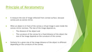
Biometry
IOL power calculation
1. Keratometry
2. A-scan biometry
3. IOL formula
Keratometry (or) ophthalmometer .
Keratometry is the measurement of a patients corneal curvature .
1. It provides on objective,quantitative measurement of corneal
astigmatism,measuring the curvature in each meridian as well as the axis.
2. Keratometry is also helpful in determining the appropriate fit of contact
lens .
3. To measurement the corneal dioptric power.
4. The measurement of the curvature of the anterior corneal surface by using
the first Purkinje image.
5. Determines corneal curvature by measuring the size of a reflected “mire”.
6. Doubling of image avoids problems from eye movements.
7. Keratometer measures only the central 3mm of the corneal diameter.
Principle of Keratometry
 It measure the size of image reflected from corneal surface, because
cornea acts as convex mirror.
 When an object is in front of the cornea a virtual image is seen inside the
convex mirror (cornea). The size of the image depends on,
1. The distance of the object and
2. The curvature of the cornea for a fixed distance of the object the
size of the image depends on the curvature of the cornea.
 Similarly for a given size of the image distance of the object is different
depending on the curvature of the cornea.
 The object used is an illuminated circle with plus and minus rings as shown in
figure.
 The two prisms inside the instrument give two additional one displaced
horizontally and another displaced vertically. Three images are seen as in
figure.
 While taking the reading the pluses and minuses coincide. This is achieved by
moving the keratometer with the object forward or backward in front of the
eye.
 When coincidence takes place the size of the images of fixed value. The
distance of the object is different for different curvatures. The instrument is
calibrated. As the drum rotate the distance varies.
Bausch and Lomb keratometer
Optics of Bausch and Lomb keratometer
 When the instrument is correctly aligned, the operator sees three images of
the instrument’s mires.
 The first is produced by light passing through aperture C and the vertically
displacing prism
 The second is produced by light passing through aperture D and the
horizontally displacing prism, and the third by light passing through aperture
A and B
 Back and forth movement of the vertically doubling prism results in
movement of the vertically displaced image, while movement of the
horizontally doubling prism results in movement of the horizontally displaced
image
 The central image formed by the light passing through A and B is unaffected
by movement of either prism
 The aperture A and B act like a Schiener disc and double the central image of
the mire when the intermediate image, produced by the objective lens does
not coincide with the focal point of the eyepiece lens
 This system is designed to assist the operator in judging when the microscope
is out of focus.
Mires of keratometer
 The images of the mire as seen through the doubling system of the
keratometer are shown in the figure for the conditions where
1. The vertical doubling is correct and the horizontal doubling is insufficient
2. The vertical doubling is too great and the horizontal doubling is correct
3. The vertical and horizontal degrees of doubling are correct
4. The mires are viewed after reflection by an astigmatic cornea, the axes of
which do not coincide with that of the keratometer
A-scan Biometry
 Ophthalmic ultrasound uses the reflection of high frequency sound waves to
define the outlines of ocular and orbital structures and to measure the
distance between them.
 A-scan biometry is to determine the power of the intra ocular lens, that
replaces the natural lens during cataract surgery.
 A-Scan biometry is also called as axial length measurement scan. This
measurement is combined with Keratometric readings to obtain the IOL
power.
 Error of 0.4mm in the measurement of axial length may result in a one
diopter change in calculated IOL power.
Physical principles
 The A-scan probe contains a ultrasonic transducer that projects a thin
sound beam that travels through liquid or tissue.
 Ultrasound waves do not travel through air.
 The frequencies most often employed for diagnostic work are
between 2.5 MHZ and 20 MHZ. Higher the frequency greater the
resolution.
 Although increasing the frequency increases the resolution,it
simultaneously decreases the depth of penetration of the sound.
 When the sound beam encounters the interface of a substance that is
dissimilar from the substance it is traveling through,part of the sound
beam energy is reflected , and part of the sound energy projects
through the new substance.
PROCEDURE:
 A probe is placed on the patient’s cornea.
 The probe is attached to a device that delivers adjustable sound waves.
 The measurements are displayed as spikes on the screen of an oscilloscope
(Visual monitor).
 The appearance of the spikes and the distance between them can be
correlated to structures within the eye and the distance between them.
 The probe lightly touches the cornea and is
positioned, such that the barrel of the
probe is aligned with the optical axis or
visual axis of the eye.
 The operator aims the probe towards the
macula of the eye.
 Alignment with the optical axis will be
indicated by high lens spikes and a high
retina spike on the scan graph.
 The gain should be adjusted high enough such that the spikes can be
maintained above the threshold level needed for an automated
acquisition of the scan if this feature is used.
 The gain should be low enough to allow the operator to visually
maximize the spike height during probe alignment.
Biometry technique
 Contact
 Applanation method
 Hand help method
 Immersion
Values are 0.14 to 0.36mm longer with immersion technique than with
contact method.
Contact technique
 A-scan biometry by applanation requires that the ultrasound probe to
be placed directly on the corneal surface. This can be done at the slit
lamp
 A-scan biometry can alo be done holding the ultrasound probe by
hand.
Immersion Technique:-
 Also called water bath method, the patients is supine and ultrasound
probe is suspended in fluid filled scleral cup placed over the eye.
Instrument Setting
1. Measurement Mode
2. Gates
3. Gain
4. Eye Type
Measurement mode:-
 Automatic
 Semi-automatic
 Manual
Gates
 Gates are electronic markers on the screen that provide
measurement of distance between 2 or more anatomic interfaces .
Gain Setting
 Initially high gain setting should be used to assess the overall
appearance of the echogram , then gain should be reduced to a
medium level to improve resolution of spikes
 Error can occur when the gain is set too high or too low.
 Very high gain short reading
 Very low gain long reading
Eye type:-
1. Phakic
2. Aphakic
3. Pseudophakic
Scan of phakic eye
1. It shows anterior lens spike(B),the
posterior lens spike(C),and the
retina spike (E), the probe tip /
cornea spike is represented by(A).
2. These spikes should be tall and
steeply rising.The retina spike
should not have smaller spikes
immediately in front of it.(D)
3. The retina spike should be followed
by tall scleral spike (F) and spikes
from the orbital fat layer of the
orbit(G).A scan without orbital fat
spikes may indicate that the beam
is striking the optic nerve instead
of the macula.
Scan of an aphakic eye
1. It will either have no lens spikes,(or) it will have
one lens spike (A) that represents an intact
posterior lens capsule. ( C )
2. Be sure to use the aphakic mode of the A-scan
instrument.
3. The velocity of sound will be different because
the beam is not passing through the lens.
4. A velocity of sound of 1532m/s is typically used
for aphakic measurements.
Scan of an pseudophakic eye
 If one is pseudophakic, A-scan and K-readings should be done for both the
eyes before calculating the IOL power for the eye required.
 Since both eyes have similar measurements in most people, this provides a
double check of the measurement.
 It is some times necessary to replace an IOL that was inserted many months or
years ago. Even if you have IOL specifications and measurement information
from the previous surgery, its nice to have the confirmation of a current
measurement.
Characteristics of a good scan
1. Corneal echo is seen as a tall single peak
2. Aqueous chamber should not provide any echo
3. Anterior and posterior lens capsule produces tall echo
4. Vitreous cavity should produce few to no echoes
5. Retina produces tall, sharply rising echoes with no staircase at the origin
6. Oribital fat produces medium to low echoes
IOL formula
 Depending upon the basis of their derivation:
 Theoretical formula
 Regression formula
 These are grouped into various generations.
Theoretical formula
 Derived from geometric optics as applied on the schematic eyes, using theoretical constants.
 Based on 3 variables:-
 AL
 K- reading
 Estimated post-operative ACD
Regression formula
 Based on regression analysis of the actual post-operative results of implant power as a function
of the variables of corneal power and AL.
 Third generation
Holladay I
SRK/T
Hoffer Q
 Forth generation
Holladay-II
Regression formulae
SRK-I formula (Sanders, Retzlaff and Kraff)
P=A-2.5L-0.9K
 Tends to predict too small value in short eyes and too long value in long eyes
SRK-II formula
P=A-2.5L-0.9K
 A constant is modified on the basis of axial length as follow:-
 If L is <20mm :A+3.0
 If L is 20-20.99 :A+2.0
 If L is 21-21.99 :A+1.0
 If L is 22-24.5 :A
 If L is >24.5 :A-0.5
Modified SRK-II formula
 Based on axial length, A constant is modified as
 If L is <20mm :A+1.5
 If L is 20-21 :A+1.0
 If L is 21-22 :A+0.5
 If L is 22-24.5 :A
 If L is 24.5-26 :A-1.0
 If L is >26mm :A-1.5
SRK/T formula
 Nonlinear theoretical optical formula optimized for post op AC depth, retinal
thickness and corneal refractive index
 Significantly more accurate for extremely long eyes (>28mm)
Optical biometers
 The introduction of optical biometer has significantly improved the accuracy
of AL measurement from 0.12mm in immersion ultrasound to 0.02mm in
optical methods.
 commercially available optical biometer include:-
1. IOL master
2. Lens star
IOL Master
 IOL MasterTM (Zeiss Humphrey System) is a
combined biometric instrument that
measures quickly and precisely parameters
of human eye needed for IOL power
calculation by a noncontact technique.
 It also incorporates the software to calculate
IOL power from various formulae.
Working principle
it is a noncontact optical device that measures the various parameters based on
following principles:
 AL measurements- It is based on a patented interference optical method
known as ‘Partial Coherence Interferometry (PCI)’. This technique relies on a
laser Doppler technique to measure the echo delay and intensity of infrared
light reflected back from the tissue interfaces- cornea and RPE. The
instrument is calibrated against the ultra high resolution of 40MHz. An
internal algorithm approximates the distance to the viteroretinal interphase
for the equipment of an immersion A-scan ultrasonic AL.
 Corneal curvature (K)- It is determined by measuring the distance between
reflected light images as in conventional keratometry
 ACD- it is determined as the distance between the optical sections of the
crystalline lens and the cornea produced by lateral slit illumination.
 White-to-white – It is determined from the image of iris.
 Calculation of IOL power- it is done by software incorporating internationally
accepted calculation formulae
Advantages of the IOL Master
 Patient comfort, as the technique involves noncontact measurements.
 User-friendly, as the operater can learn the technique very quickly
 Single instruments is required for measuring AL, corneal curvature (K), and
ACD.
 Cross-infection risk is not there, as the technique is noncontact
 It incorporates 5 IOL power calculating formulae in an integrated manner,
these are- Haigis, Hoffer, Holladay,SRK-II, and SRK/T formula.
Lens Star
 LenStar LS900 (hag-streit diagnostics)
provides highly accurate laser optic
measurements for every section of the
eye and is the first optical biometer that
can measure the thickness of the
crystalline lens.
 With its integrated Olsen formula and
the optional Toric Planner, the LenStar
provides user with latest technology in
IOL prediction for any patient.
Working principle
 It is a noncontact optical device which measures multiple parameters on the
following parameters:-
 Central corneal thickness- it uses optical coherence biometry to measure
central corneal thickness (CCT), with stunning reproducibility of ± 2μm.
 Keratometry/Topography- LenStar’s unique dual zone keratometry, featuring
32 marker points, provides perfect spherical equivalent, magnitude of
astigmatism and axis position. With the optional T-Cone topography add-on,
LenStar provides full topography maps of the central 6mm optical zone.
 White-to-white- Based on high-resolution colour photography of the eye,
every white-to-white measurement can be reviewed and adjusted by the user.
 Pupillometry- Measurements of the pupil diameter in ambient light
conditions can be used as a n indicator for the patient’s suitable apodized
premium IOLs, as well as for laser refractive procedures.
 Lens thickness- Accurate measurement of LT is the key to optimal IOL
prediction accuracy when using the latest IOL calculation formulae, Olsen or
Holladay II.
 ACD- It is measured by optical coherence biometry which provides more
precision and reproducibility. This allows ACD to be measured on phakic as
well as on pseudophakic eyes.
 Axial length- it uses a superluminescent diode as the laser source which
enables measurements of the AL of the patient’s eye, precisely on the
patient’s visual axis and even in the presence of dense media.
THANK YOU

Biometry.pptx

  • 1.
  • 2.
    IOL power calculation 1.Keratometry 2. A-scan biometry 3. IOL formula
  • 3.
    Keratometry (or) ophthalmometer. Keratometry is the measurement of a patients corneal curvature . 1. It provides on objective,quantitative measurement of corneal astigmatism,measuring the curvature in each meridian as well as the axis. 2. Keratometry is also helpful in determining the appropriate fit of contact lens . 3. To measurement the corneal dioptric power. 4. The measurement of the curvature of the anterior corneal surface by using the first Purkinje image. 5. Determines corneal curvature by measuring the size of a reflected “mire”. 6. Doubling of image avoids problems from eye movements. 7. Keratometer measures only the central 3mm of the corneal diameter.
  • 4.
    Principle of Keratometry It measure the size of image reflected from corneal surface, because cornea acts as convex mirror.  When an object is in front of the cornea a virtual image is seen inside the convex mirror (cornea). The size of the image depends on, 1. The distance of the object and 2. The curvature of the cornea for a fixed distance of the object the size of the image depends on the curvature of the cornea.  Similarly for a given size of the image distance of the object is different depending on the curvature of the cornea.
  • 5.
     The objectused is an illuminated circle with plus and minus rings as shown in figure.  The two prisms inside the instrument give two additional one displaced horizontally and another displaced vertically. Three images are seen as in figure.  While taking the reading the pluses and minuses coincide. This is achieved by moving the keratometer with the object forward or backward in front of the eye.  When coincidence takes place the size of the images of fixed value. The distance of the object is different for different curvatures. The instrument is calibrated. As the drum rotate the distance varies.
  • 6.
    Bausch and Lombkeratometer
  • 8.
    Optics of Bauschand Lomb keratometer  When the instrument is correctly aligned, the operator sees three images of the instrument’s mires.  The first is produced by light passing through aperture C and the vertically displacing prism  The second is produced by light passing through aperture D and the horizontally displacing prism, and the third by light passing through aperture A and B  Back and forth movement of the vertically doubling prism results in movement of the vertically displaced image, while movement of the horizontally doubling prism results in movement of the horizontally displaced image
  • 9.
     The centralimage formed by the light passing through A and B is unaffected by movement of either prism  The aperture A and B act like a Schiener disc and double the central image of the mire when the intermediate image, produced by the objective lens does not coincide with the focal point of the eyepiece lens  This system is designed to assist the operator in judging when the microscope is out of focus.
  • 10.
  • 11.
     The imagesof the mire as seen through the doubling system of the keratometer are shown in the figure for the conditions where 1. The vertical doubling is correct and the horizontal doubling is insufficient 2. The vertical doubling is too great and the horizontal doubling is correct 3. The vertical and horizontal degrees of doubling are correct 4. The mires are viewed after reflection by an astigmatic cornea, the axes of which do not coincide with that of the keratometer
  • 12.
    A-scan Biometry  Ophthalmicultrasound uses the reflection of high frequency sound waves to define the outlines of ocular and orbital structures and to measure the distance between them.  A-scan biometry is to determine the power of the intra ocular lens, that replaces the natural lens during cataract surgery.  A-Scan biometry is also called as axial length measurement scan. This measurement is combined with Keratometric readings to obtain the IOL power.  Error of 0.4mm in the measurement of axial length may result in a one diopter change in calculated IOL power.
  • 13.
    Physical principles  TheA-scan probe contains a ultrasonic transducer that projects a thin sound beam that travels through liquid or tissue.  Ultrasound waves do not travel through air.  The frequencies most often employed for diagnostic work are between 2.5 MHZ and 20 MHZ. Higher the frequency greater the resolution.
  • 14.
     Although increasingthe frequency increases the resolution,it simultaneously decreases the depth of penetration of the sound.  When the sound beam encounters the interface of a substance that is dissimilar from the substance it is traveling through,part of the sound beam energy is reflected , and part of the sound energy projects through the new substance.
  • 15.
    PROCEDURE:  A probeis placed on the patient’s cornea.  The probe is attached to a device that delivers adjustable sound waves.  The measurements are displayed as spikes on the screen of an oscilloscope (Visual monitor).  The appearance of the spikes and the distance between them can be correlated to structures within the eye and the distance between them.
  • 16.
     The probelightly touches the cornea and is positioned, such that the barrel of the probe is aligned with the optical axis or visual axis of the eye.  The operator aims the probe towards the macula of the eye.  Alignment with the optical axis will be indicated by high lens spikes and a high retina spike on the scan graph.
  • 17.
     The gainshould be adjusted high enough such that the spikes can be maintained above the threshold level needed for an automated acquisition of the scan if this feature is used.  The gain should be low enough to allow the operator to visually maximize the spike height during probe alignment.
  • 18.
    Biometry technique  Contact Applanation method  Hand help method  Immersion Values are 0.14 to 0.36mm longer with immersion technique than with contact method.
  • 19.
    Contact technique  A-scanbiometry by applanation requires that the ultrasound probe to be placed directly on the corneal surface. This can be done at the slit lamp  A-scan biometry can alo be done holding the ultrasound probe by hand.
  • 20.
    Immersion Technique:-  Alsocalled water bath method, the patients is supine and ultrasound probe is suspended in fluid filled scleral cup placed over the eye.
  • 21.
    Instrument Setting 1. MeasurementMode 2. Gates 3. Gain 4. Eye Type
  • 22.
    Measurement mode:-  Automatic Semi-automatic  Manual
  • 23.
    Gates  Gates areelectronic markers on the screen that provide measurement of distance between 2 or more anatomic interfaces .
  • 24.
    Gain Setting  Initiallyhigh gain setting should be used to assess the overall appearance of the echogram , then gain should be reduced to a medium level to improve resolution of spikes
  • 25.
     Error canoccur when the gain is set too high or too low.  Very high gain short reading  Very low gain long reading
  • 26.
    Eye type:- 1. Phakic 2.Aphakic 3. Pseudophakic
  • 27.
    Scan of phakiceye 1. It shows anterior lens spike(B),the posterior lens spike(C),and the retina spike (E), the probe tip / cornea spike is represented by(A). 2. These spikes should be tall and steeply rising.The retina spike should not have smaller spikes immediately in front of it.(D) 3. The retina spike should be followed by tall scleral spike (F) and spikes from the orbital fat layer of the orbit(G).A scan without orbital fat spikes may indicate that the beam is striking the optic nerve instead of the macula.
  • 28.
    Scan of anaphakic eye 1. It will either have no lens spikes,(or) it will have one lens spike (A) that represents an intact posterior lens capsule. ( C ) 2. Be sure to use the aphakic mode of the A-scan instrument. 3. The velocity of sound will be different because the beam is not passing through the lens. 4. A velocity of sound of 1532m/s is typically used for aphakic measurements.
  • 29.
    Scan of anpseudophakic eye  If one is pseudophakic, A-scan and K-readings should be done for both the eyes before calculating the IOL power for the eye required.  Since both eyes have similar measurements in most people, this provides a double check of the measurement.  It is some times necessary to replace an IOL that was inserted many months or years ago. Even if you have IOL specifications and measurement information from the previous surgery, its nice to have the confirmation of a current measurement.
  • 30.
    Characteristics of agood scan 1. Corneal echo is seen as a tall single peak 2. Aqueous chamber should not provide any echo 3. Anterior and posterior lens capsule produces tall echo 4. Vitreous cavity should produce few to no echoes 5. Retina produces tall, sharply rising echoes with no staircase at the origin 6. Oribital fat produces medium to low echoes
  • 31.
    IOL formula  Dependingupon the basis of their derivation:  Theoretical formula  Regression formula  These are grouped into various generations.
  • 32.
    Theoretical formula  Derivedfrom geometric optics as applied on the schematic eyes, using theoretical constants.  Based on 3 variables:-  AL  K- reading  Estimated post-operative ACD Regression formula  Based on regression analysis of the actual post-operative results of implant power as a function of the variables of corneal power and AL.
  • 35.
     Third generation HolladayI SRK/T Hoffer Q  Forth generation Holladay-II
  • 36.
    Regression formulae SRK-I formula(Sanders, Retzlaff and Kraff) P=A-2.5L-0.9K  Tends to predict too small value in short eyes and too long value in long eyes
  • 37.
    SRK-II formula P=A-2.5L-0.9K  Aconstant is modified on the basis of axial length as follow:-  If L is <20mm :A+3.0  If L is 20-20.99 :A+2.0  If L is 21-21.99 :A+1.0  If L is 22-24.5 :A  If L is >24.5 :A-0.5
  • 38.
    Modified SRK-II formula Based on axial length, A constant is modified as  If L is <20mm :A+1.5  If L is 20-21 :A+1.0  If L is 21-22 :A+0.5  If L is 22-24.5 :A  If L is 24.5-26 :A-1.0  If L is >26mm :A-1.5
  • 39.
    SRK/T formula  Nonlineartheoretical optical formula optimized for post op AC depth, retinal thickness and corneal refractive index  Significantly more accurate for extremely long eyes (>28mm)
  • 40.
    Optical biometers  Theintroduction of optical biometer has significantly improved the accuracy of AL measurement from 0.12mm in immersion ultrasound to 0.02mm in optical methods.  commercially available optical biometer include:- 1. IOL master 2. Lens star
  • 41.
    IOL Master  IOLMasterTM (Zeiss Humphrey System) is a combined biometric instrument that measures quickly and precisely parameters of human eye needed for IOL power calculation by a noncontact technique.  It also incorporates the software to calculate IOL power from various formulae.
  • 42.
    Working principle it isa noncontact optical device that measures the various parameters based on following principles:  AL measurements- It is based on a patented interference optical method known as ‘Partial Coherence Interferometry (PCI)’. This technique relies on a laser Doppler technique to measure the echo delay and intensity of infrared light reflected back from the tissue interfaces- cornea and RPE. The instrument is calibrated against the ultra high resolution of 40MHz. An internal algorithm approximates the distance to the viteroretinal interphase for the equipment of an immersion A-scan ultrasonic AL.  Corneal curvature (K)- It is determined by measuring the distance between reflected light images as in conventional keratometry
  • 43.
     ACD- itis determined as the distance between the optical sections of the crystalline lens and the cornea produced by lateral slit illumination.  White-to-white – It is determined from the image of iris.  Calculation of IOL power- it is done by software incorporating internationally accepted calculation formulae
  • 44.
    Advantages of theIOL Master  Patient comfort, as the technique involves noncontact measurements.  User-friendly, as the operater can learn the technique very quickly  Single instruments is required for measuring AL, corneal curvature (K), and ACD.  Cross-infection risk is not there, as the technique is noncontact  It incorporates 5 IOL power calculating formulae in an integrated manner, these are- Haigis, Hoffer, Holladay,SRK-II, and SRK/T formula.
  • 45.
    Lens Star  LenStarLS900 (hag-streit diagnostics) provides highly accurate laser optic measurements for every section of the eye and is the first optical biometer that can measure the thickness of the crystalline lens.  With its integrated Olsen formula and the optional Toric Planner, the LenStar provides user with latest technology in IOL prediction for any patient.
  • 46.
    Working principle  Itis a noncontact optical device which measures multiple parameters on the following parameters:-  Central corneal thickness- it uses optical coherence biometry to measure central corneal thickness (CCT), with stunning reproducibility of ± 2μm.  Keratometry/Topography- LenStar’s unique dual zone keratometry, featuring 32 marker points, provides perfect spherical equivalent, magnitude of astigmatism and axis position. With the optional T-Cone topography add-on, LenStar provides full topography maps of the central 6mm optical zone.  White-to-white- Based on high-resolution colour photography of the eye, every white-to-white measurement can be reviewed and adjusted by the user.  Pupillometry- Measurements of the pupil diameter in ambient light conditions can be used as a n indicator for the patient’s suitable apodized premium IOLs, as well as for laser refractive procedures.
  • 47.
     Lens thickness-Accurate measurement of LT is the key to optimal IOL prediction accuracy when using the latest IOL calculation formulae, Olsen or Holladay II.  ACD- It is measured by optical coherence biometry which provides more precision and reproducibility. This allows ACD to be measured on phakic as well as on pseudophakic eyes.  Axial length- it uses a superluminescent diode as the laser source which enables measurements of the AL of the patient’s eye, precisely on the patient’s visual axis and even in the presence of dense media.
  • 49.