SlideShare a Scribd company logo
1 of 77
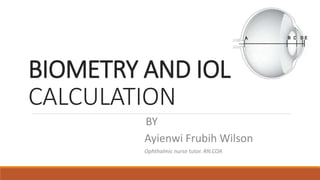
BY
Ayienwi Frubih Wilson
Ophthalmic nurse tutor. RN.COA
BIOMETRY AND IOL
CALCULATION
Outline of presentation
PRE TEST
I- INTRODUCTION
A.DEFINITION OF OCULAR BIOMETRY
II-ANATOMY OF THE EYE
Key Components Related To Ocular Biometry
III- TECHNIQUES IN OCULAR BIOMETRY
IV- MEASUREMENT OF AXIAL LENGTH (AL)
V-MEASUREMENT OF CORNEA POWER
VI-FORMULA FOR IOL POWER CALCULATION
VII- BIOMETRY IN PEDIATRIC CASES
VII- QUESTIONS
PRE-TEST
1). which ocular biometry technique is most suitable for
measuring the corneal radius of curvature and corneal
astigmatism?
a) A-scan ultrasound
b) Keratometry
c) B-scan ultrasound
d) Applanation tonometry
2). what does A-scan ultrasound measure?
a) Corneal curvature
b) Anterior chamber depth
c) Axial length
d) Lens power
3). which type of error can occur if the eye is not properly
aligned during optical biometry?
a) Spherical aberration
b) Iris capture
c) Astigmatism
d) Parallax error
4). A patient's axial length measures 23 mm in one eye and 25
mm in the other eye. What condition might this difference
suggest?
a) Amblyopia (lazy eye)
b) Presbyopia
c) Anisometropia
d) Cataracts
INTRODUCTION
Ocular biometry :
This is the process of measuring the power of the cornea
(keratometry) and the Axial length of the eye, by using this data
to determine the ideal intraocular lens power
Anatomy of the eye
Key components related to Ocular Biometry
A)AXIAL Length
B)ACD
C)Lens thickness
D)Cornea
curvature
Axial Length:
Distance from the corneal surface to the retina at the back of the eye.
Range: In adults, is typically between 22 to 24 millimeters.
Anterior Chamber Depth:
The distance between the corneal endothelium and the front surface of the lens.
Range: In adults, is around 2.5 to 3.5 millimeters.
Lens Thickness:
In adults, lens thickness is typically between 4 to 5 millimeters.
TECHNIQUES IN OCULAR BIOMETRY
Techniques in Ocular Biometry
Ocular biometry involves various techniques for measuring
different eye structures and parameters.
Some of the commonly used techniques:
1). A-Scan Ultrasound (contact and immersion)
2). B-Scan Ultrasound
3). Optical Biometry (IOL Master,lenstar)
4). Keratometry
MEASUREMENT OF AXIAL LENGTH (AL)
Techniques of Axial Length Measurement
1).A-Scan Ultrasound Biometry
A).Contact
B).Immersion
2). `B-Scan
3). Optical Biometry
A).Lenstar
B).IOL MASTER 500/700
A-scan Biometry
A scan: Amplitude Scan; utilizes ultrasound waves of 10 - 12 MHz frequency.
In A-scan, thin, parallel sound beam is emitted from the probe tip, with an echo bouncing
back into the probe tip as the sound beam strikes each interface.
An interface is the junction between any two media of different densities and velocities.
Techniques of A-Scan
Contact
Applanation Method
Immersion
Applanation/contact A-scan Biometry
A-scan biometry by applanation requires that the ultrasound probe be placed directly on the corneal surface
PROCEDURE
◦A probe is placed on the patient’s cornea.
◦The probe is attached to a device that delivers
adjustable sound waves
◦The measurements are displayed as spikes on the
screen of an oscilloscope (Visual monitor)
◦The appearance of the spikes and the distance
between them can be correlated to structures within
the eye and the distance between them
A:Initial spike
(probe tip and
cornea)
B: Anterior lens
capsule
C: Posterior lens
capsule
D: Retina
e: Sclera
f: Orbital fat
A
B
C D E
F
When echoes B through D are high and steeply rising, the ultrasound beam is most likely on
visual axis.
A
B
C D E
Errors resulting from Probe positioning
Spike height is affected by the difference in
density & by the angle of incidence, which is
determined by the probe orientation to the
visual axis
If the probe is held nonparallel, part of the
echo is diverted at an angle away from the
probe tip, and is not received by the
machine.Induces a Paralax Error.
The sound beam is directed at an angle through the lens
rather than through the center of both the front and back
surfaces. When the beam is not going through the center of
the lens, it is not on the visual axis or center of the macula
Posterior lens spike too short
A perfect high, steeply rising retinal spike
may be impossible when macular
pathology is present (eg, macular edema,
macular degeneration, epiretinal
membranes, posterior staphylomas).
Retinal Spikes Not steeply rising
This common error is caused when the beam is not
perpendicular to the macular surface
No Orbital Fat
If no scleral or orbital fat
echoes visible, then ultrasound
beam is mostlikely aligned with
optic nerve
Summary
limitations of Contact Aplanation biometry
Variable corneal compression
Broad sound beam without precise localization
Limited resolution.
Potential for incorrect measurement distance
Corneal Compression
If pressure is applied on the cornea, the axial length measurement
may be falsely too short.
It can be monitored by observing the anterior chamber depth, read
out by an instrument.
Most eyes will have an ACD readings between
2.5 to 4.0mm.
The corneal compression error factor can be avoided by using the
immersion technique
Error caused by
1 mm Corneal Compression
Average eye 2.5 D
Long eye 1.75 D
Short eye 3.75 D
Immersion A-scan Biometry
The immersion technique is accomplished by placing a small
scleral shell between the patient's lids, filling it with saline, and
immersing the probe into the fluid, being careful to avoid contact
with the cornea.
More accurate than contact method because corneal
compression is avoided.
Eyes measured with the immersion method are, on average, 0.1-
0.3 mm longer
Scan produced by Immersion
Immersion graph contact graph
1 2 3 4 5 6 1 2 3 4 5
Immersion A-scan Biometry
When the ultrasound beam is properly aligned with the center of the
macula, all five spikes will be steeply rising and of maximum height.
Both the peaks of corneal spike should be equal in height ideally
A scan in special cases
1). Inadequate Patient Fixation
Low Vision
Nystagmus
2).Posterior Staphyloma
Blepharospasm
Strabismus
A circumscribed outpouching of the
wall of the globe which could result in
long AL
Cont…
3). Macular Lesions
RD
Edema
Tumor
An elevated macular lesion may prevent
the display of a distinct retinal spike and
often causes a shortened AL.
A wide space between Retina spike and
Sclera.
Cont…
4. Dense Cataract
Strong sound attenuation
Maximum gain setting may be required
Factors to Consider for type of eye and IOL material
•The type of material or the type of eye is important because the velocity of
sound is a function of the material that the sound is passing through.
•A-Scan does not actually measure length, they measure how long it takes a
sound beam to bounce off an object ( Anterior lens, Posterior lens, and
Retina) & return to the probe.
•The instrument is pre programmed with the velocity of sound factors for the
aqueous, the lens material, and the vitreous
Cont…
Type of EYE
Phakic (with natural lens)
Aphakic (without lens)
Pseudophakic (with artificial lens)
Silicone Oil Filled
Lens Material:
Acrylic
PMMA
Silicone IOL
Cont…
Lens material Velocity
Phakic 1641
Aqueous 1532
Acrylic 2120
Pmma 2660
Silicone 980
PMMA protocol : (Polymethylmethacrylate)
Sound travels faster through PMMA than it does through the
natural lens.
If a pseudophakic mode is not available, use the aphakic mode
and add a standard compensating factor of 0.4mm to the
resultant axial length.
Compensations When the eye is
pseudophakic
Silicon protocol:
Sound travels much slower through a silicon lens than it does through the
natural lens.
If not taken in to account could result in a –3.0D post - op refractive error.
If your biometer does not have a pseudophakic silicon mode, use the aphakic
mode and subtract a compensation factor of 0.8mm.
Acrylic protocol:
Sound travels faster through acrylic than it does through the natural lens.
If biometer does not have an acrylic mode, use the aphakic mode and add a compensation
factor of 0.2mm.
Cont…
Compensation Factors
Lens material Velocity Compensation on
Axial Length
Phakic 1641
Acrylic 2120 +0.2 mm
Pmma 2660 +0.4 mm
Silicone 980 -0.8 mm
Important Terms
Gain: The gain setting on A-scan is measured in decibels and affects amplification
and resolution of spikes. In cases of dense opacities(cataract) the gain can be
adjusted to amplify the signals.
Error can occur when the gain is set too high or too low
oVery high gain short reading
oVery low gain long reading
Important Terms …
Gates are electronic calipers on the display screen that measure distance
between two points.
Proper gate placement is on the ascending edge of each appropriate spike.
Error can occur when the gates are not appropriately placed.
POTENTIAL SOURCES OF ERROR IN AL
MEASURES
SHORT MEASUREMENT
Corneal compression in contact A-Scan
Corneal gate to right of corneal spike
Retinal Gate incorrectly positioned at
spike in the vitreous cavity
Gain set too high
Macular swelling
Retinal detachment
Misalignment of sound beam
Lens measured too thin
LONG MEASUREMENT
Air bubble in fluid bath (immersion method)
Fluid bridge contact method
Retinal gate at right of spike
Gain set too low
Posterior staphyloma eccentric to macular
Misalignment of sound beam
Lens measured too thick
OPTICAL BIOMETERS
Optical biometers are noncontact instruments that use infrared laser light (780 nm) and partial
coherence interferometry to measure multiple parameters, such as; AL, corneal curvature,
anterior chamber depth, lens thickness, and horizontal white-to-white distance (corneal
diameter).
These devices require the patient to fixate on a target, which gives an AL along the visual axis.
IOL MASTER
Principle of functioning:
Based on partial coherence interferometry (PCI)’.
Diode laser (780nm) measures echo delay and intensity of infrared light reflected
back from tissue interfaces– cornea & RPE
LENSTAR
PRINCIPLE OF LENSTAR
Based on ‘low coherence optical reflectometry (LCOR)’. Superluminescent Diode
laser of 820nm is used.
IOL MASTER 700
PRINCIPLE OF FUNCTIONING
Based on swept source OCT technology. It provides an image-based measurement, allowing to
view the complete longitudinal section of eyeball.
LENSTAR/IOL MASTER
LENSTAR IOL MASTER 500
NO OF POINTS TESTED – 32 POINTS IN TWO CIRCLES (16
EACH)
NO OF POINTS TESTED – 6 POINTS IN HEXAGONAL
PATTERN
ZONE OF CORNEA TESTED – INNER CIRCLE DIAMETER –
1.65MM
OUTER CIRCLE DIAMETER – 2.3MM
ZONE OF CORNEA TESTED – DIAMETER OF 2.3MM
BETTER IN TERMS OF MEASURING TRUE CENTRAL
CORNEAL POWER(1.65mm)
MEASURES 2.3mm
MEASURES ACD USING OPTICAL BIOMETRY MEASURES ACD USING SLIT IMAGERY
KERATOMETRY
Measurement of Cornea Curvature
Used to measure the corneal curvature
It is use for measurement the corneal dioptric power D
It provides an objective, quantitative measurement of corneal
astigmatism, measuring the curvature in each meridian as well as the axis.
The average keratometry value (K) → 43.0 -44.0D
Formula for corneal power
Keratometer:
Determines corneal curvature by measuring the size of a reflected Image
Surface power formula: D = ......... n - 1
R
D = the dioptric power of the cornea
n = the refractive index of the cornea used (1.3375)
R = the radius of curvature of the cornea in meters
Keratometer measures only the central 3mm of the corneal diameter.
Types of keratometer:
Manual keratometer
Auto keratometer, keratometers incorporated in IOL master and lenstar
Topography – placido disc based or elevation based topography
Manual Keratometry
Two Types:
Bausch And Lomb
Javal Schiotz
Principle of Functioning:
In order to find the refracting power of the cornea, we need to reflect an object of a
known size at a known distance off the corneal surface.
Then determine the size of the reflecting image with measuring telescope and
calculate the refractive power of the cornea.
AUTOMATED KERATOMETER
Principle- Focuses the reflected corneal image on to an electronic photosensitive
device, which instantly records the size and computes the radius of curvature.
Zone of measurement- central 3mm zone
Source of keratometry errors
• Unfocused eye piece
• Failure to calibrate unit
• Poor patient fixation
• Dry eye
• Drooping eye lids
• Irregular cornea
NB: Repeat Keratometery If
• Corneal curvature more than 47D or less than 40D.
• The difference in corneal cylinder is more than one diopter
between eyes.
Difficult Situations
• Post Refractive Surgery
• Corneal Transplantation
• Corneal Scar
• Keratoconus etc.
FORMULA FOR
IOL POWER CALCULATION
Formulae
On the basis of their derivation ,the various formulae for calculating IOL power have been grouped into
THEORETICAL FORMULAE
Derived from the geometric optics as applied to the schematic eyes, using theoretical constants.
Based on 3 variables – AL, K reading and estimated postoperative ACD.
REGRESSION FORMULAE
Based on regression analysis of the actual postop results of implant power as a function of the variables
of corneal power and AL
Grouped into various generation
1st ,2nd ,3rd and 4th
First generation
Theoretical Regression
1. Binkhorst
2. Colenbrander-Hoffer
3. Gilll’s
4. Clayman
5. Fyodorov
SRK I
FIRST GENERATION
Most are based on regression formula developed by Sander ,Retzlaff & Kraff
Known as SRK formula.
P = A - 2.5(L) - 0.9(K)
P=lens implant power for emetropia
L= Axial length (mm)
K=average keratometric reading (diaopters)
A= lens constant
Tends to predict too small value in short eyes and too large value in long eyes.
SECOND GENERATION
THEORETICAL REGRESSION
MODIFIED BINKHORST SRK-II
SECOND GENERATION FORMULA
SRK formula –works well for average eyes.
less accurate for long, short eyes.
SRK II formula Modification of SRK
A-constant is modified on the basis of AL
P = A1 – 2.5L – 0.9K A1 = A + 3 AL < 20mm
A1 = A + 2 AL 20-21
A1 = A + 1 AL 21-22
A1 = A AL 22-24.5
A1 = A – 0.5 AL >24.5
THIRD GENERATION
THIRD
GENERATION
HOLLADAY I SRK/T HOFFER Q
THIRD GENERATION FORMULA
SRK/T -very long eyes >26mm
Holladay I -long eyes 24-26 mm
HofferQ -Short eyes<22mm
FOURTH GENERATION
HOLLADAY - II
STATE OF AL/CHOICE OF FORMULA
CIRCUMSTANCE OF AL CHOICE OF FORMULA
AL < 20 MM HOLLADAY II/ HOFFER Q
20- 22 MM HOFFER Q
22- 24.5 MM SRK/ T; HOLLADAY
24.5-26 MM HOLLADAY I
> 26 MM SRK / T ; HOLLADAY I
NEWER GENERATION: ONLINE CALCULATORS
FOR TORIC IOL:
◦ http://eyecryltoriccalculator.com/
◦ http://www.ascrs.org/barrett-toric-calculator
◦ https://www.myalcon-iolcalc.com/#/calculator
FOR ERV(Extended Range of Vision) IOL:
◦ https://www.amoeasy.com/toric2(bd1lbizjpta1ma==)/toric.h tm
CONSIDERATIONS IF IMPLANTING IN THE SULCUS
IOL POWER POWER CONSIDERATION
>=28.5 D Decrease by 1.5 D
+17 To 28 D Decrease by 1.0 D
+9 To 17 D Decrease by 0.5 D
<+ 9 D No change
Biometry In Pediatric Cases
It has shorter axial length, steeper cornea with higher keratometry value and
smaller anterior chamber depth.
The formular to use for IOL calculation should be carefully picked( Holladay II or
Hoffer Q)
Errors in axial length measurement affect IOL power calculation the most, it
increases to 3.75 D per mm in children thus AL should be measured by
immersion technique.
Keratometry: hand held keratometer should be used.
Considerations in IOL Power
As myopia increases rapidly in pediatric age group, the goal should be
under correction, The younger the age, more is the under correction.
20% undercorrection if the child is less than 2 years 10% undercorrection for age 2–8
years.
Example:
Age :4
calculated IOL power: +20.0 D
Power to implant -(10%) of calculated power: +18.0 D
POTENTIAL ERRORS IN BIOMETRY
•Error caused by Corneal Compression
•Difference in AL measurement between both eyes more than 0.3 mm
(anisometropia)
•Difference Power of lens in both eyes more than 1.00 D always check for refraction if
available.
•K reading differences should not be more than 1.50 D if not check for any corneal
pathology (dystrophy, Keratoconus, pterygium…etc )
Possible causes of Bad outcomes Post Surgery
•Inaccuracy of formula used
•Inaccuracy AL measurment
•Inaccuracy Keratometry measurment
•Wrong A constant of IOL chosen
•Predisposal Eye condition (Staphyloma, Retinal Detachment, Choroidal Detachment…)
•Wrong IOL brought in OR
•Mixing right eye value to left eye
Consequences of Bad Biometry
•Bad visual acuity post operatively
•Need of spectacles after surgery for far vision
•Leaving the patient aphakia
•May need re-surgery for secondary implantation or IOL removal
•Bad reputation of the hospital
Benefits of good Biometry
•Good visual acuity post operatively
•Meet the patient’s need
• (Mostly emmetropic target)
• (Slightly myopic to give a comfortable near vision also)
• (Far and near vision if doing mono vision but anisometropia must be kept below 3.00
D)
•Patient’s satisfaction
•Medical staff satisfaction
•Good reputation of the hospital
Clear vision begins with Precise
Ocular Biometry
The Key to unlocking a world of
visual clarity
OCULAR BIOMETRY AND IOL.pptx

More Related Content

What's hot

Non contact tonometer ppt
Non contact tonometer pptNon contact tonometer ppt
Non contact tonometer pptsatish kumar
 
Role of bandage contact lens in corneal wound healing
Role of bandage contact lens in corneal wound healingRole of bandage contact lens in corneal wound healing
Role of bandage contact lens in corneal wound healingRabindraAdhikary
 
Optics of RGP contact lens
Optics of RGP contact lensOptics of RGP contact lens
Optics of RGP contact lensPabita Dhungel
 
AMBLYOPIA TREATMENT STUDIES PEDIG
AMBLYOPIA TREATMENT STUDIES PEDIGAMBLYOPIA TREATMENT STUDIES PEDIG
AMBLYOPIA TREATMENT STUDIES PEDIGSivateja Challa
 
History of contact lenses
History of contact lenses History of contact lenses
History of contact lenses YashuChaudhary5
 
The Low Vision Examination
The Low Vision ExaminationThe Low Vision Examination
The Low Vision ExaminationHossein Mirzaie
 
Diplopia charting
Diplopia chartingDiplopia charting
Diplopia chartingMantu Akon
 
Iol power calculation normal and post lasik eyes
Iol power calculation normal and post lasik eyesIol power calculation normal and post lasik eyes
Iol power calculation normal and post lasik eyesDINESH and SONALEE
 
Anterior segment OCT & UBM
Anterior segment OCT & UBMAnterior segment OCT & UBM
Anterior segment OCT & UBMDinesh Madduri
 
School Sight Testing Program
School Sight Testing ProgramSchool Sight Testing Program
School Sight Testing ProgramDr. A Huq
 
250+ High Frequency MCQs in Optometry and Ophthalmology
250+ High Frequency MCQs in Optometry and Ophthalmology250+ High Frequency MCQs in Optometry and Ophthalmology
250+ High Frequency MCQs in Optometry and OphthalmologyRabindraAdhikary
 
Types of pediatric contact lens [autosaved]
Types of pediatric contact lens [autosaved]Types of pediatric contact lens [autosaved]
Types of pediatric contact lens [autosaved]Bipin Koirala
 

What's hot (20)

Hvid
Hvid Hvid
Hvid
 
Non contact tonometer ppt
Non contact tonometer pptNon contact tonometer ppt
Non contact tonometer ppt
 
Biometry
Biometry Biometry
Biometry
 
2019 Prescribing Eyeglasses in Children
2019 Prescribing Eyeglasses in Children2019 Prescribing Eyeglasses in Children
2019 Prescribing Eyeglasses in Children
 
Role of bandage contact lens in corneal wound healing
Role of bandage contact lens in corneal wound healingRole of bandage contact lens in corneal wound healing
Role of bandage contact lens in corneal wound healing
 
Low vision chart
Low vision chart Low vision chart
Low vision chart
 
Optics of RGP contact lens
Optics of RGP contact lensOptics of RGP contact lens
Optics of RGP contact lens
 
Exophthalmometer
ExophthalmometerExophthalmometer
Exophthalmometer
 
AMBLYOPIA TREATMENT STUDIES PEDIG
AMBLYOPIA TREATMENT STUDIES PEDIGAMBLYOPIA TREATMENT STUDIES PEDIG
AMBLYOPIA TREATMENT STUDIES PEDIG
 
History of contact lenses
History of contact lenses History of contact lenses
History of contact lenses
 
The Low Vision Examination
The Low Vision ExaminationThe Low Vision Examination
The Low Vision Examination
 
corneal Pachymetry
 corneal Pachymetry corneal Pachymetry
corneal Pachymetry
 
Diplopia charting
Diplopia chartingDiplopia charting
Diplopia charting
 
Iol power calculation normal and post lasik eyes
Iol power calculation normal and post lasik eyesIol power calculation normal and post lasik eyes
Iol power calculation normal and post lasik eyes
 
Anterior segment OCT & UBM
Anterior segment OCT & UBMAnterior segment OCT & UBM
Anterior segment OCT & UBM
 
School Sight Testing Program
School Sight Testing ProgramSchool Sight Testing Program
School Sight Testing Program
 
250+ High Frequency MCQs in Optometry and Ophthalmology
250+ High Frequency MCQs in Optometry and Ophthalmology250+ High Frequency MCQs in Optometry and Ophthalmology
250+ High Frequency MCQs in Optometry and Ophthalmology
 
Types of pediatric contact lens [autosaved]
Types of pediatric contact lens [autosaved]Types of pediatric contact lens [autosaved]
Types of pediatric contact lens [autosaved]
 
Presbyopic cl
Presbyopic clPresbyopic cl
Presbyopic cl
 
GDx
GDxGDx
GDx
 

Similar to OCULAR BIOMETRY AND IOL.pptx

biometry-160212135415 (1).pdf
biometry-160212135415 (1).pdfbiometry-160212135415 (1).pdf
biometry-160212135415 (1).pdfssuser0f453c
 
biometry for ON.ppt
biometry for ON.pptbiometry for ON.ppt
biometry for ON.pptmikaelgirum
 
National Ocular Biometry Course (NOBC) 2015 An echoslide presentation
National Ocular Biometry Course (NOBC) 2015 An echoslide presentation National Ocular Biometry Course (NOBC) 2015 An echoslide presentation
National Ocular Biometry Course (NOBC) 2015 An echoslide presentation Anis Suzanna Mohamad
 
Biometry Yonas.res.ppt
Biometry Yonas.res.pptBiometry Yonas.res.ppt
Biometry Yonas.res.pptabokoo1
 
Biometry Yonas.res.ppt
Biometry Yonas.res.pptBiometry Yonas.res.ppt
Biometry Yonas.res.pptabokoo1
 
A SCAN, B SCAN & BIOMETRY---19.pptx
A SCAN, B SCAN & BIOMETRY---19.pptxA SCAN, B SCAN & BIOMETRY---19.pptx
A SCAN, B SCAN & BIOMETRY---19.pptxLavanyaMadabushi
 
Ocular Biometry- IOL calculation methods
Ocular Biometry- IOL calculation methodsOcular Biometry- IOL calculation methods
Ocular Biometry- IOL calculation methodsDrMadhumita Prasad
 
Biometry instruments & equipment
Biometry instruments & equipmentBiometry instruments & equipment
Biometry instruments & equipmentDevdutta Nayak
 
introduction into Biometry and it's formulas.pptx
introduction into Biometry and it's formulas.pptxintroduction into Biometry and it's formulas.pptx
introduction into Biometry and it's formulas.pptxQusaiAbusleem1
 
Optical Coherence Tomography (OCT)
Optical Coherence Tomography (OCT)Optical Coherence Tomography (OCT)
Optical Coherence Tomography (OCT)ShrutiDagar1
 
A scan biometry | How to Use A-scan? Types of A-Scan Biometry?
A scan biometry | How to Use A-scan? Types of A-Scan Biometry?A scan biometry | How to Use A-scan? Types of A-Scan Biometry?
A scan biometry | How to Use A-scan? Types of A-Scan Biometry?Naeem Ahmad
 
Michael Duplessie Eye Department, Eye surgery and eye care
Michael Duplessie Eye Department, Eye surgery and eye careMichael Duplessie Eye Department, Eye surgery and eye care
Michael Duplessie Eye Department, Eye surgery and eye careMichael Duplessie
 
Anterior Chamber : Anatomy , Aqueous Production & Drainage
Anterior Chamber : Anatomy , Aqueous Production & Drainage Anterior Chamber : Anatomy , Aqueous Production & Drainage
Anterior Chamber : Anatomy , Aqueous Production & Drainage Aayush Chandan
 
Optical Biometry Measurements For Future Iol’S
Optical Biometry Measurements For Future Iol’SOptical Biometry Measurements For Future Iol’S
Optical Biometry Measurements For Future Iol’Smeikocat
 
Optical coherence-tomography-120421562843883-4
Optical coherence-tomography-120421562843883-4Optical coherence-tomography-120421562843883-4
Optical coherence-tomography-120421562843883-4kebaplik
 

Similar to OCULAR BIOMETRY AND IOL.pptx (20)

biometry-160212135415 (1).pdf
biometry-160212135415 (1).pdfbiometry-160212135415 (1).pdf
biometry-160212135415 (1).pdf
 
biometry for ON.ppt
biometry for ON.pptbiometry for ON.ppt
biometry for ON.ppt
 
National Ocular Biometry Course (NOBC) 2015 An echoslide presentation
National Ocular Biometry Course (NOBC) 2015 An echoslide presentation National Ocular Biometry Course (NOBC) 2015 An echoslide presentation
National Ocular Biometry Course (NOBC) 2015 An echoslide presentation
 
Biometry Yonas.res.ppt
Biometry Yonas.res.pptBiometry Yonas.res.ppt
Biometry Yonas.res.ppt
 
Biometry Yonas.res.ppt
Biometry Yonas.res.pptBiometry Yonas.res.ppt
Biometry Yonas.res.ppt
 
A SCAN, B SCAN & BIOMETRY---19.pptx
A SCAN, B SCAN & BIOMETRY---19.pptxA SCAN, B SCAN & BIOMETRY---19.pptx
A SCAN, B SCAN & BIOMETRY---19.pptx
 
Ocular Biometry- IOL calculation methods
Ocular Biometry- IOL calculation methodsOcular Biometry- IOL calculation methods
Ocular Biometry- IOL calculation methods
 
Biometry.pptx
Biometry.pptxBiometry.pptx
Biometry.pptx
 
A scan biometry
A scan biometryA scan biometry
A scan biometry
 
Biometry instruments & equipment
Biometry instruments & equipmentBiometry instruments & equipment
Biometry instruments & equipment
 
introduction into Biometry and it's formulas.pptx
introduction into Biometry and it's formulas.pptxintroduction into Biometry and it's formulas.pptx
introduction into Biometry and it's formulas.pptx
 
oct-ujjval solanki
oct-ujjval solankioct-ujjval solanki
oct-ujjval solanki
 
High Precision Biometry
High Precision BiometryHigh Precision Biometry
High Precision Biometry
 
Nikolakopoulos fundamentals and principals of oct
Nikolakopoulos fundamentals and principals of octNikolakopoulos fundamentals and principals of oct
Nikolakopoulos fundamentals and principals of oct
 
Optical Coherence Tomography (OCT)
Optical Coherence Tomography (OCT)Optical Coherence Tomography (OCT)
Optical Coherence Tomography (OCT)
 
A scan biometry | How to Use A-scan? Types of A-Scan Biometry?
A scan biometry | How to Use A-scan? Types of A-Scan Biometry?A scan biometry | How to Use A-scan? Types of A-Scan Biometry?
A scan biometry | How to Use A-scan? Types of A-Scan Biometry?
 
Michael Duplessie Eye Department, Eye surgery and eye care
Michael Duplessie Eye Department, Eye surgery and eye careMichael Duplessie Eye Department, Eye surgery and eye care
Michael Duplessie Eye Department, Eye surgery and eye care
 
Anterior Chamber : Anatomy , Aqueous Production & Drainage
Anterior Chamber : Anatomy , Aqueous Production & Drainage Anterior Chamber : Anatomy , Aqueous Production & Drainage
Anterior Chamber : Anatomy , Aqueous Production & Drainage
 
Optical Biometry Measurements For Future Iol’S
Optical Biometry Measurements For Future Iol’SOptical Biometry Measurements For Future Iol’S
Optical Biometry Measurements For Future Iol’S
 
Optical coherence-tomography-120421562843883-4
Optical coherence-tomography-120421562843883-4Optical coherence-tomography-120421562843883-4
Optical coherence-tomography-120421562843883-4
 

More from Ayienwi Wilson

keratometer french.pptx
keratometer french.pptxkeratometer french.pptx
keratometer french.pptxAyienwi Wilson
 
TYPES OF INTRAOCULAR LENSES.pptx
 TYPES OF INTRAOCULAR  LENSES.pptx TYPES OF INTRAOCULAR  LENSES.pptx
TYPES OF INTRAOCULAR LENSES.pptxAyienwi Wilson
 
Corneal Cross-Linking .pptx
Corneal Cross-Linking .pptxCorneal Cross-Linking .pptx
Corneal Cross-Linking .pptxAyienwi Wilson
 
Ophthalmic Sutures and needles.pptx
Ophthalmic Sutures and needles.pptxOphthalmic Sutures and needles.pptx
Ophthalmic Sutures and needles.pptxAyienwi Wilson
 
DIFFERENT TYPES OF CATARACT LENSES.pptx
DIFFERENT TYPES OF CATARACT LENSES.pptxDIFFERENT TYPES OF CATARACT LENSES.pptx
DIFFERENT TYPES OF CATARACT LENSES.pptxAyienwi Wilson
 

More from Ayienwi Wilson (6)

keratometer french.pptx
keratometer french.pptxkeratometer french.pptx
keratometer french.pptx
 
TYPES OF INTRAOCULAR LENSES.pptx
 TYPES OF INTRAOCULAR  LENSES.pptx TYPES OF INTRAOCULAR  LENSES.pptx
TYPES OF INTRAOCULAR LENSES.pptx
 
Corneal Cross-Linking .pptx
Corneal Cross-Linking .pptxCorneal Cross-Linking .pptx
Corneal Cross-Linking .pptx
 
Ophthalmic Sutures and needles.pptx
Ophthalmic Sutures and needles.pptxOphthalmic Sutures and needles.pptx
Ophthalmic Sutures and needles.pptx
 
DIFFERENT TYPES OF CATARACT LENSES.pptx
DIFFERENT TYPES OF CATARACT LENSES.pptxDIFFERENT TYPES OF CATARACT LENSES.pptx
DIFFERENT TYPES OF CATARACT LENSES.pptx
 
DRY EYE SYNDROME.pptx
DRY EYE SYNDROME.pptxDRY EYE SYNDROME.pptx
DRY EYE SYNDROME.pptx
 

Recently uploaded

Russian Call Girls Hyderabad Indira 9907093804 Independent Escort Service Hyd...
Russian Call Girls Hyderabad Indira 9907093804 Independent Escort Service Hyd...Russian Call Girls Hyderabad Indira 9907093804 Independent Escort Service Hyd...
Russian Call Girls Hyderabad Indira 9907093804 Independent Escort Service Hyd...delhimodelshub1
 
Models Call Girls Electronic City | 7001305949 At Low Cost Cash Payment Booking
Models Call Girls Electronic City | 7001305949 At Low Cost Cash Payment BookingModels Call Girls Electronic City | 7001305949 At Low Cost Cash Payment Booking
Models Call Girls Electronic City | 7001305949 At Low Cost Cash Payment Bookingnarwatsonia7
 
Russian Call Girls in Chandigarh Ojaswi ❤️🍑 9907093804 👄🫦 Independent Escort ...
Russian Call Girls in Chandigarh Ojaswi ❤️🍑 9907093804 👄🫦 Independent Escort ...Russian Call Girls in Chandigarh Ojaswi ❤️🍑 9907093804 👄🫦 Independent Escort ...
Russian Call Girls in Chandigarh Ojaswi ❤️🍑 9907093804 👄🫦 Independent Escort ...High Profile Call Girls Chandigarh Aarushi
 
Call Girls in Mohali Surbhi ❤️🍑 9907093804 👄🫦 Independent Escort Service Mohali
Call Girls in Mohali Surbhi ❤️🍑 9907093804 👄🫦 Independent Escort Service MohaliCall Girls in Mohali Surbhi ❤️🍑 9907093804 👄🫦 Independent Escort Service Mohali
Call Girls in Mohali Surbhi ❤️🍑 9907093804 👄🫦 Independent Escort Service MohaliHigh Profile Call Girls Chandigarh Aarushi
 
Hi,Fi Call Girl In Marathahalli - 7001305949 with real photos and phone numbers
Hi,Fi Call Girl In Marathahalli - 7001305949 with real photos and phone numbersHi,Fi Call Girl In Marathahalli - 7001305949 with real photos and phone numbers
Hi,Fi Call Girl In Marathahalli - 7001305949 with real photos and phone numbersnarwatsonia7
 
Russian Call Girls Hyderabad Saloni 9907093804 Independent Escort Service Hyd...
Russian Call Girls Hyderabad Saloni 9907093804 Independent Escort Service Hyd...Russian Call Girls Hyderabad Saloni 9907093804 Independent Escort Service Hyd...
Russian Call Girls Hyderabad Saloni 9907093804 Independent Escort Service Hyd...delhimodelshub1
 
Low Rate Call Girls In Bommanahalli Just Call 7001305949
Low Rate Call Girls In Bommanahalli Just Call 7001305949Low Rate Call Girls In Bommanahalli Just Call 7001305949
Low Rate Call Girls In Bommanahalli Just Call 7001305949ps5894268
 
Call Girls Hyderabad Krisha 9907093804 Independent Escort Service Hyderabad
Call Girls Hyderabad Krisha 9907093804 Independent Escort Service HyderabadCall Girls Hyderabad Krisha 9907093804 Independent Escort Service Hyderabad
Call Girls Hyderabad Krisha 9907093804 Independent Escort Service Hyderabaddelhimodelshub1
 
Book Call Girls in Hosur - 7001305949 | 24x7 Service Available Near Me
Book Call Girls in Hosur - 7001305949 | 24x7 Service Available Near MeBook Call Girls in Hosur - 7001305949 | 24x7 Service Available Near Me
Book Call Girls in Hosur - 7001305949 | 24x7 Service Available Near Menarwatsonia7
 
Russian Escorts Aishbagh Road * 9548273370 Naughty Call Girls Service in Lucknow
Russian Escorts Aishbagh Road * 9548273370 Naughty Call Girls Service in LucknowRussian Escorts Aishbagh Road * 9548273370 Naughty Call Girls Service in Lucknow
Russian Escorts Aishbagh Road * 9548273370 Naughty Call Girls Service in Lucknowgragteena
 
Call Girls Dilsukhnagar 7001305949 all area service COD available Any Time
Call Girls Dilsukhnagar 7001305949 all area service COD available Any TimeCall Girls Dilsukhnagar 7001305949 all area service COD available Any Time
Call Girls Dilsukhnagar 7001305949 all area service COD available Any Timedelhimodelshub1
 
Call Girls Service Bommasandra - Call 7001305949 Rs-3500 with A/C Room Cash o...
Call Girls Service Bommasandra - Call 7001305949 Rs-3500 with A/C Room Cash o...Call Girls Service Bommasandra - Call 7001305949 Rs-3500 with A/C Room Cash o...
Call Girls Service Bommasandra - Call 7001305949 Rs-3500 with A/C Room Cash o...narwatsonia7
 
Call Girl Hyderabad Madhuri 9907093804 Independent Escort Service Hyderabad
Call Girl Hyderabad Madhuri 9907093804 Independent Escort Service HyderabadCall Girl Hyderabad Madhuri 9907093804 Independent Escort Service Hyderabad
Call Girl Hyderabad Madhuri 9907093804 Independent Escort Service Hyderabaddelhimodelshub1
 

Recently uploaded (20)

Russian Call Girls South Delhi 9711199171 discount on your booking
Russian Call Girls South Delhi 9711199171 discount on your bookingRussian Call Girls South Delhi 9711199171 discount on your booking
Russian Call Girls South Delhi 9711199171 discount on your booking
 
Russian Call Girls Hyderabad Indira 9907093804 Independent Escort Service Hyd...
Russian Call Girls Hyderabad Indira 9907093804 Independent Escort Service Hyd...Russian Call Girls Hyderabad Indira 9907093804 Independent Escort Service Hyd...
Russian Call Girls Hyderabad Indira 9907093804 Independent Escort Service Hyd...
 
Models Call Girls Electronic City | 7001305949 At Low Cost Cash Payment Booking
Models Call Girls Electronic City | 7001305949 At Low Cost Cash Payment BookingModels Call Girls Electronic City | 7001305949 At Low Cost Cash Payment Booking
Models Call Girls Electronic City | 7001305949 At Low Cost Cash Payment Booking
 
Russian Call Girls in Chandigarh Ojaswi ❤️🍑 9907093804 👄🫦 Independent Escort ...
Russian Call Girls in Chandigarh Ojaswi ❤️🍑 9907093804 👄🫦 Independent Escort ...Russian Call Girls in Chandigarh Ojaswi ❤️🍑 9907093804 👄🫦 Independent Escort ...
Russian Call Girls in Chandigarh Ojaswi ❤️🍑 9907093804 👄🫦 Independent Escort ...
 
Call Girl Guwahati Aashi 👉 7001305949 👈 🔝 Independent Escort Service Guwahati
Call Girl Guwahati Aashi 👉 7001305949 👈 🔝 Independent Escort Service GuwahatiCall Girl Guwahati Aashi 👉 7001305949 👈 🔝 Independent Escort Service Guwahati
Call Girl Guwahati Aashi 👉 7001305949 👈 🔝 Independent Escort Service Guwahati
 
Call Girl Lucknow Gauri 🔝 8923113531 🔝 🎶 Independent Escort Service Lucknow
Call Girl Lucknow Gauri 🔝 8923113531  🔝 🎶 Independent Escort Service LucknowCall Girl Lucknow Gauri 🔝 8923113531  🔝 🎶 Independent Escort Service Lucknow
Call Girl Lucknow Gauri 🔝 8923113531 🔝 🎶 Independent Escort Service Lucknow
 
Call Girls in Mohali Surbhi ❤️🍑 9907093804 👄🫦 Independent Escort Service Mohali
Call Girls in Mohali Surbhi ❤️🍑 9907093804 👄🫦 Independent Escort Service MohaliCall Girls in Mohali Surbhi ❤️🍑 9907093804 👄🫦 Independent Escort Service Mohali
Call Girls in Mohali Surbhi ❤️🍑 9907093804 👄🫦 Independent Escort Service Mohali
 
Call Girl Dehradun Aashi 🔝 7001305949 🔝 💃 Independent Escort Service Dehradun
Call Girl Dehradun Aashi 🔝 7001305949 🔝 💃 Independent Escort Service DehradunCall Girl Dehradun Aashi 🔝 7001305949 🔝 💃 Independent Escort Service Dehradun
Call Girl Dehradun Aashi 🔝 7001305949 🔝 💃 Independent Escort Service Dehradun
 
Hi,Fi Call Girl In Marathahalli - 7001305949 with real photos and phone numbers
Hi,Fi Call Girl In Marathahalli - 7001305949 with real photos and phone numbersHi,Fi Call Girl In Marathahalli - 7001305949 with real photos and phone numbers
Hi,Fi Call Girl In Marathahalli - 7001305949 with real photos and phone numbers
 
Russian Call Girls Hyderabad Saloni 9907093804 Independent Escort Service Hyd...
Russian Call Girls Hyderabad Saloni 9907093804 Independent Escort Service Hyd...Russian Call Girls Hyderabad Saloni 9907093804 Independent Escort Service Hyd...
Russian Call Girls Hyderabad Saloni 9907093804 Independent Escort Service Hyd...
 
Low Rate Call Girls In Bommanahalli Just Call 7001305949
Low Rate Call Girls In Bommanahalli Just Call 7001305949Low Rate Call Girls In Bommanahalli Just Call 7001305949
Low Rate Call Girls In Bommanahalli Just Call 7001305949
 
Call Girls Hyderabad Krisha 9907093804 Independent Escort Service Hyderabad
Call Girls Hyderabad Krisha 9907093804 Independent Escort Service HyderabadCall Girls Hyderabad Krisha 9907093804 Independent Escort Service Hyderabad
Call Girls Hyderabad Krisha 9907093804 Independent Escort Service Hyderabad
 
College Call Girls Dehradun Kavya 🔝 7001305949 🔝 📍 Independent Escort Service...
College Call Girls Dehradun Kavya 🔝 7001305949 🔝 📍 Independent Escort Service...College Call Girls Dehradun Kavya 🔝 7001305949 🔝 📍 Independent Escort Service...
College Call Girls Dehradun Kavya 🔝 7001305949 🔝 📍 Independent Escort Service...
 
Book Call Girls in Hosur - 7001305949 | 24x7 Service Available Near Me
Book Call Girls in Hosur - 7001305949 | 24x7 Service Available Near MeBook Call Girls in Hosur - 7001305949 | 24x7 Service Available Near Me
Book Call Girls in Hosur - 7001305949 | 24x7 Service Available Near Me
 
Russian Escorts Aishbagh Road * 9548273370 Naughty Call Girls Service in Lucknow
Russian Escorts Aishbagh Road * 9548273370 Naughty Call Girls Service in LucknowRussian Escorts Aishbagh Road * 9548273370 Naughty Call Girls Service in Lucknow
Russian Escorts Aishbagh Road * 9548273370 Naughty Call Girls Service in Lucknow
 
Model Call Girl in Subhash Nagar Delhi reach out to us at 🔝9953056974🔝
Model Call Girl in Subhash Nagar Delhi reach out to us at 🔝9953056974🔝Model Call Girl in Subhash Nagar Delhi reach out to us at 🔝9953056974🔝
Model Call Girl in Subhash Nagar Delhi reach out to us at 🔝9953056974🔝
 
Call Girls Dilsukhnagar 7001305949 all area service COD available Any Time
Call Girls Dilsukhnagar 7001305949 all area service COD available Any TimeCall Girls Dilsukhnagar 7001305949 all area service COD available Any Time
Call Girls Dilsukhnagar 7001305949 all area service COD available Any Time
 
Call Girls Service Bommasandra - Call 7001305949 Rs-3500 with A/C Room Cash o...
Call Girls Service Bommasandra - Call 7001305949 Rs-3500 with A/C Room Cash o...Call Girls Service Bommasandra - Call 7001305949 Rs-3500 with A/C Room Cash o...
Call Girls Service Bommasandra - Call 7001305949 Rs-3500 with A/C Room Cash o...
 
Call Girl Hyderabad Madhuri 9907093804 Independent Escort Service Hyderabad
Call Girl Hyderabad Madhuri 9907093804 Independent Escort Service HyderabadCall Girl Hyderabad Madhuri 9907093804 Independent Escort Service Hyderabad
Call Girl Hyderabad Madhuri 9907093804 Independent Escort Service Hyderabad
 
VIP Call Girls Lucknow Isha 🔝 9719455033 🔝 🎶 Independent Escort Service Lucknow
VIP Call Girls Lucknow Isha 🔝 9719455033 🔝 🎶 Independent Escort Service LucknowVIP Call Girls Lucknow Isha 🔝 9719455033 🔝 🎶 Independent Escort Service Lucknow
VIP Call Girls Lucknow Isha 🔝 9719455033 🔝 🎶 Independent Escort Service Lucknow
 

OCULAR BIOMETRY AND IOL.pptx

  • 1. BY Ayienwi Frubih Wilson Ophthalmic nurse tutor. RN.COA BIOMETRY AND IOL CALCULATION
  • 2. Outline of presentation PRE TEST I- INTRODUCTION A.DEFINITION OF OCULAR BIOMETRY II-ANATOMY OF THE EYE Key Components Related To Ocular Biometry III- TECHNIQUES IN OCULAR BIOMETRY IV- MEASUREMENT OF AXIAL LENGTH (AL) V-MEASUREMENT OF CORNEA POWER VI-FORMULA FOR IOL POWER CALCULATION VII- BIOMETRY IN PEDIATRIC CASES VII- QUESTIONS
  • 3. PRE-TEST 1). which ocular biometry technique is most suitable for measuring the corneal radius of curvature and corneal astigmatism? a) A-scan ultrasound b) Keratometry c) B-scan ultrasound d) Applanation tonometry 2). what does A-scan ultrasound measure? a) Corneal curvature b) Anterior chamber depth c) Axial length d) Lens power 3). which type of error can occur if the eye is not properly aligned during optical biometry? a) Spherical aberration b) Iris capture c) Astigmatism d) Parallax error 4). A patient's axial length measures 23 mm in one eye and 25 mm in the other eye. What condition might this difference suggest? a) Amblyopia (lazy eye) b) Presbyopia c) Anisometropia d) Cataracts
  • 4. INTRODUCTION Ocular biometry : This is the process of measuring the power of the cornea (keratometry) and the Axial length of the eye, by using this data to determine the ideal intraocular lens power
  • 6. Key components related to Ocular Biometry A)AXIAL Length B)ACD C)Lens thickness D)Cornea curvature
  • 7. Axial Length: Distance from the corneal surface to the retina at the back of the eye. Range: In adults, is typically between 22 to 24 millimeters. Anterior Chamber Depth: The distance between the corneal endothelium and the front surface of the lens. Range: In adults, is around 2.5 to 3.5 millimeters. Lens Thickness: In adults, lens thickness is typically between 4 to 5 millimeters.
  • 9. Techniques in Ocular Biometry Ocular biometry involves various techniques for measuring different eye structures and parameters. Some of the commonly used techniques: 1). A-Scan Ultrasound (contact and immersion) 2). B-Scan Ultrasound 3). Optical Biometry (IOL Master,lenstar) 4). Keratometry
  • 10.
  • 11. MEASUREMENT OF AXIAL LENGTH (AL)
  • 12. Techniques of Axial Length Measurement 1).A-Scan Ultrasound Biometry A).Contact B).Immersion 2). `B-Scan 3). Optical Biometry A).Lenstar B).IOL MASTER 500/700
  • 13. A-scan Biometry A scan: Amplitude Scan; utilizes ultrasound waves of 10 - 12 MHz frequency. In A-scan, thin, parallel sound beam is emitted from the probe tip, with an echo bouncing back into the probe tip as the sound beam strikes each interface. An interface is the junction between any two media of different densities and velocities.
  • 15. Applanation/contact A-scan Biometry A-scan biometry by applanation requires that the ultrasound probe be placed directly on the corneal surface
  • 16. PROCEDURE ◦A probe is placed on the patient’s cornea. ◦The probe is attached to a device that delivers adjustable sound waves ◦The measurements are displayed as spikes on the screen of an oscilloscope (Visual monitor) ◦The appearance of the spikes and the distance between them can be correlated to structures within the eye and the distance between them
  • 17. A:Initial spike (probe tip and cornea) B: Anterior lens capsule C: Posterior lens capsule D: Retina e: Sclera f: Orbital fat A B C D E F
  • 18. When echoes B through D are high and steeply rising, the ultrasound beam is most likely on visual axis. A B C D E
  • 19. Errors resulting from Probe positioning Spike height is affected by the difference in density & by the angle of incidence, which is determined by the probe orientation to the visual axis If the probe is held nonparallel, part of the echo is diverted at an angle away from the probe tip, and is not received by the machine.Induces a Paralax Error.
  • 20. The sound beam is directed at an angle through the lens rather than through the center of both the front and back surfaces. When the beam is not going through the center of the lens, it is not on the visual axis or center of the macula Posterior lens spike too short
  • 21. A perfect high, steeply rising retinal spike may be impossible when macular pathology is present (eg, macular edema, macular degeneration, epiretinal membranes, posterior staphylomas).
  • 22. Retinal Spikes Not steeply rising This common error is caused when the beam is not perpendicular to the macular surface
  • 23. No Orbital Fat If no scleral or orbital fat echoes visible, then ultrasound beam is mostlikely aligned with optic nerve
  • 25. limitations of Contact Aplanation biometry Variable corneal compression Broad sound beam without precise localization Limited resolution. Potential for incorrect measurement distance
  • 26. Corneal Compression If pressure is applied on the cornea, the axial length measurement may be falsely too short. It can be monitored by observing the anterior chamber depth, read out by an instrument. Most eyes will have an ACD readings between 2.5 to 4.0mm. The corneal compression error factor can be avoided by using the immersion technique
  • 27. Error caused by 1 mm Corneal Compression Average eye 2.5 D Long eye 1.75 D Short eye 3.75 D
  • 28. Immersion A-scan Biometry The immersion technique is accomplished by placing a small scleral shell between the patient's lids, filling it with saline, and immersing the probe into the fluid, being careful to avoid contact with the cornea. More accurate than contact method because corneal compression is avoided. Eyes measured with the immersion method are, on average, 0.1- 0.3 mm longer
  • 29.
  • 30. Scan produced by Immersion Immersion graph contact graph 1 2 3 4 5 6 1 2 3 4 5
  • 31. Immersion A-scan Biometry When the ultrasound beam is properly aligned with the center of the macula, all five spikes will be steeply rising and of maximum height. Both the peaks of corneal spike should be equal in height ideally
  • 32. A scan in special cases 1). Inadequate Patient Fixation Low Vision Nystagmus 2).Posterior Staphyloma Blepharospasm Strabismus A circumscribed outpouching of the wall of the globe which could result in long AL
  • 33. Cont… 3). Macular Lesions RD Edema Tumor An elevated macular lesion may prevent the display of a distinct retinal spike and often causes a shortened AL. A wide space between Retina spike and Sclera.
  • 34. Cont… 4. Dense Cataract Strong sound attenuation Maximum gain setting may be required
  • 35. Factors to Consider for type of eye and IOL material •The type of material or the type of eye is important because the velocity of sound is a function of the material that the sound is passing through. •A-Scan does not actually measure length, they measure how long it takes a sound beam to bounce off an object ( Anterior lens, Posterior lens, and Retina) & return to the probe. •The instrument is pre programmed with the velocity of sound factors for the aqueous, the lens material, and the vitreous
  • 36. Cont… Type of EYE Phakic (with natural lens) Aphakic (without lens) Pseudophakic (with artificial lens) Silicone Oil Filled Lens Material: Acrylic PMMA Silicone IOL
  • 37. Cont… Lens material Velocity Phakic 1641 Aqueous 1532 Acrylic 2120 Pmma 2660 Silicone 980
  • 38. PMMA protocol : (Polymethylmethacrylate) Sound travels faster through PMMA than it does through the natural lens. If a pseudophakic mode is not available, use the aphakic mode and add a standard compensating factor of 0.4mm to the resultant axial length. Compensations When the eye is pseudophakic
  • 39. Silicon protocol: Sound travels much slower through a silicon lens than it does through the natural lens. If not taken in to account could result in a –3.0D post - op refractive error. If your biometer does not have a pseudophakic silicon mode, use the aphakic mode and subtract a compensation factor of 0.8mm. Acrylic protocol: Sound travels faster through acrylic than it does through the natural lens. If biometer does not have an acrylic mode, use the aphakic mode and add a compensation factor of 0.2mm. Cont…
  • 40. Compensation Factors Lens material Velocity Compensation on Axial Length Phakic 1641 Acrylic 2120 +0.2 mm Pmma 2660 +0.4 mm Silicone 980 -0.8 mm
  • 41. Important Terms Gain: The gain setting on A-scan is measured in decibels and affects amplification and resolution of spikes. In cases of dense opacities(cataract) the gain can be adjusted to amplify the signals. Error can occur when the gain is set too high or too low oVery high gain short reading oVery low gain long reading
  • 42. Important Terms … Gates are electronic calipers on the display screen that measure distance between two points. Proper gate placement is on the ascending edge of each appropriate spike. Error can occur when the gates are not appropriately placed.
  • 43. POTENTIAL SOURCES OF ERROR IN AL MEASURES SHORT MEASUREMENT Corneal compression in contact A-Scan Corneal gate to right of corneal spike Retinal Gate incorrectly positioned at spike in the vitreous cavity Gain set too high Macular swelling Retinal detachment Misalignment of sound beam Lens measured too thin LONG MEASUREMENT Air bubble in fluid bath (immersion method) Fluid bridge contact method Retinal gate at right of spike Gain set too low Posterior staphyloma eccentric to macular Misalignment of sound beam Lens measured too thick
  • 44. OPTICAL BIOMETERS Optical biometers are noncontact instruments that use infrared laser light (780 nm) and partial coherence interferometry to measure multiple parameters, such as; AL, corneal curvature, anterior chamber depth, lens thickness, and horizontal white-to-white distance (corneal diameter). These devices require the patient to fixate on a target, which gives an AL along the visual axis.
  • 45. IOL MASTER Principle of functioning: Based on partial coherence interferometry (PCI)’. Diode laser (780nm) measures echo delay and intensity of infrared light reflected back from tissue interfaces– cornea & RPE
  • 46. LENSTAR PRINCIPLE OF LENSTAR Based on ‘low coherence optical reflectometry (LCOR)’. Superluminescent Diode laser of 820nm is used.
  • 47. IOL MASTER 700 PRINCIPLE OF FUNCTIONING Based on swept source OCT technology. It provides an image-based measurement, allowing to view the complete longitudinal section of eyeball.
  • 48. LENSTAR/IOL MASTER LENSTAR IOL MASTER 500 NO OF POINTS TESTED – 32 POINTS IN TWO CIRCLES (16 EACH) NO OF POINTS TESTED – 6 POINTS IN HEXAGONAL PATTERN ZONE OF CORNEA TESTED – INNER CIRCLE DIAMETER – 1.65MM OUTER CIRCLE DIAMETER – 2.3MM ZONE OF CORNEA TESTED – DIAMETER OF 2.3MM BETTER IN TERMS OF MEASURING TRUE CENTRAL CORNEAL POWER(1.65mm) MEASURES 2.3mm MEASURES ACD USING OPTICAL BIOMETRY MEASURES ACD USING SLIT IMAGERY
  • 50. Measurement of Cornea Curvature Used to measure the corneal curvature It is use for measurement the corneal dioptric power D It provides an objective, quantitative measurement of corneal astigmatism, measuring the curvature in each meridian as well as the axis. The average keratometry value (K) → 43.0 -44.0D
  • 51. Formula for corneal power Keratometer: Determines corneal curvature by measuring the size of a reflected Image Surface power formula: D = ......... n - 1 R D = the dioptric power of the cornea n = the refractive index of the cornea used (1.3375) R = the radius of curvature of the cornea in meters Keratometer measures only the central 3mm of the corneal diameter.
  • 52. Types of keratometer: Manual keratometer Auto keratometer, keratometers incorporated in IOL master and lenstar Topography – placido disc based or elevation based topography
  • 53. Manual Keratometry Two Types: Bausch And Lomb Javal Schiotz Principle of Functioning: In order to find the refracting power of the cornea, we need to reflect an object of a known size at a known distance off the corneal surface. Then determine the size of the reflecting image with measuring telescope and calculate the refractive power of the cornea.
  • 54. AUTOMATED KERATOMETER Principle- Focuses the reflected corneal image on to an electronic photosensitive device, which instantly records the size and computes the radius of curvature. Zone of measurement- central 3mm zone
  • 55. Source of keratometry errors • Unfocused eye piece • Failure to calibrate unit • Poor patient fixation • Dry eye • Drooping eye lids • Irregular cornea NB: Repeat Keratometery If • Corneal curvature more than 47D or less than 40D. • The difference in corneal cylinder is more than one diopter between eyes.
  • 56. Difficult Situations • Post Refractive Surgery • Corneal Transplantation • Corneal Scar • Keratoconus etc.
  • 57. FORMULA FOR IOL POWER CALCULATION
  • 58. Formulae On the basis of their derivation ,the various formulae for calculating IOL power have been grouped into THEORETICAL FORMULAE Derived from the geometric optics as applied to the schematic eyes, using theoretical constants. Based on 3 variables – AL, K reading and estimated postoperative ACD. REGRESSION FORMULAE Based on regression analysis of the actual postop results of implant power as a function of the variables of corneal power and AL Grouped into various generation 1st ,2nd ,3rd and 4th
  • 59. First generation Theoretical Regression 1. Binkhorst 2. Colenbrander-Hoffer 3. Gilll’s 4. Clayman 5. Fyodorov SRK I
  • 60. FIRST GENERATION Most are based on regression formula developed by Sander ,Retzlaff & Kraff Known as SRK formula. P = A - 2.5(L) - 0.9(K) P=lens implant power for emetropia L= Axial length (mm) K=average keratometric reading (diaopters) A= lens constant Tends to predict too small value in short eyes and too large value in long eyes.
  • 62. SECOND GENERATION FORMULA SRK formula –works well for average eyes. less accurate for long, short eyes. SRK II formula Modification of SRK A-constant is modified on the basis of AL P = A1 – 2.5L – 0.9K A1 = A + 3 AL < 20mm A1 = A + 2 AL 20-21 A1 = A + 1 AL 21-22 A1 = A AL 22-24.5 A1 = A – 0.5 AL >24.5
  • 64. THIRD GENERATION FORMULA SRK/T -very long eyes >26mm Holladay I -long eyes 24-26 mm HofferQ -Short eyes<22mm
  • 66. STATE OF AL/CHOICE OF FORMULA CIRCUMSTANCE OF AL CHOICE OF FORMULA AL < 20 MM HOLLADAY II/ HOFFER Q 20- 22 MM HOFFER Q 22- 24.5 MM SRK/ T; HOLLADAY 24.5-26 MM HOLLADAY I > 26 MM SRK / T ; HOLLADAY I
  • 67. NEWER GENERATION: ONLINE CALCULATORS FOR TORIC IOL: ◦ http://eyecryltoriccalculator.com/ ◦ http://www.ascrs.org/barrett-toric-calculator ◦ https://www.myalcon-iolcalc.com/#/calculator FOR ERV(Extended Range of Vision) IOL: ◦ https://www.amoeasy.com/toric2(bd1lbizjpta1ma==)/toric.h tm
  • 68. CONSIDERATIONS IF IMPLANTING IN THE SULCUS IOL POWER POWER CONSIDERATION >=28.5 D Decrease by 1.5 D +17 To 28 D Decrease by 1.0 D +9 To 17 D Decrease by 0.5 D <+ 9 D No change
  • 69. Biometry In Pediatric Cases It has shorter axial length, steeper cornea with higher keratometry value and smaller anterior chamber depth. The formular to use for IOL calculation should be carefully picked( Holladay II or Hoffer Q) Errors in axial length measurement affect IOL power calculation the most, it increases to 3.75 D per mm in children thus AL should be measured by immersion technique. Keratometry: hand held keratometer should be used.
  • 70. Considerations in IOL Power As myopia increases rapidly in pediatric age group, the goal should be under correction, The younger the age, more is the under correction. 20% undercorrection if the child is less than 2 years 10% undercorrection for age 2–8 years. Example: Age :4 calculated IOL power: +20.0 D Power to implant -(10%) of calculated power: +18.0 D
  • 71.
  • 72. POTENTIAL ERRORS IN BIOMETRY •Error caused by Corneal Compression •Difference in AL measurement between both eyes more than 0.3 mm (anisometropia) •Difference Power of lens in both eyes more than 1.00 D always check for refraction if available. •K reading differences should not be more than 1.50 D if not check for any corneal pathology (dystrophy, Keratoconus, pterygium…etc )
  • 73. Possible causes of Bad outcomes Post Surgery •Inaccuracy of formula used •Inaccuracy AL measurment •Inaccuracy Keratometry measurment •Wrong A constant of IOL chosen •Predisposal Eye condition (Staphyloma, Retinal Detachment, Choroidal Detachment…) •Wrong IOL brought in OR •Mixing right eye value to left eye
  • 74. Consequences of Bad Biometry •Bad visual acuity post operatively •Need of spectacles after surgery for far vision •Leaving the patient aphakia •May need re-surgery for secondary implantation or IOL removal •Bad reputation of the hospital
  • 75. Benefits of good Biometry •Good visual acuity post operatively •Meet the patient’s need • (Mostly emmetropic target) • (Slightly myopic to give a comfortable near vision also) • (Far and near vision if doing mono vision but anisometropia must be kept below 3.00 D) •Patient’s satisfaction •Medical staff satisfaction •Good reputation of the hospital
  • 76. Clear vision begins with Precise Ocular Biometry The Key to unlocking a world of visual clarity