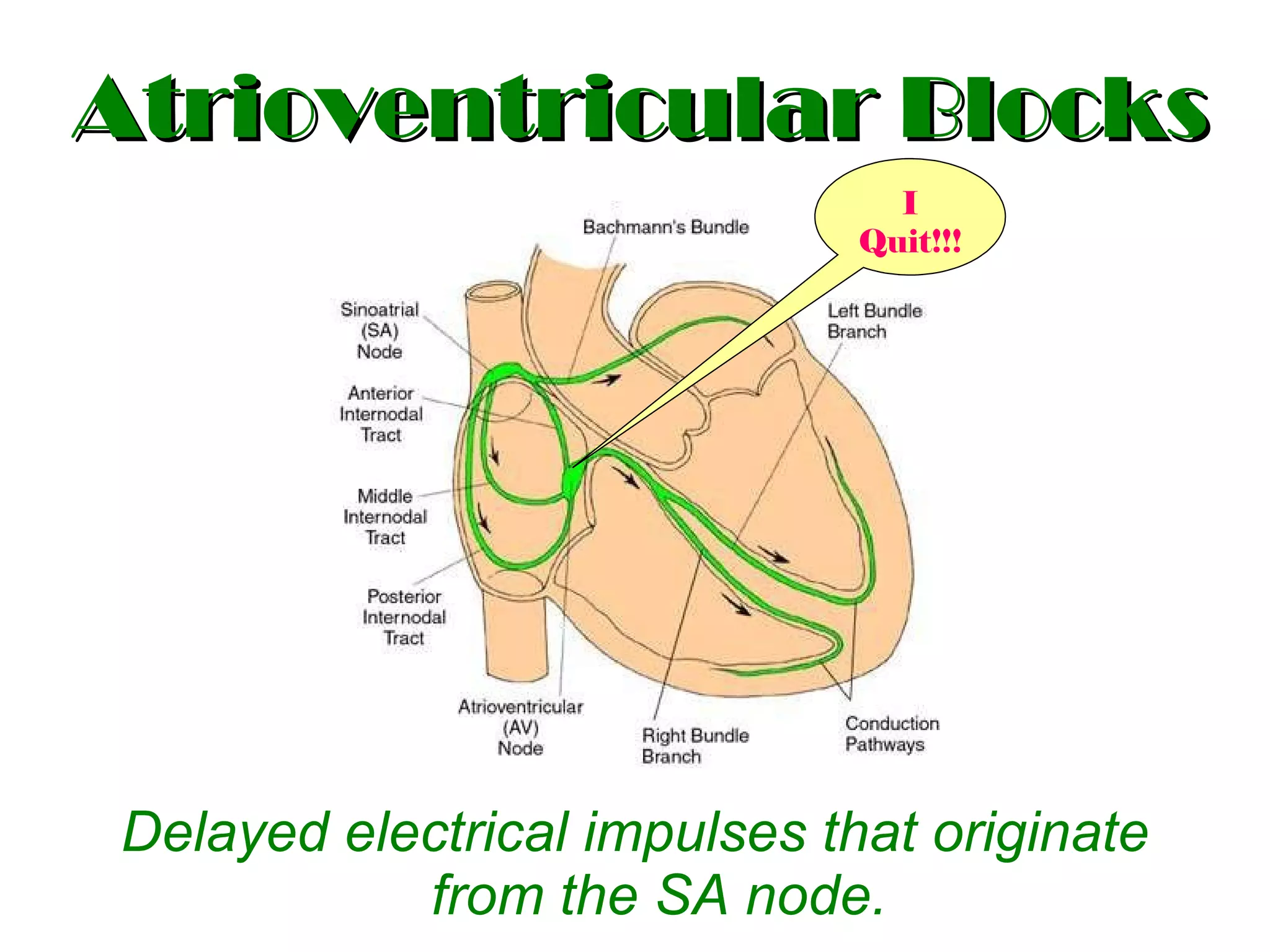
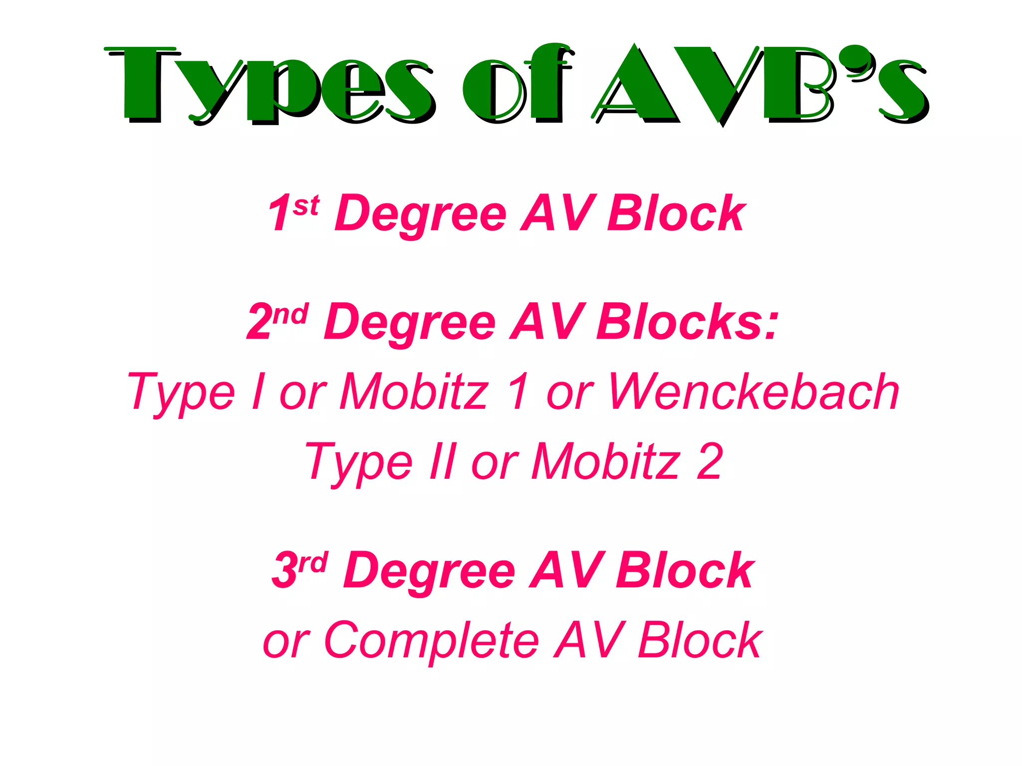
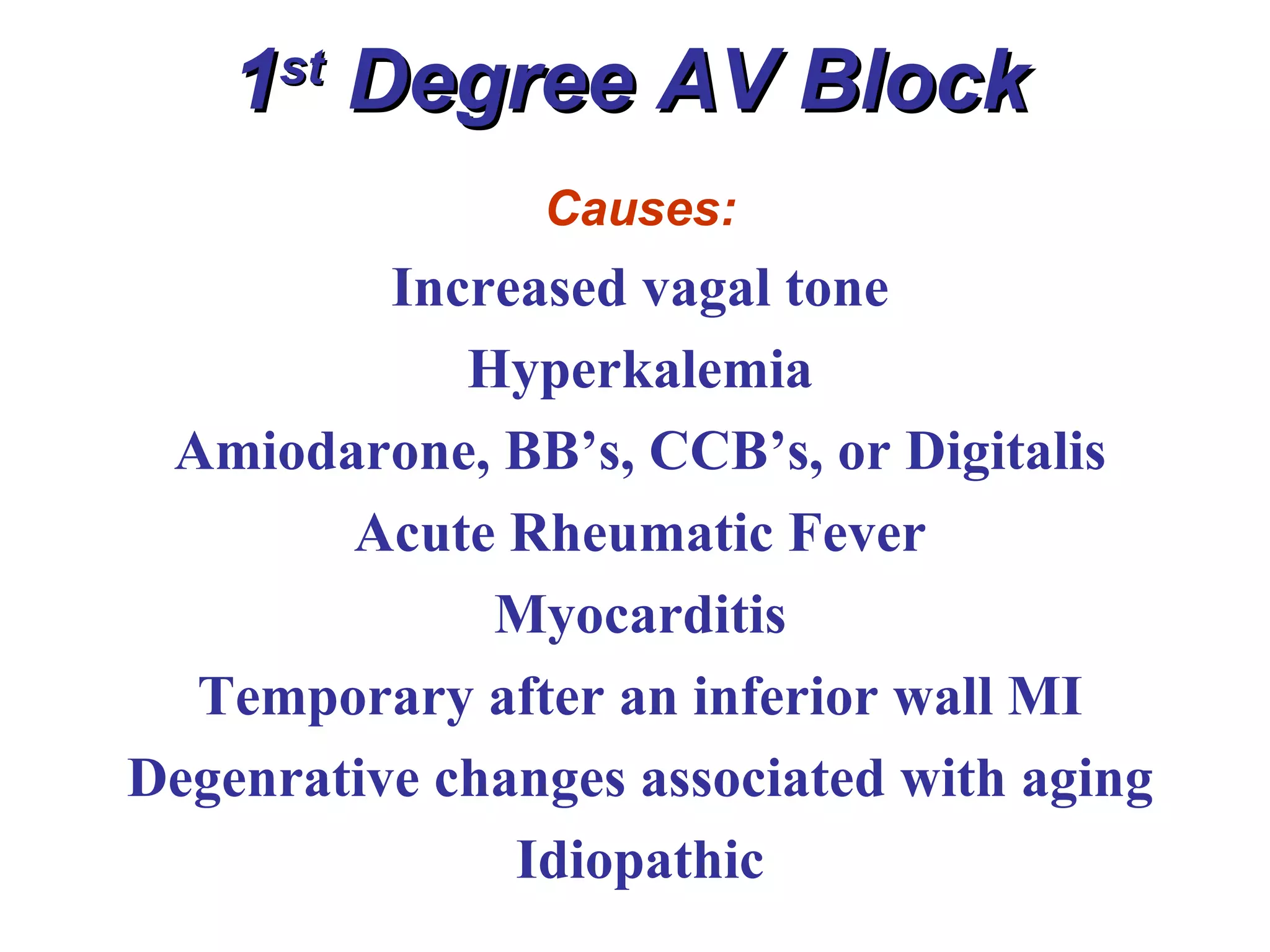
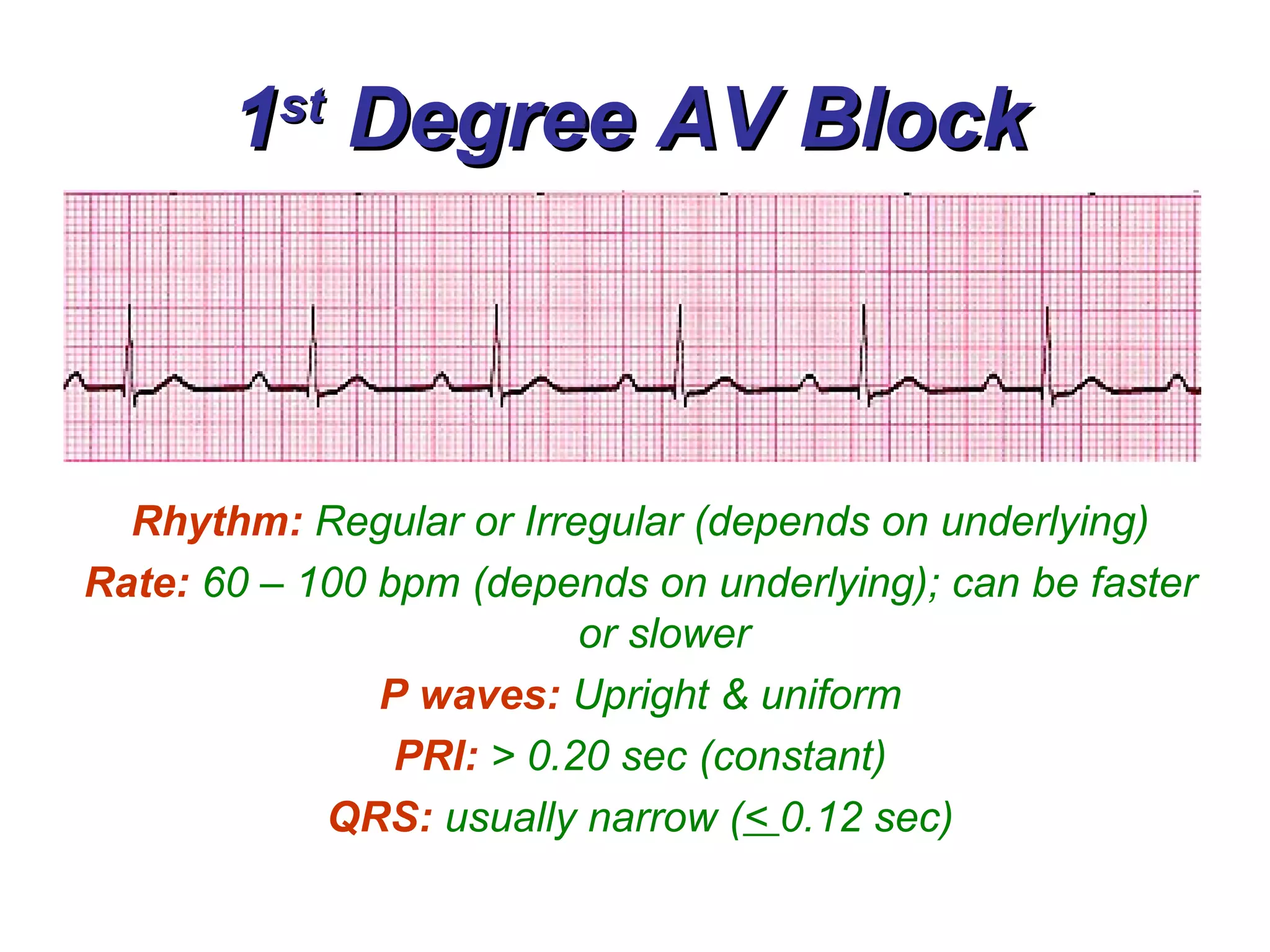
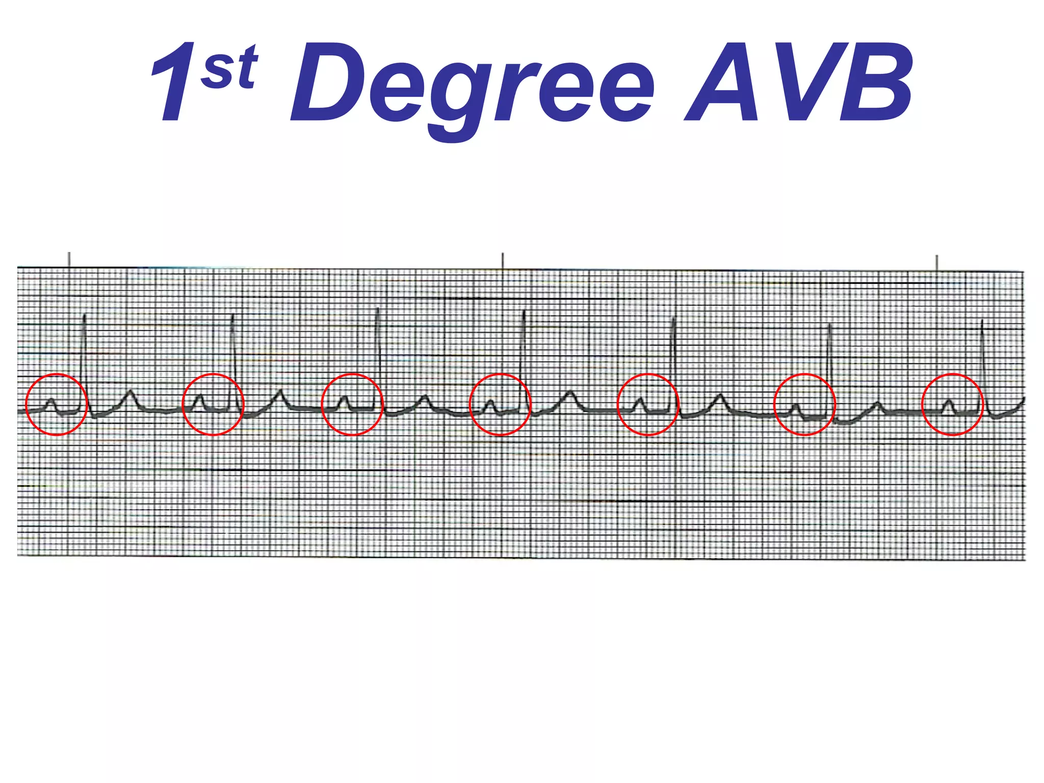
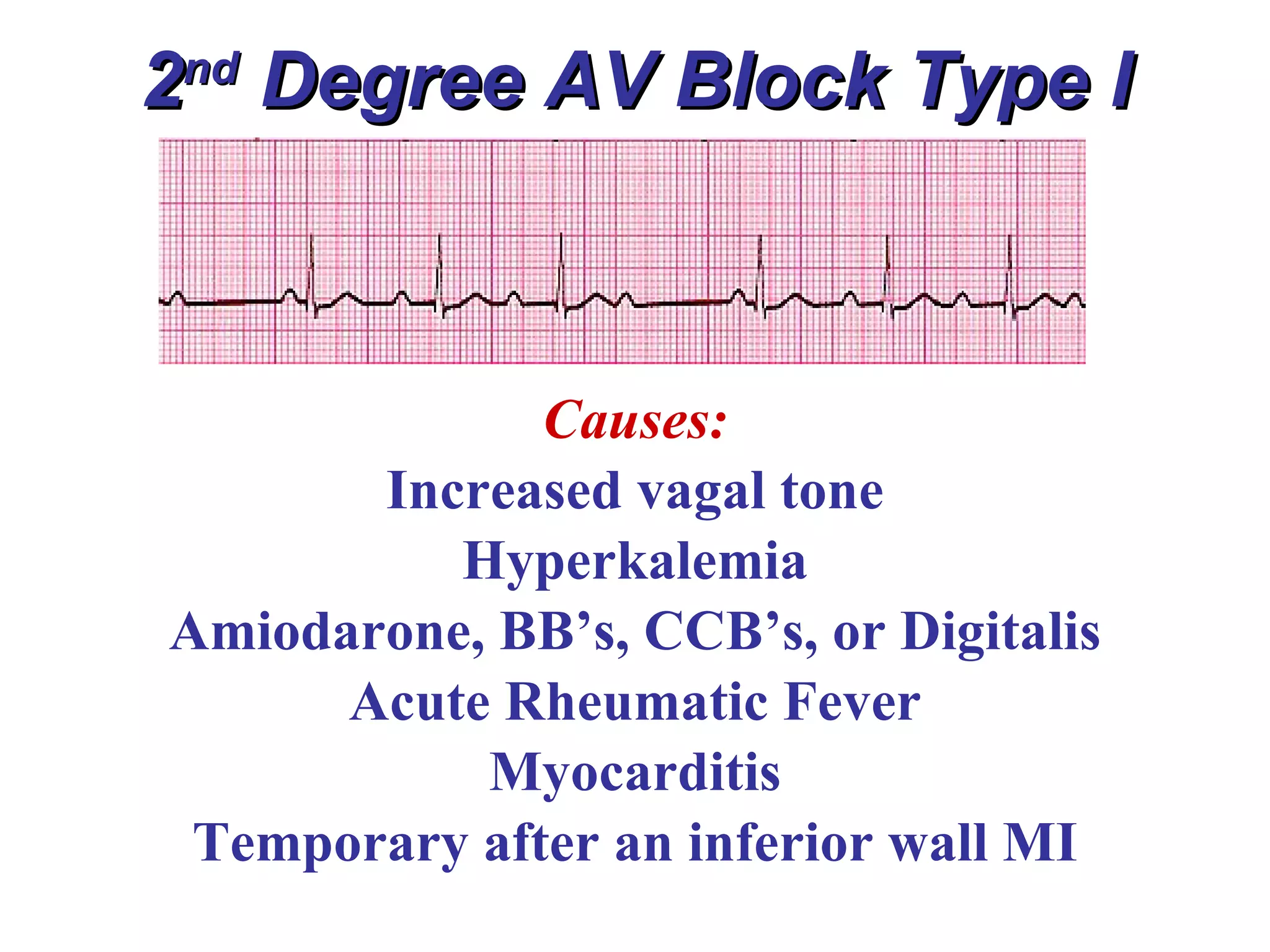
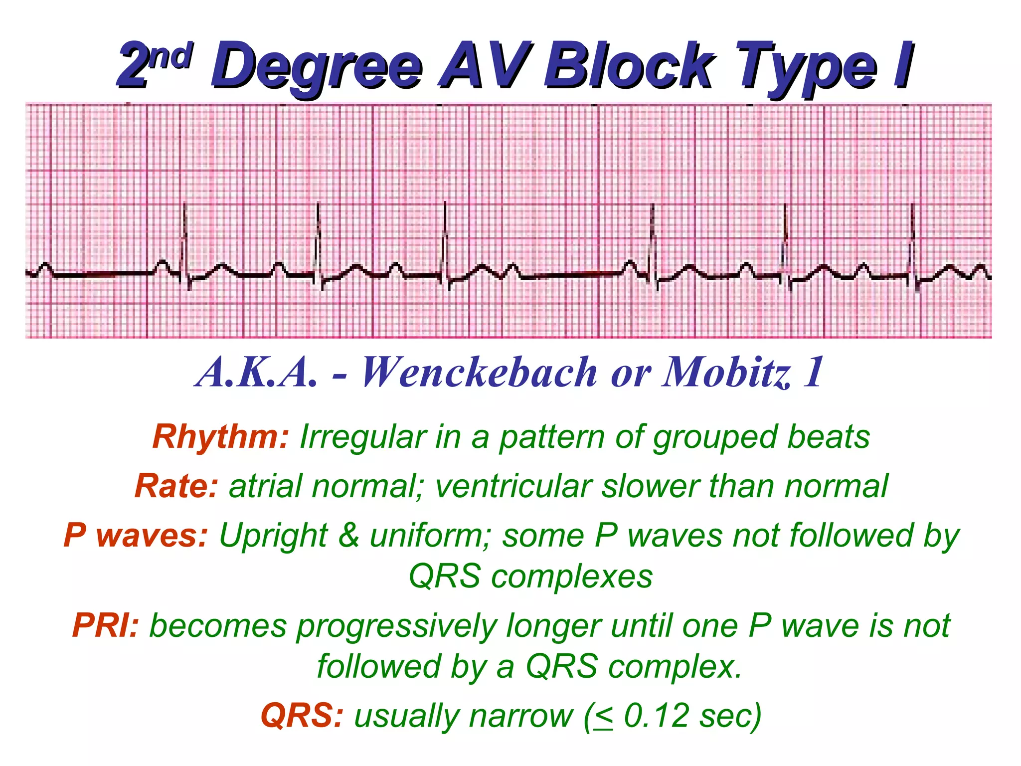
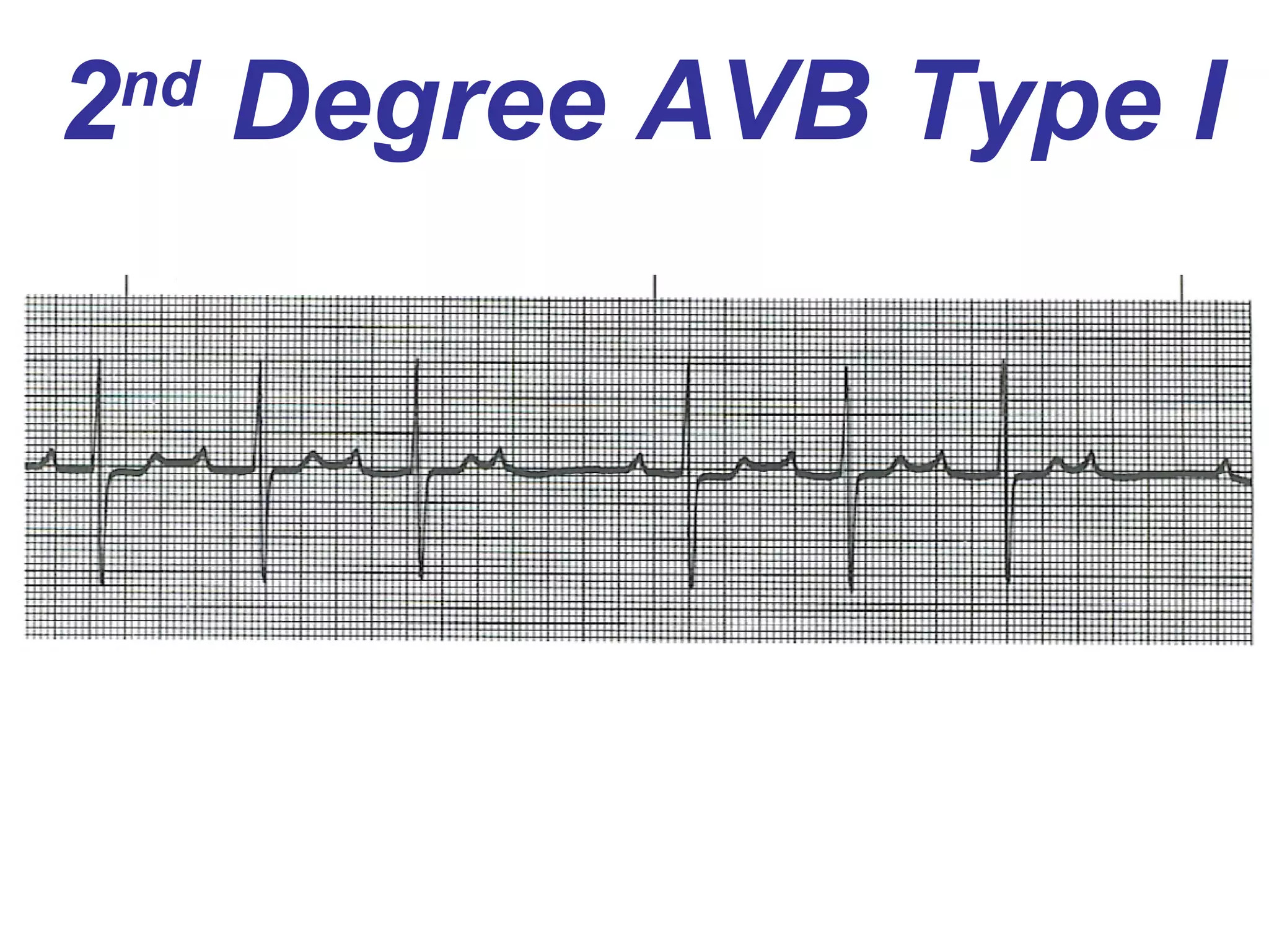
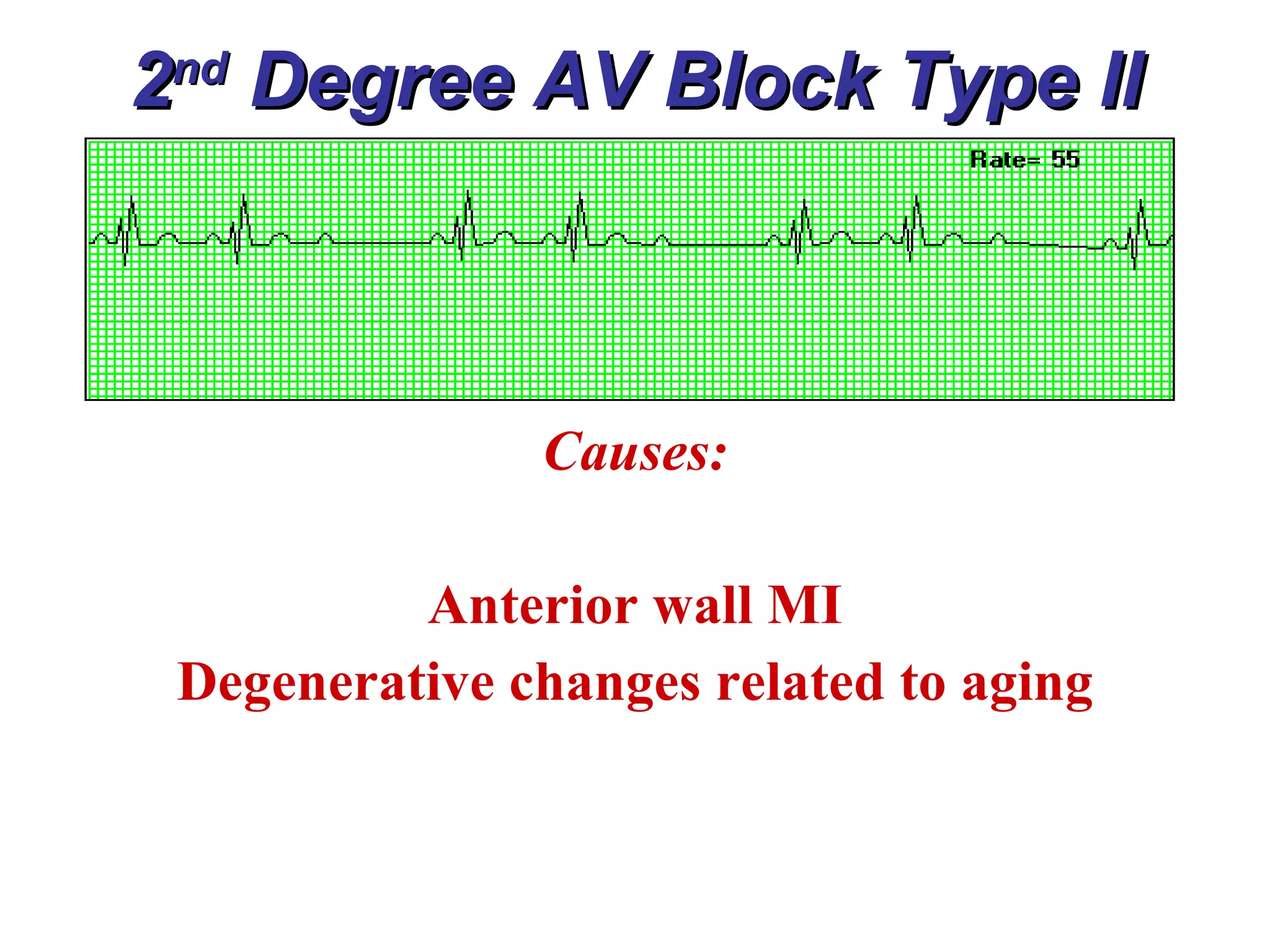
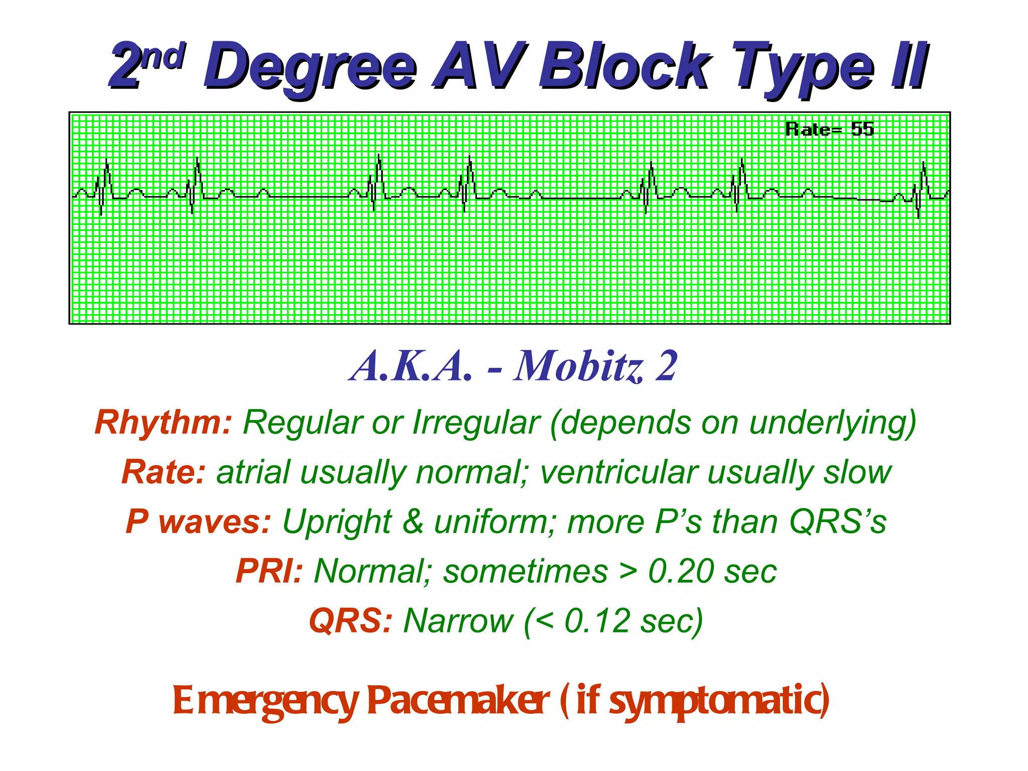
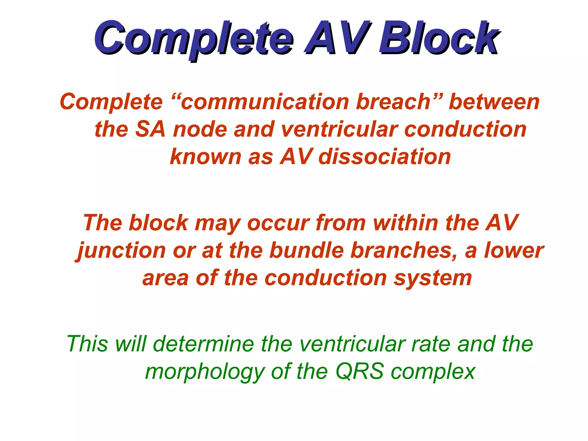
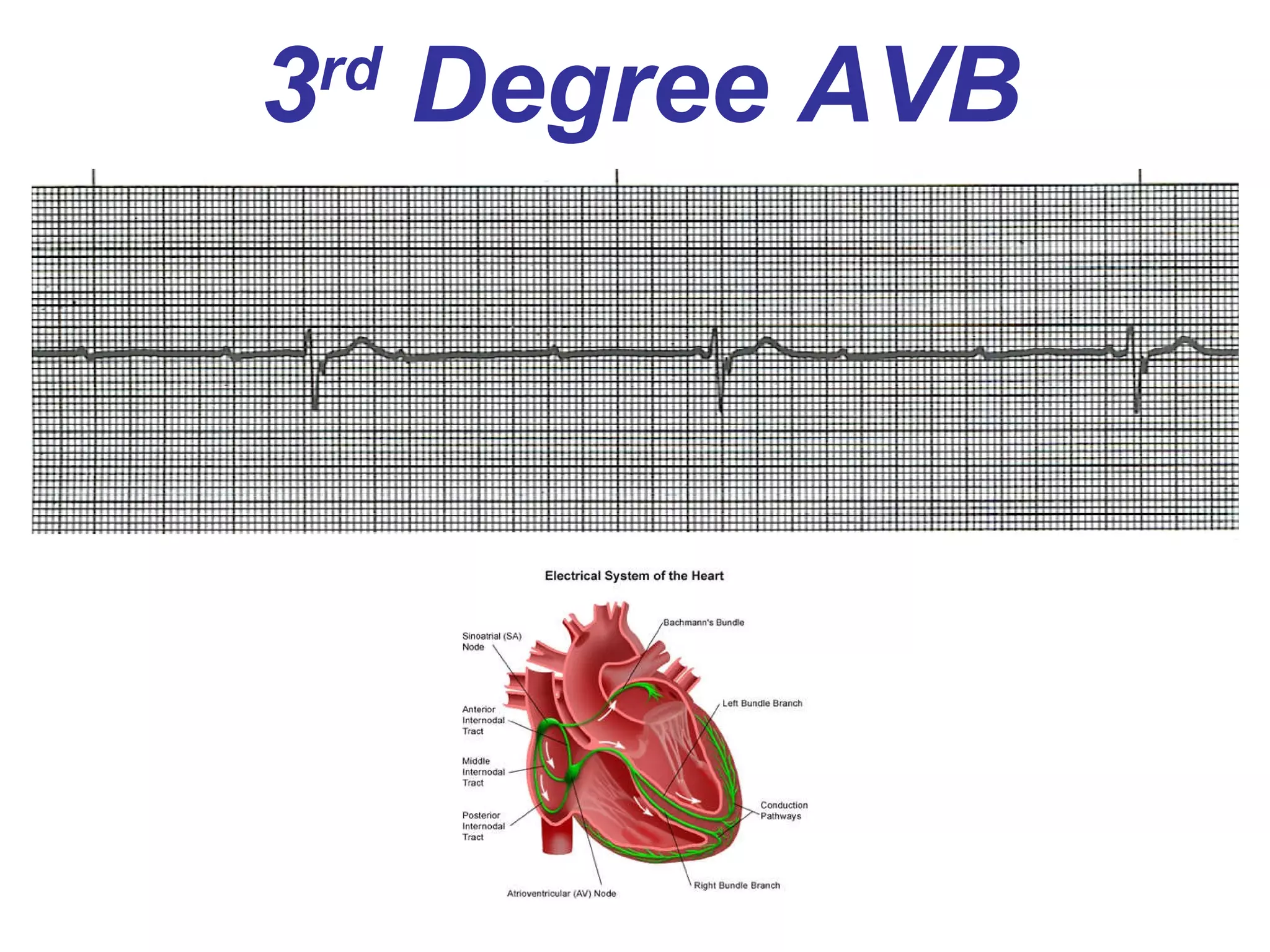
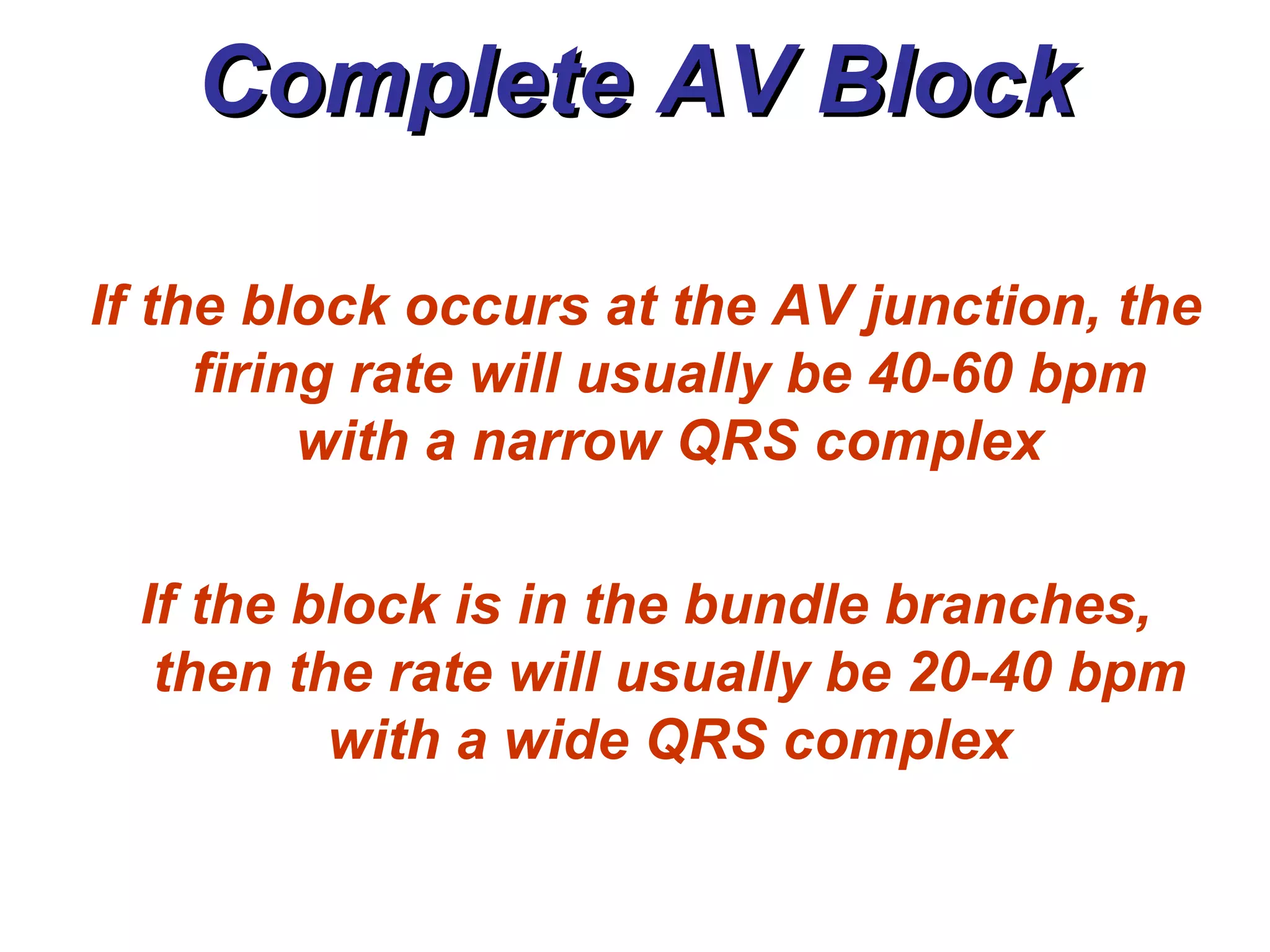
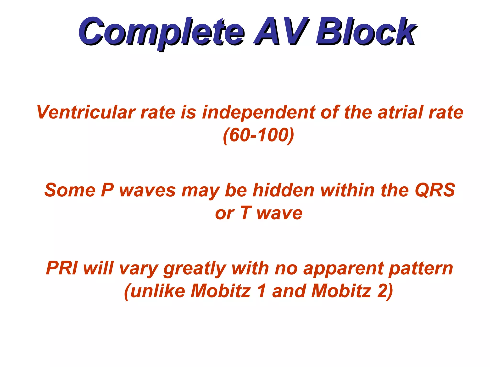
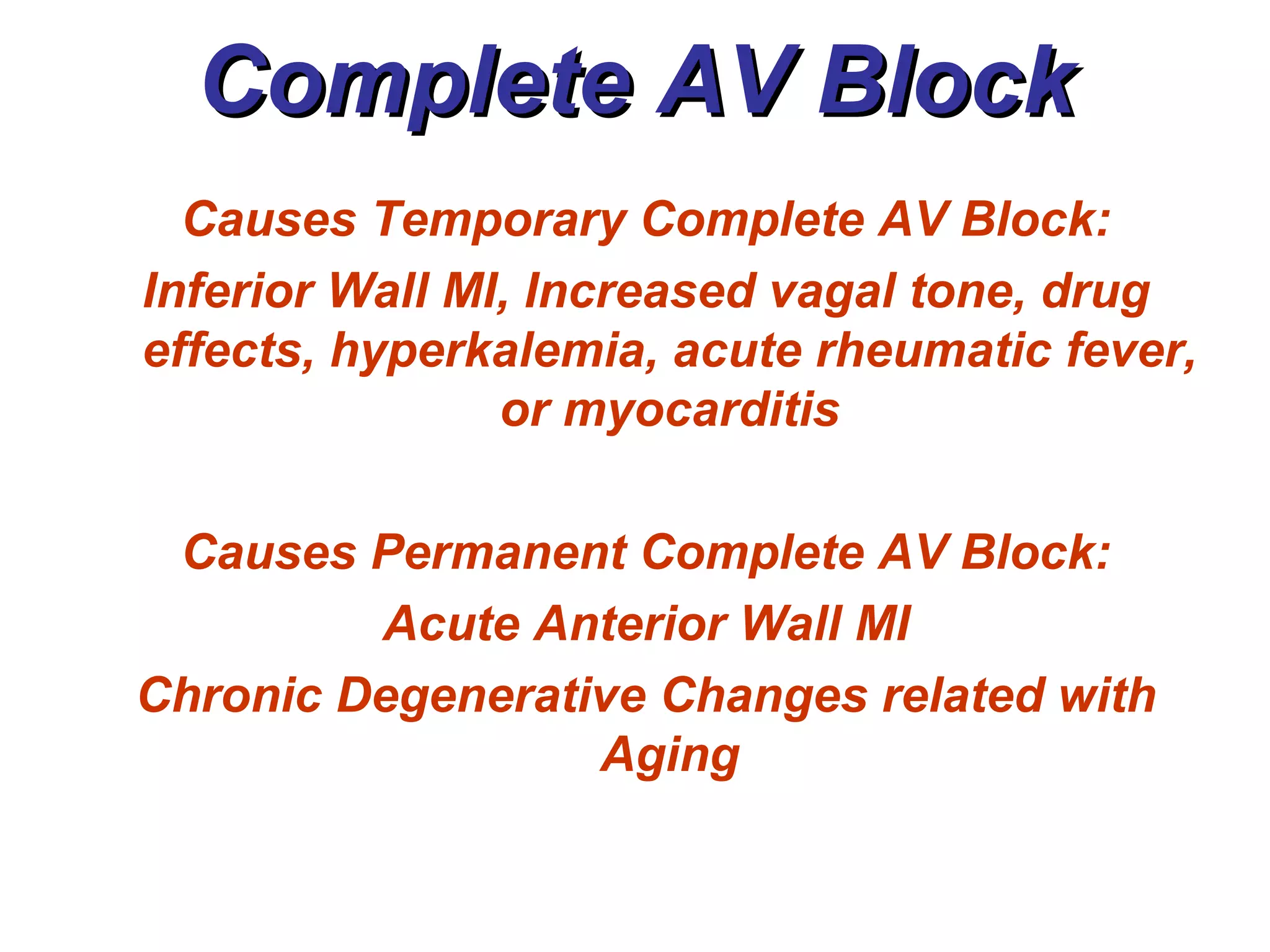
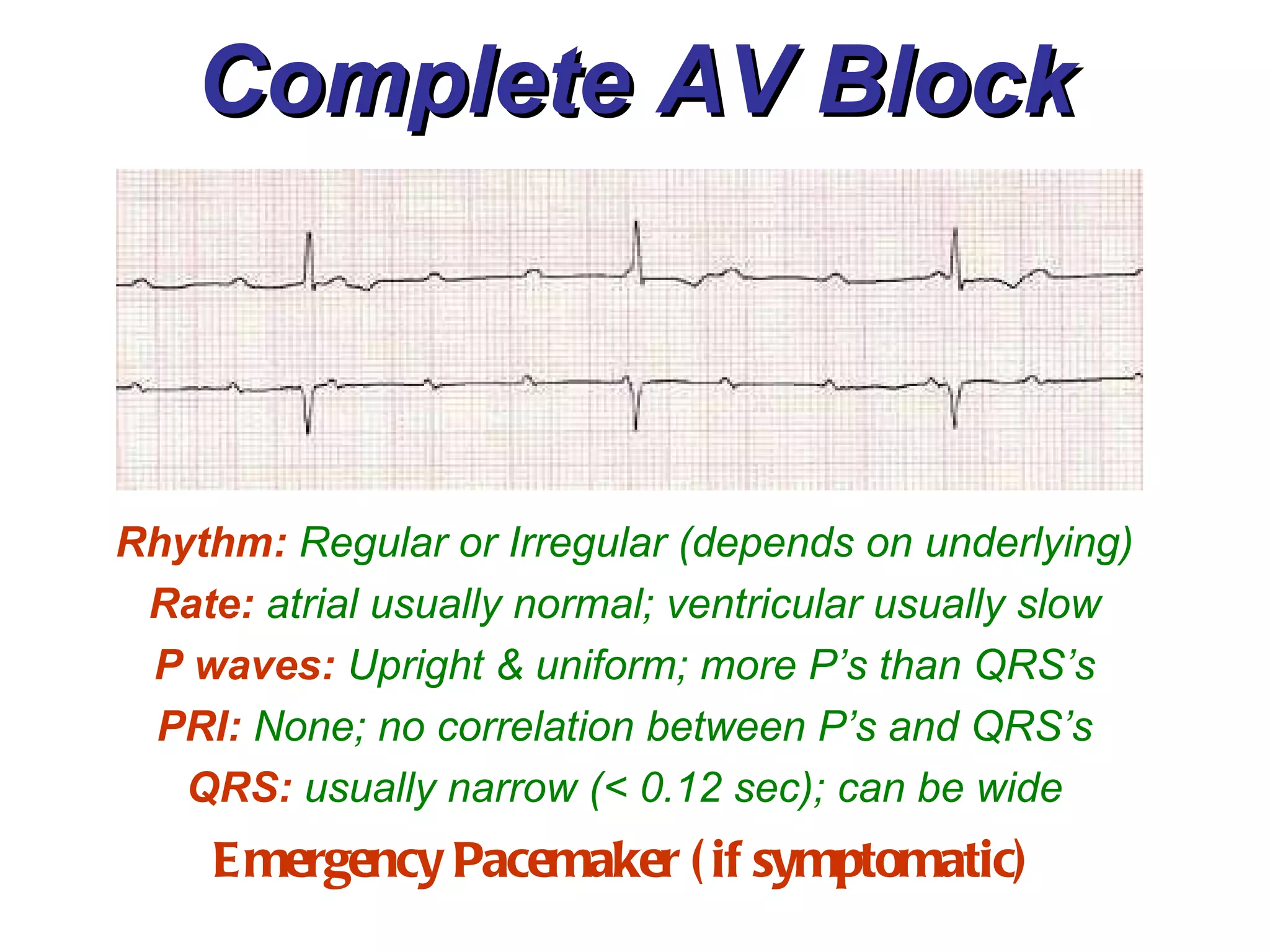
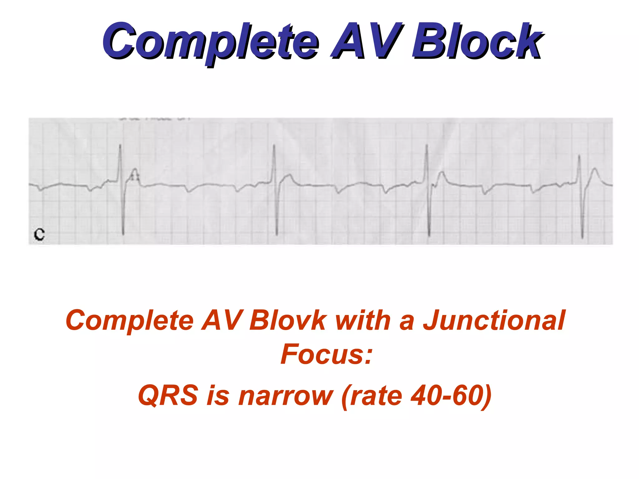
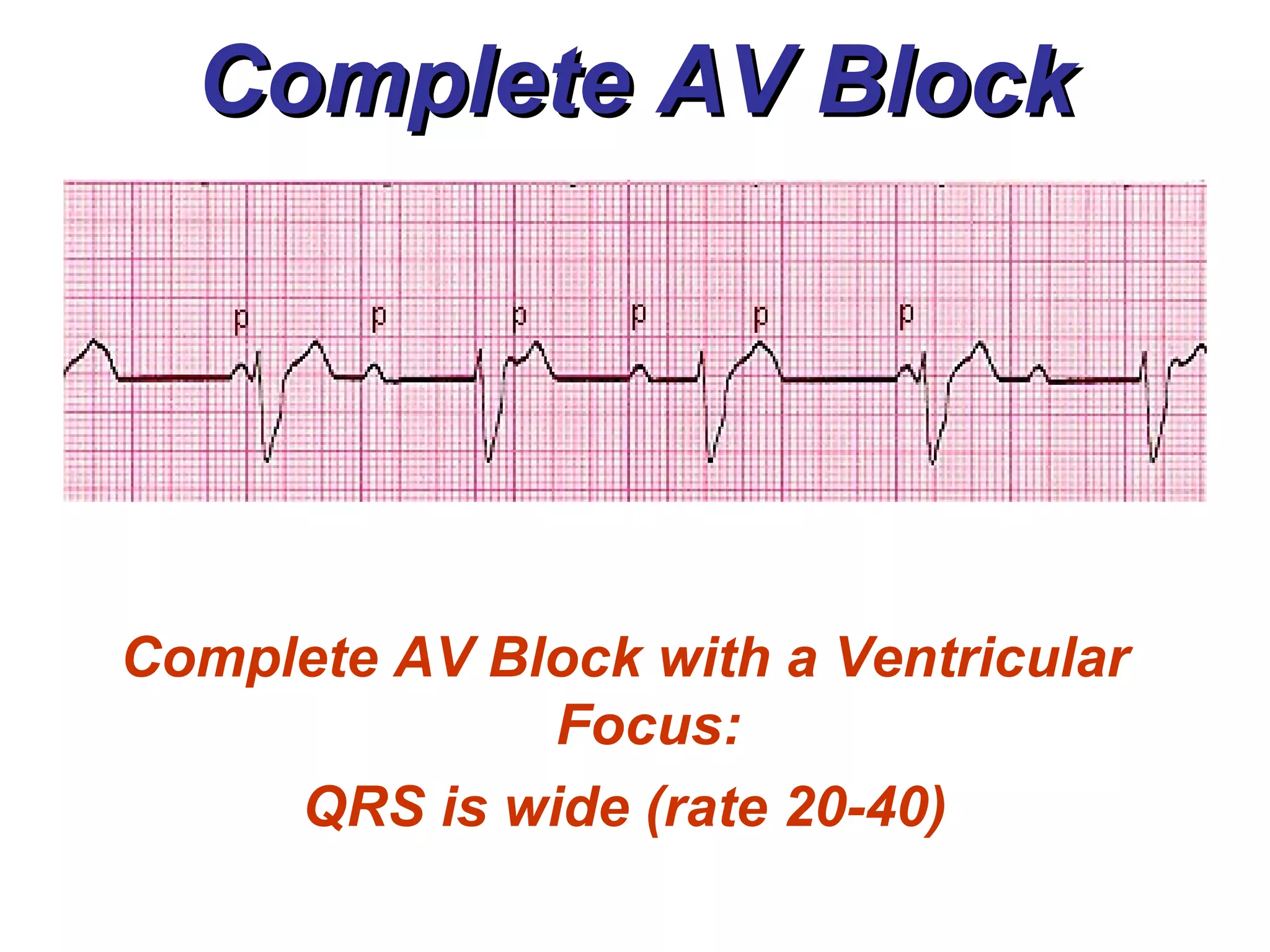
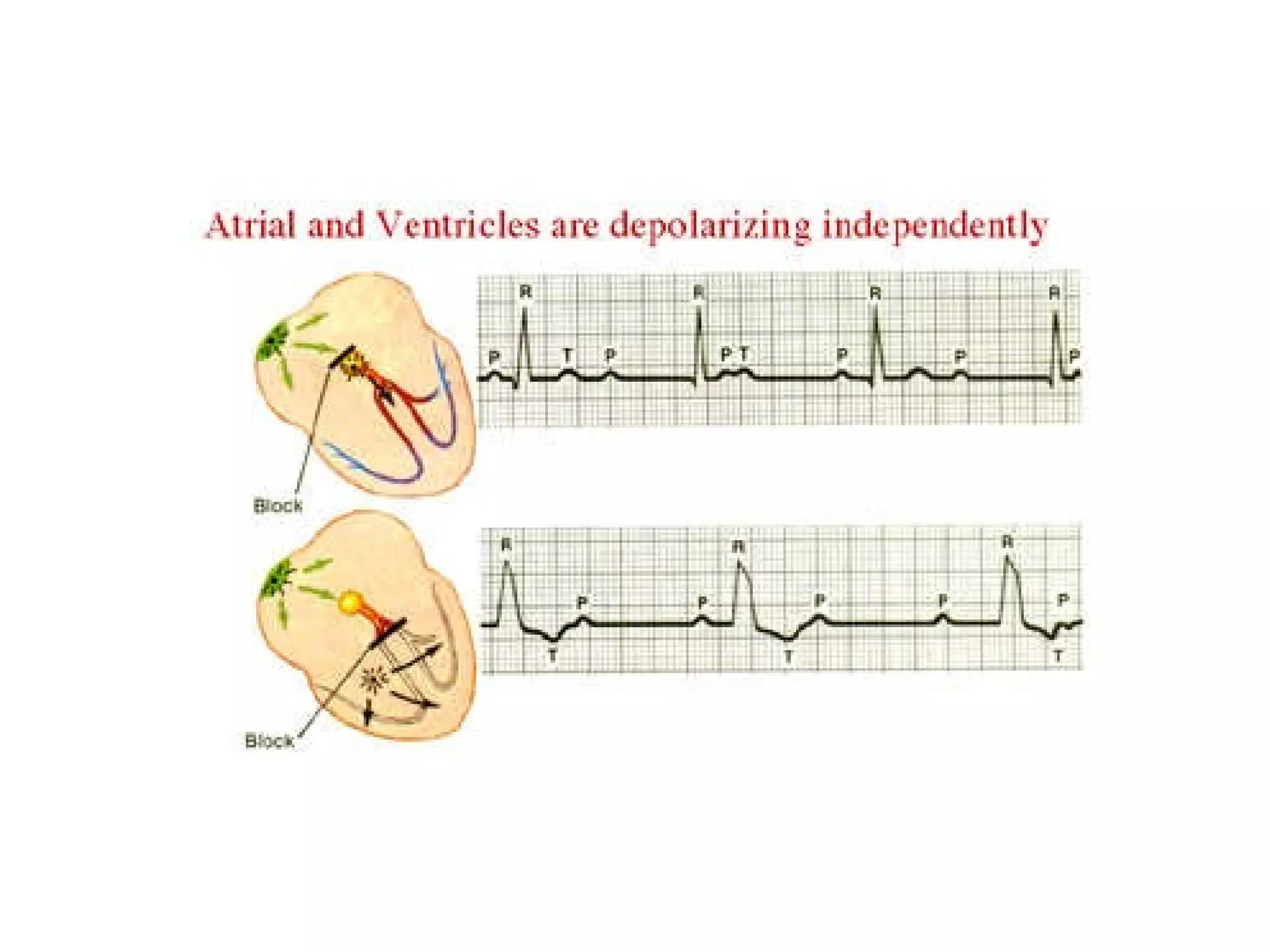
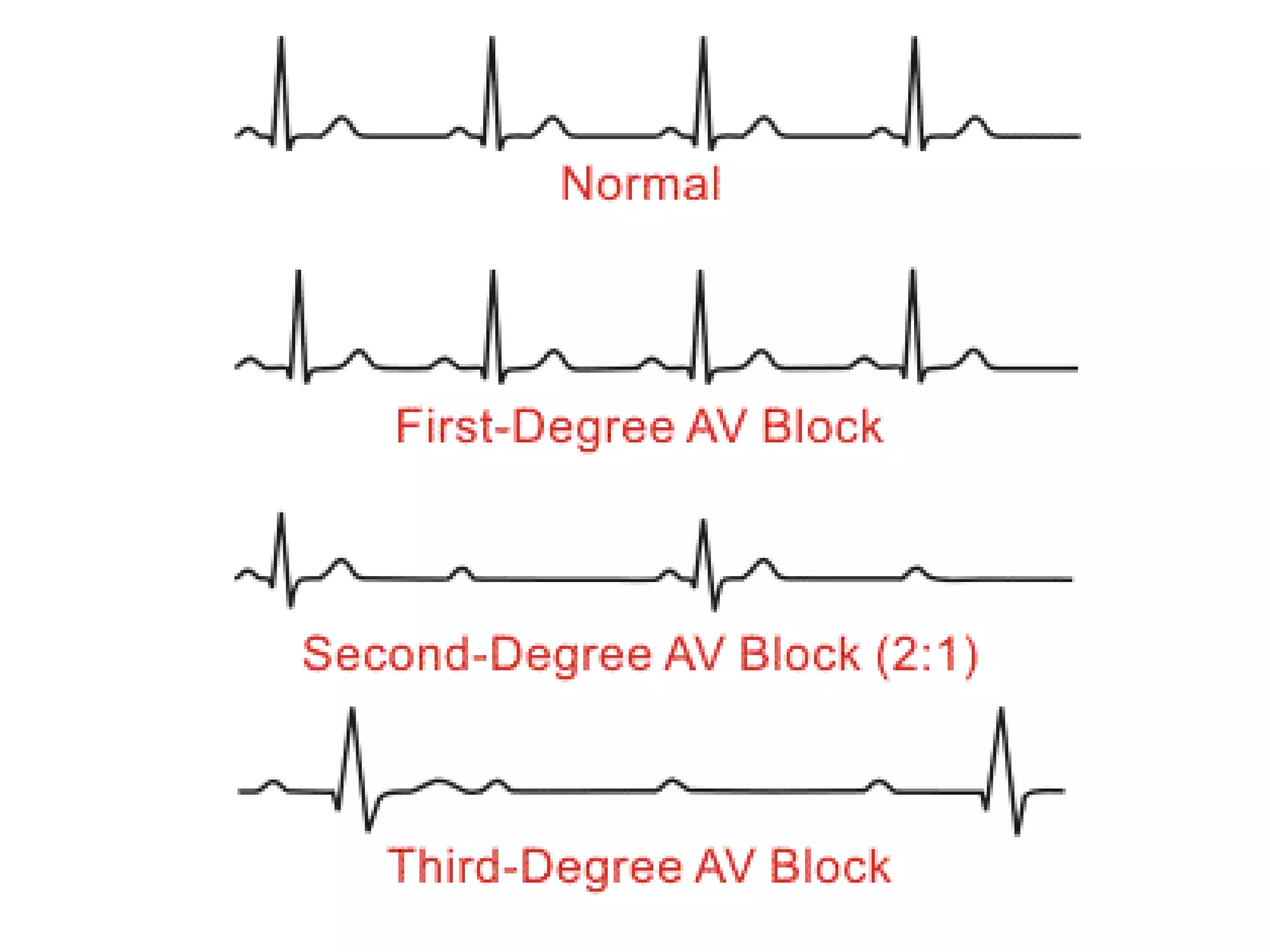
Atrioventricular Blocks Delayed electrical impulses that originate from the SA node. I Quit!!!
AV Blocks Causes: Underlying heart conditions Certain drugs Congenital anomalies Conditions that cause disruption in the cardiac conduction system
AV Blocks Conditions that cause TEMPRORARY disruption in the cardiac conduction system: MI of the inferior wall Digoxin Toxicity Acute Myocarditis Calcium Channel Blockers Beta-adrenergic Blockers Cardiac Surgery
AV Blocks Conditions that cause PERMANENT disruption in the cardiac conduction system: Changes Associated with Aging MI of the anteroseptal wall Congenital Abnormalities Cardiomyopathy Cardiac Surgery
Types of AVB’s 1 st  Degree AV Block  2 nd  Degree AV Blocks: Type I or Mobitz 1 or Wenckebach Type II or Mobitz 2 3 rd  Degree AV Block or Complete AV Block
1 st  Degree AV Block Causes: Increased vagal tone Hyperkalemia Amiodarone, BB’s, CCB’s, or Digitalis Acute Rheumatic Fever Myocarditis Temporary after an inferior wall MI Degenrative changes associated with aging Idiopathic
1 st  Degree AV Block Rhythm:   Regular or Irregular (depends on underlying) Rate:   60 – 100 bpm (depends on underlying); can be faster or slower P waves:   Upright & uniform PRI:   > 0.20 sec (constant) QRS:   usually narrow ( <  0.12 sec)
1 st  Degree AVB
2 nd  Degree AV Block Type I Causes: Increased vagal tone Hyperkalemia Amiodarone, BB’s, CCB’s, or Digitalis Acute Rheumatic Fever Myocarditis Temporary after an inferior wall MI
2 nd  Degree AV Block Type I A.K.A. - Wenckebach or Mobitz 1 Rhythm:   Irregular in a pattern of grouped beats Rate:   atrial normal; ventricular slower than normal P waves:   Upright & uniform; some P waves not followed by QRS complexes PRI:   becomes progressively longer until one P wave is not followed by a QRS complex. QRS:   usually narrow ( <  0.12 sec)
2 nd  Degree AVB Type I
2 nd  Degree AV Block Type II Causes: Anterior wall MI Degenerative changes related to aging
2 nd  Degree AV Block Type II A.K.A. - Mobitz 2 Rhythm:   Regular or Irregular (depends on underlying) Rate:   atrial usually normal; ventricular usually slow P waves:   Upright & uniform; more P’s than QRS’s PRI:   Normal; sometimes > 0.20 sec QRS:   Narrow (< 0.12 sec) Emergency Pacemaker (if symptomatic)
Complete AV Block Complete “communication breach” between the SA node and ventricular conduction known as AV dissociation The block may occur from within the AV junction or at the bundle branches, a lower area of the conduction system  This will determine the ventricular rate and the morphology of the QRS complex
3 rd  Degree AVB
Complete AV Block If the block occurs at the AV junction, the firing rate will usually be 40-60 bpm with a narrow QRS complex If the block is in the bundle branches, then the rate will usually be 20-40 bpm with a wide QRS complex
Complete AV Block Ventricular rate is independent of the atrial rate (60-100) Some P waves may be hidden within the QRS or T wave PRI will vary greatly with no apparent pattern (unlike Mobitz 1 and Mobitz 2)
Complete AV Block Causes Temporary Complete AV Block: Inferior Wall MI, Increased vagal tone, drug effects, hyperkalemia, acute rheumatic fever, or myocarditis Causes Permanent Complete AV Block: Acute Anterior Wall MI Chronic Degenerative Changes related with Aging
Complete AV Block Emergency Pacemaker (if symptomatic) Rhythm:  Regular or Irregular (depends on underlying) Rate:  atrial usually normal; ventricular usually slow P waves:  Upright & uniform; more P’s than QRS’s PRI:  None; no correlation between P’s and QRS’s QRS:  usually narrow (< 0.12 sec); can be wide
Complete AV Block Complete AV Blovk with a Junctional Focus: QRS is narrow (rate 40-60)
Complete AV Block Complete AV Block with a Ventricular Focus: QRS is wide (rate 20-40)
 
 
TIME TO WORKOUT!!!
References Beverage, D. Haworth, K., Labus, D. Mayer, B. H., & Munson, C. (2005).  ECG interpretation made incredibly easy,  (3 rd  ed.). Ambler, PA: Lippincott, Williams, & Wilkins. Chernecky, C., et al. (2002).  Real world nursing survival guide: ECG’s & the heart.  United States of America: W. B. Saunders Company. Huff, J. (2006).  ECG workout: Exercises in arrhythmia interpretation  (5 th  ed.). United States of America: Lippincott, Williams & Wilkins. Walraven, G. (1999).  Basic arrhythmias  (5 th  ed.). United States of America: Prentice-Hall, Inc. www.madsci.com/manu/ekg_rhy.htm

Atrioventricular Blocks - BMH/Tele

  • 1.
    Atrioventricular Blocks Delayedelectrical impulses that originate from the SA node. I Quit!!!
  • 2.
    AV Blocks Causes:Underlying heart conditions Certain drugs Congenital anomalies Conditions that cause disruption in the cardiac conduction system
  • 3.
    AV Blocks Conditionsthat cause TEMPRORARY disruption in the cardiac conduction system: MI of the inferior wall Digoxin Toxicity Acute Myocarditis Calcium Channel Blockers Beta-adrenergic Blockers Cardiac Surgery
  • 4.
    AV Blocks Conditionsthat cause PERMANENT disruption in the cardiac conduction system: Changes Associated with Aging MI of the anteroseptal wall Congenital Abnormalities Cardiomyopathy Cardiac Surgery
  • 5.
    Types of AVB’s1 st Degree AV Block 2 nd Degree AV Blocks: Type I or Mobitz 1 or Wenckebach Type II or Mobitz 2 3 rd Degree AV Block or Complete AV Block
  • 6.
    1 st Degree AV Block Causes: Increased vagal tone Hyperkalemia Amiodarone, BB’s, CCB’s, or Digitalis Acute Rheumatic Fever Myocarditis Temporary after an inferior wall MI Degenrative changes associated with aging Idiopathic
  • 7.
    1 st Degree AV Block Rhythm: Regular or Irregular (depends on underlying) Rate: 60 – 100 bpm (depends on underlying); can be faster or slower P waves: Upright & uniform PRI: > 0.20 sec (constant) QRS: usually narrow ( < 0.12 sec)
  • 8.
    1 st Degree AVB
  • 9.
    2 nd Degree AV Block Type I Causes: Increased vagal tone Hyperkalemia Amiodarone, BB’s, CCB’s, or Digitalis Acute Rheumatic Fever Myocarditis Temporary after an inferior wall MI
  • 10.
    2 nd Degree AV Block Type I A.K.A. - Wenckebach or Mobitz 1 Rhythm: Irregular in a pattern of grouped beats Rate: atrial normal; ventricular slower than normal P waves: Upright & uniform; some P waves not followed by QRS complexes PRI: becomes progressively longer until one P wave is not followed by a QRS complex. QRS: usually narrow ( < 0.12 sec)
  • 11.
    2 nd Degree AVB Type I
  • 12.
    2 nd Degree AV Block Type II Causes: Anterior wall MI Degenerative changes related to aging
  • 13.
    2 nd Degree AV Block Type II A.K.A. - Mobitz 2 Rhythm: Regular or Irregular (depends on underlying) Rate: atrial usually normal; ventricular usually slow P waves: Upright & uniform; more P’s than QRS’s PRI: Normal; sometimes > 0.20 sec QRS: Narrow (< 0.12 sec) Emergency Pacemaker (if symptomatic)
  • 14.
    Complete AV BlockComplete “communication breach” between the SA node and ventricular conduction known as AV dissociation The block may occur from within the AV junction or at the bundle branches, a lower area of the conduction system This will determine the ventricular rate and the morphology of the QRS complex
  • 15.
    3 rd Degree AVB
  • 16.
    Complete AV BlockIf the block occurs at the AV junction, the firing rate will usually be 40-60 bpm with a narrow QRS complex If the block is in the bundle branches, then the rate will usually be 20-40 bpm with a wide QRS complex
  • 17.
    Complete AV BlockVentricular rate is independent of the atrial rate (60-100) Some P waves may be hidden within the QRS or T wave PRI will vary greatly with no apparent pattern (unlike Mobitz 1 and Mobitz 2)
  • 18.
    Complete AV BlockCauses Temporary Complete AV Block: Inferior Wall MI, Increased vagal tone, drug effects, hyperkalemia, acute rheumatic fever, or myocarditis Causes Permanent Complete AV Block: Acute Anterior Wall MI Chronic Degenerative Changes related with Aging
  • 19.
    Complete AV BlockEmergency Pacemaker (if symptomatic) Rhythm: Regular or Irregular (depends on underlying) Rate: atrial usually normal; ventricular usually slow P waves: Upright & uniform; more P’s than QRS’s PRI: None; no correlation between P’s and QRS’s QRS: usually narrow (< 0.12 sec); can be wide
  • 20.
    Complete AV BlockComplete AV Blovk with a Junctional Focus: QRS is narrow (rate 40-60)
  • 21.
    Complete AV BlockComplete AV Block with a Ventricular Focus: QRS is wide (rate 20-40)
  • 22.
  • 23.
  • 24.
  • 25.
    References Beverage, D.Haworth, K., Labus, D. Mayer, B. H., & Munson, C. (2005). ECG interpretation made incredibly easy, (3 rd ed.). Ambler, PA: Lippincott, Williams, & Wilkins. Chernecky, C., et al. (2002). Real world nursing survival guide: ECG’s & the heart. United States of America: W. B. Saunders Company. Huff, J. (2006). ECG workout: Exercises in arrhythmia interpretation (5 th ed.). United States of America: Lippincott, Williams & Wilkins. Walraven, G. (1999). Basic arrhythmias (5 th ed.). United States of America: Prentice-Hall, Inc. www.madsci.com/manu/ekg_rhy.htm