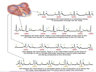
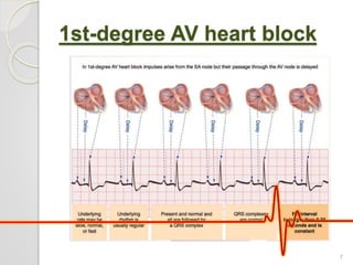
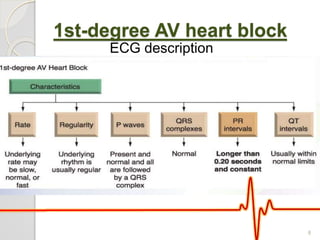
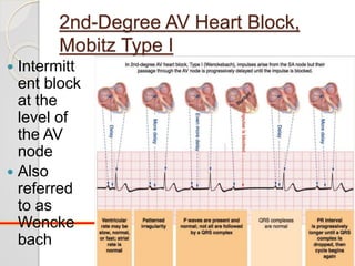
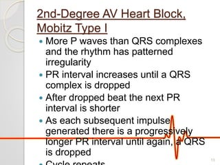
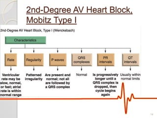
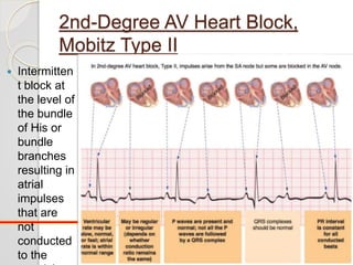
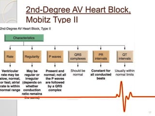
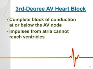
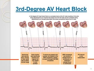
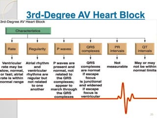
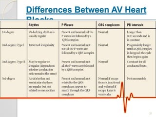
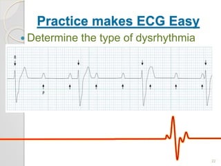
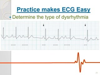
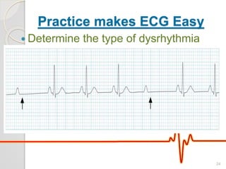
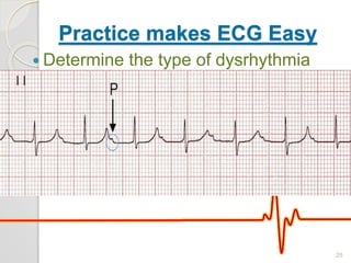
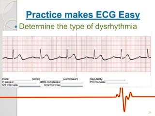
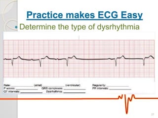
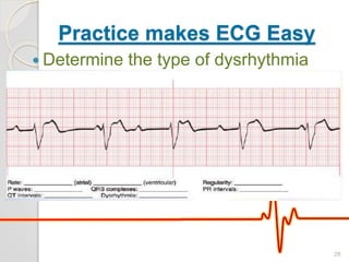
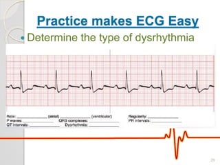
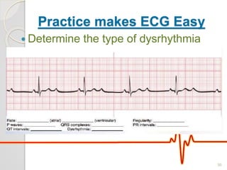
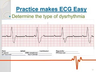
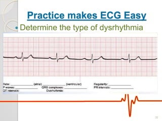
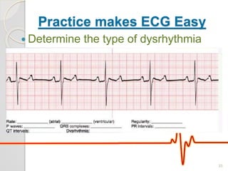
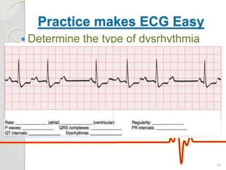
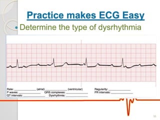
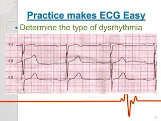
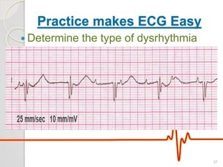
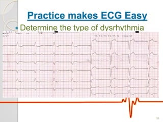
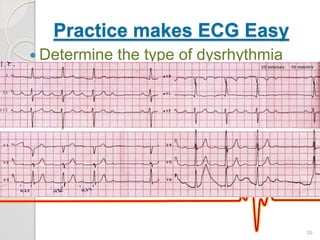
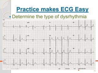
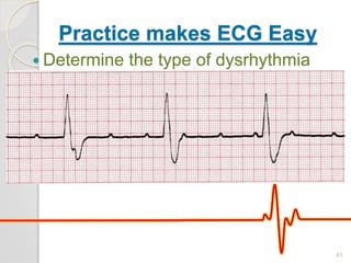
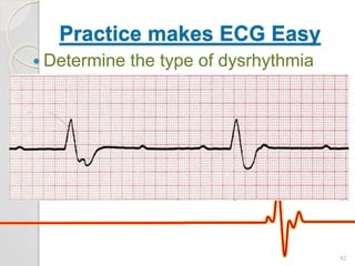
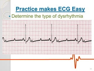
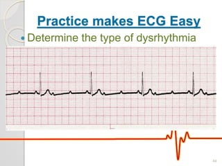
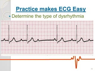
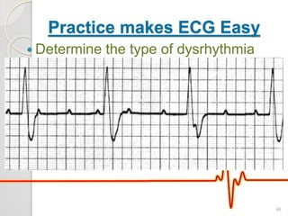
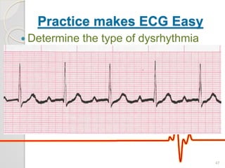
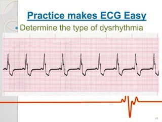
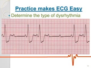
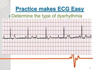
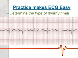
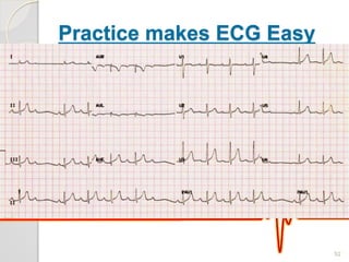
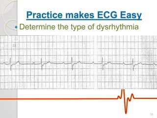
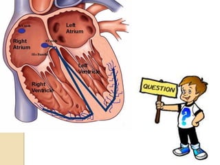
Atrio-Ventricular Block
ECG Analysis
Shubhendu Sharma
1
AV Block
 Partial delays or complete
interruptions in the cardiac conduction
pathway between the atria and
ventricles
 The degree of block defines the type
and classification of heart block
2
AV Block
 Common causes:
◦ Ischemia
◦ Myocardial necrosis
◦ Degenerative disease of the conduction
system
◦ Congenital anomalies
◦ Drugs (especially digitalis preparations)
3
AV Block
 1st-degree AV heart block
 2nd-degree AV heart block, Mobitz
type I (Wenckebach)
 2nd-degree AV heart block, Mobitz
type II
 3rd-degree AV heart block
4
AV Block
5
1st-degree AV heart block
 Not a true block
 Is a consistent delay of conduction at
the level of the AV node
6
1st-degree AV heart block
7
1st-degree AV heart block
ECG description
8
2nd-Degree AV Heart Block,
Mobitz Type I
 Intermitt
ent block
at the
level of
the AV
node
 Also
referred
to as
Wencke
bach
12
2nd-Degree AV Heart Block,
Mobitz Type I
 More P waves than QRS complexes
and the rhythm has patterned
irregularity
 PR interval increases until a QRS
complex is dropped
 After dropped beat the next PR
interval is shorter
 As each subsequent impulse
generated there is a progressively
longer PR interval until again, a QRS
is dropped
13
2nd-Degree AV Heart Block,
Mobitz Type I
14
2nd-Degree AV Heart Block,
Mobitz Type II
 Intermitten
t block at
the level of
the bundle
of His or
bundle
branches
resulting in
atrial
impulses
that are
not
conducted
to the 16
2nd-Degree AV Heart Block,
Mobitz Type II
17
3rd-Degree AV Heart Block
 Complete block of conduction
at or below the AV node
 Impulses from atria cannot
reach ventricles
18
3rd-Degree AV Heart Block
19
3rd-Degree AV Heart Block
20
Differences Between AV Heart
Blocks
21
Practice makes ECG Easy
 Determine the type of dysrhythmia
22
Practice makes ECG Easy
 Determine the type of dysrhythmia
23
Practice makes ECG Easy
 Determine the type of dysrhythmia
24
Practice makes ECG Easy
 Determine the type of dysrhythmia
25
Practice makes ECG Easy
 Determine the type of dysrhythmia
26
Practice makes ECG Easy
 Determine the type of dysrhythmia
27
Practice makes ECG Easy
 Determine the type of dysrhythmia
28
Practice makes ECG Easy
 Determine the type of dysrhythmia
29
Practice makes ECG Easy
 Determine the type of dysrhythmia
30
Practice makes ECG Easy
 Determine the type of dysrhythmia
31
Practice makes ECG Easy
 Determine the type of dysrhythmia
32
Practice makes ECG Easy
 Determine the type of dysrhythmia
33
Practice makes ECG Easy
 Determine the type of dysrhythmia
34
Practice makes ECG Easy
 Determine the type of dysrhythmia
35
Practice makes ECG Easy
 Determine the type of dysrhythmia
36
Practice makes ECG Easy
 Determine the type of dysrhythmia
37
Practice makes ECG Easy
 Determine the type of dysrhythmia
38
Practice makes ECG Easy
 Determine the type of dysrhythmia
39
Practice makes ECG Easy
 Determine the type of dysrhythmia
40
Practice makes ECG Easy
 Determine the type of dysrhythmia
41
Practice makes ECG Easy
 Determine the type of dysrhythmia
42
Practice makes ECG Easy
 Determine the type of dysrhythmia
43
Practice makes ECG Easy
 Determine the type of dysrhythmia
44
Practice makes ECG Easy
 Determine the type of dysrhythmia
45
Practice makes ECG Easy
 Determine the type of dysrhythmia
46
Practice makes ECG Easy
 Determine the type of dysrhythmia
47
Practice makes ECG Easy
 Determine the type of dysrhythmia
48
Practice makes ECG Easy
 Determine the type of dysrhythmia
49
Practice makes ECG Easy
 Determine the type of dysrhythmia
50
Practice makes ECG Easy
 Determine the type of dysrhythmia
51
Practice makes ECG Easy
 Determine the type of dysrhythmia
52
Practice makes ECG Easy
 Determine the type of dysrhythmia
53
54
55

AV block ecg analysis

Editor's Notes

  • #7 The normal PR interval is 0.12 to 0.20 seconds in duration.
  • #8 The normal PR interval is 0.12 to 0.20 seconds in duration.
  • #23 3
  • #24 2.1
  • #25 Mobitz 2
  • #26 Severe 1 degree
  • #27 Atrial rate 56 BPM, Ventricular rate 56 BPM, regular rhythm, P waves are upright and normal and each is followed by a QRS complex, QRS complexes are normal at 0.08 seconds in duration, PRI has a longer than normal duration 0.30 seconds, QT 0.40 seconds. Sinus bradycardia with 1st-degree AV heart block.
  • #28 Answer: Atrial rate 60 BPM, Ventricular rate 50 BPM, patterned irregularity, normal and upright P waves but not all are followed by a QRS complex, normal QRS complexes at 0.08 seconds, PRI is progressively longer until a QRS complex is dropped and then the cycle starts over again, QT 0.40 seconds. 2nd-degree AV heart block, type I.
  • #29 Answer: 50 BPM (ventricular) 84 BPM (atrial), regular rhythm, P waves are upright and normal but there are more P waves than QRS complexes, QRS complexes are bizarre-looking and wide at 0.20 seconds in duration, PRI is immeasurable (as the P waves are not associated with the QRS complexes), the P waves appear to march through the QRS complexes, QT 0.48 seconds. 3rd-degree AV heart block.
  • #30 Answer: Atrial rate 58 BPM, Ventricular rate 60 BPM, regular rhythm, P waves are upright and normal and each is followed by a QRS complex, QRS complexes are normal at 0.12 seconds in duration, PRI has a longer than normal duration 0.30 seconds and constant, QT 0.40 seconds. Sinus bradycardia with 1st-degree AV heart block.
  • #31 Answer: Atrial rate 94 BPM, Ventricular rate 48 BPM, regular rhythm, P waves are upright and normal but there are two P waves preceding each QRS (in other words, not all the P waves are followed by a QRS complex), QRS complexes are normal at 0.08 seconds in duration, PRI 0.16 seconds and is constant for the conducted beats, QT 0.36 seconds. 2nd-degree AV heart block, type II.
  • #32 Answer: Atrial rate 75 BPM, Ventricular rate 36 BPM, regular rhythm, P waves are upright and normal but there are more P waves than QRS complexes, QRS complexes are bizarre-looking and wide at 0.16 seconds in duration, PRI is immeasurable (as the P waves are not associated with the QRS complexes), the P waves appear to march through the QRS complexes, QT 0.36 seconds. 3rd- degree AV heart block.
  • #33 Answer: Atrial rate 45 BPM, Ventricular rate 45 BPM, regular rhythm, normal and upright P waves and each is followed by a QRS complex, QRS complexes at 0.08 seconds, PRI 0.44 seconds and constant, QT 0.48 seconds. Sinus bradycardia with 1st-degree AV heart block.
  • #34 Answer: Atrial rate 94 BPM, Ventricular rate 48 BPM, regular rhythm, P waves are upright and normal but there are two P waves preceding each QRS (in other words, not all the P waves are followed by a QRS complex), QRS (more accurately described as RS) complexes are 0.08 seconds in duration, PRI 0.24 seconds and is constant for the conducted beats, QT 0.36 seconds. 2nd-degree AV heart block, type II (there is also a 1st degree AV block).
  • #35 mobitz1
  • #36 Answer: Atrial rate 115 BPM, Ventricular rate 42 BPM, regular rhythm, P waves are upright and normal but there are more P waves than QRS complexes, QRS complexes are normal at 0.12 seconds in duration, PRI is immeasurable (as the P waves are not associated with the QRS complexes), the P waves appear to march through the QRS complexes, QT 0.44 seconds. 3rd-degree AV heart block.
  • #37 Av block 1 from mcq
  • #38 3
  • #39 av2m2
  • #40 Av m1 mcq
  • #41 Mobitz 1
  • #42 3
  • #43 3
  • #44 2 2
  • #45 2 2
  • #46 2 1
  • #47 3
  • #48 1
  • #49 Bbbbbb
  • #50 Mobiz 2
  • #51 3
  • #52 2.1
  • #53 2.1
  • #54 2 1 new