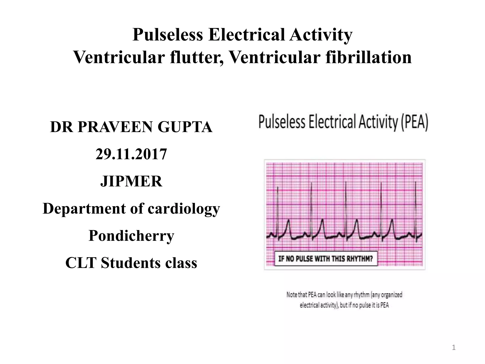
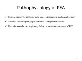
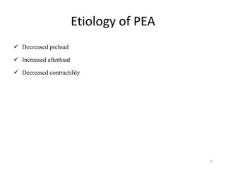
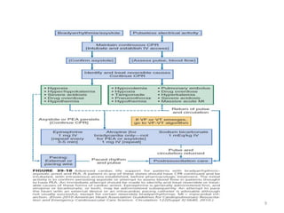
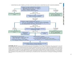
Pulseless Electrical Activity
Ventricular flutter, Ventricular fibrillation
DR PRAVEEN GUPTA
29.11.2017
JIPMER
Department of cardiology
Pondicherry
CLT Students class
1
Introduction
 Characterized by unresponsiveness and lack of palpable pulse in the presence of
organized cardiac electrical activity
 Previously known as electromechanical dissociation (EMD)
 Lack of ventricular electrical activity always implies a lack of ventricular
mechanical activity or asystole, the reverse is not always true
 Electrical activity is a necessary, but not sufficient, condition for mechanical
activity.
2
Pulseless Electrical Activity
 Meaningful” ventricular mechanical activity, generate a palpable pulse
 True PEA is a condition in which cardiac contractions are absent in the
presence of coordinated electrical activity
 Absence of peripheral pulses should not be equated with PEA, as it may be
due to severe PVD
 PEA encompasses a number of organized cardiac rhythms, including
supraventricular rhythms (sinus versus nonsinus) and ventricular rhythms
(accelerated idioventricular or escape)
3
Pathophysiology
 Occurs when a major cardiovascular, respiratory, or metabolic derangement
results in the inability of cardiac muscle to generate sufficient force in
response to electrical depolarization.
 The initial insult weakens cardiac contraction, and this situation is
exacerbated by worsening acidosis, hypoxia, and increasing vagal tone.
4
Pathophysiology of PEA
 Compromise of the inotropic state leads to inadequate mechanical activity
 Creates a vicious cycle, degeneration of the rhythm and death
 Hypoxia secondary to respiratory failure is most common cause of PEA,
5
Etiology of PEA
 Decreased preload
 Increased afterload
 Decreased contractility
6
7
Prognosis
 Females are more likely to develop PEA than males
 Prognosis is poor unless reversible cause is identified and corrected
 ECG wider QRS (>0.2 s), fare worse
 Out-of-hospital PEA likely to recover then in hospital PEA
8
9
Ventricular Flutter and Fibrillation
 Represent severe derangements of the heartbeat that can terminate fatally or
produce significant brain damage within 3 to 5 minutes
 Ventricular flutter manifested as a sine wave in appearance—regular large
oscillations occurring at a rate of 150 to 300 beats/minute
 VF is recognized by presence of irregular undulations of varying contour &
amplitude . Distinct QRS , ST , and T waves absent.
 Fine-amplitude fibrillatory waves (0.2 mV) are present with prolonged VF.
 Fine waves identify worse survival rates & confused with asystole.
10
Ventricular flutter
11
Ventricular fibrillation
12
Clinical Features
 Ventricular flutter or VF results in faintness, loss of consciousness,
seizures, apnea, and eventually death.
 Blood pressure is unobtainable, and heart sounds are usually absent.
 Atria can continue to beat at an independent rhythm for a time or in
response to impulses from the fibrillating ventricles.
 Eventually, electrical activity of the heart ceases
13
Mechanisms
 VF occurs most commonly in association with coronary artery disease
 VF, occur mostly in the morning
 Occur during antiarrhythmic drug administration, hypoxia, ischemia, or AF
that results in very rapid ventricular rates in patients with preexcitation
syndrome; after electrical shock administered during cardioversion or
accidentally by improperly grounded equipment; and during competitive
ventricular pacing to terminate VT.
14
Management
 Basic life support and advanced cardiac life support
 Immediate nonsynchronized DC shock (200 to 400 J) for VF, ventricular
flutter, and pulseless VT.
 CPR is performed only until the defibrillation equipment ready
 If the circulation is markedly inadequate despite return to sinus rhythm,
closed-chest massage should be instituted.
 After conversion to a normal rhythm, monitor rhythm continuously and to
institute measures to prevent recurrence.
15
16
References
 Emedicine
 Braunwald’s Heart Disease A Textbook of Cardiovascular Medicine, 10TH
Edition
17
18

Pulseless electrical activity, Ventricular flutter, Ventricular fibrillation

  • 1.
    Pulseless Electrical Activity Ventricularflutter, Ventricular fibrillation DR PRAVEEN GUPTA 29.11.2017 JIPMER Department of cardiology Pondicherry CLT Students class 1
  • 2.
    Introduction  Characterized byunresponsiveness and lack of palpable pulse in the presence of organized cardiac electrical activity  Previously known as electromechanical dissociation (EMD)  Lack of ventricular electrical activity always implies a lack of ventricular mechanical activity or asystole, the reverse is not always true  Electrical activity is a necessary, but not sufficient, condition for mechanical activity. 2
  • 3.
    Pulseless Electrical Activity Meaningful” ventricular mechanical activity, generate a palpable pulse  True PEA is a condition in which cardiac contractions are absent in the presence of coordinated electrical activity  Absence of peripheral pulses should not be equated with PEA, as it may be due to severe PVD  PEA encompasses a number of organized cardiac rhythms, including supraventricular rhythms (sinus versus nonsinus) and ventricular rhythms (accelerated idioventricular or escape) 3
  • 4.
    Pathophysiology  Occurs whena major cardiovascular, respiratory, or metabolic derangement results in the inability of cardiac muscle to generate sufficient force in response to electrical depolarization.  The initial insult weakens cardiac contraction, and this situation is exacerbated by worsening acidosis, hypoxia, and increasing vagal tone. 4
  • 5.
    Pathophysiology of PEA Compromise of the inotropic state leads to inadequate mechanical activity  Creates a vicious cycle, degeneration of the rhythm and death  Hypoxia secondary to respiratory failure is most common cause of PEA, 5
  • 6.
    Etiology of PEA Decreased preload  Increased afterload  Decreased contractility 6
  • 7.
  • 8.
    Prognosis  Females aremore likely to develop PEA than males  Prognosis is poor unless reversible cause is identified and corrected  ECG wider QRS (>0.2 s), fare worse  Out-of-hospital PEA likely to recover then in hospital PEA 8
  • 9.
  • 10.
    Ventricular Flutter andFibrillation  Represent severe derangements of the heartbeat that can terminate fatally or produce significant brain damage within 3 to 5 minutes  Ventricular flutter manifested as a sine wave in appearance—regular large oscillations occurring at a rate of 150 to 300 beats/minute  VF is recognized by presence of irregular undulations of varying contour & amplitude . Distinct QRS , ST , and T waves absent.  Fine-amplitude fibrillatory waves (0.2 mV) are present with prolonged VF.  Fine waves identify worse survival rates & confused with asystole. 10
  • 11.
  • 12.
  • 13.
    Clinical Features  Ventricularflutter or VF results in faintness, loss of consciousness, seizures, apnea, and eventually death.  Blood pressure is unobtainable, and heart sounds are usually absent.  Atria can continue to beat at an independent rhythm for a time or in response to impulses from the fibrillating ventricles.  Eventually, electrical activity of the heart ceases 13
  • 14.
    Mechanisms  VF occursmost commonly in association with coronary artery disease  VF, occur mostly in the morning  Occur during antiarrhythmic drug administration, hypoxia, ischemia, or AF that results in very rapid ventricular rates in patients with preexcitation syndrome; after electrical shock administered during cardioversion or accidentally by improperly grounded equipment; and during competitive ventricular pacing to terminate VT. 14
  • 15.
    Management  Basic lifesupport and advanced cardiac life support  Immediate nonsynchronized DC shock (200 to 400 J) for VF, ventricular flutter, and pulseless VT.  CPR is performed only until the defibrillation equipment ready  If the circulation is markedly inadequate despite return to sinus rhythm, closed-chest massage should be instituted.  After conversion to a normal rhythm, monitor rhythm continuously and to institute measures to prevent recurrence. 15
  • 16.
  • 17.
    References  Emedicine  Braunwald’sHeart Disease A Textbook of Cardiovascular Medicine, 10TH Edition 17
  • 18.