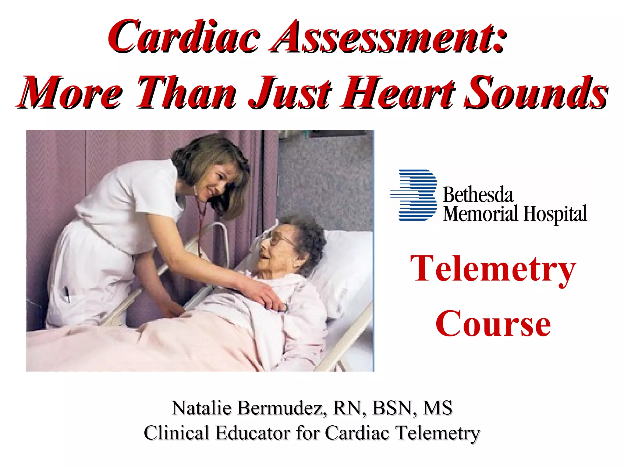
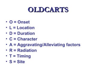
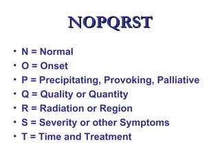
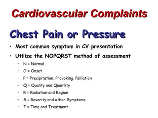
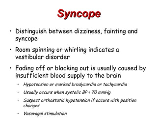
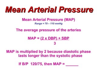
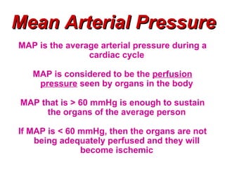
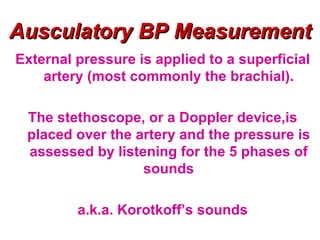
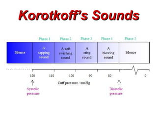
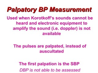
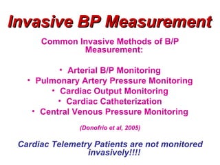
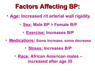
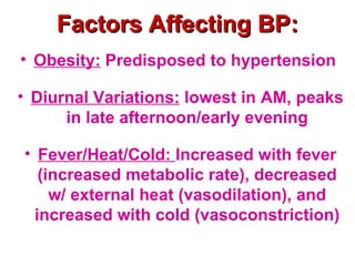
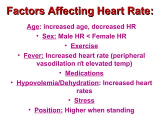
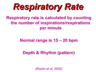
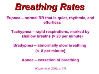
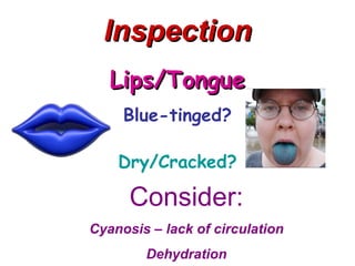
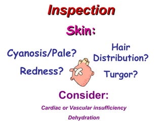
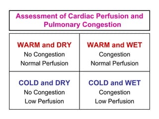
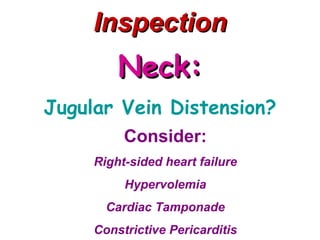
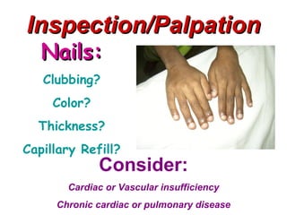
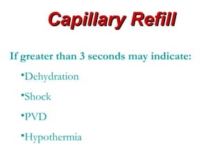
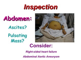
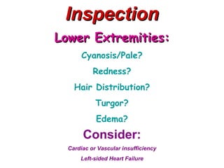
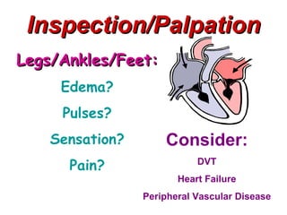
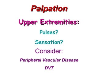
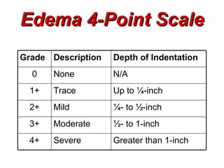
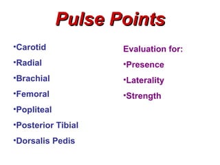
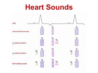
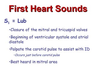
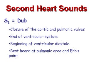
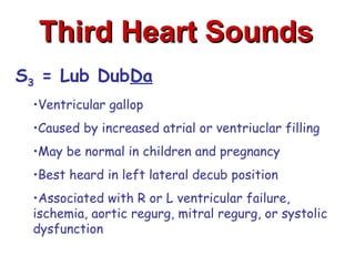
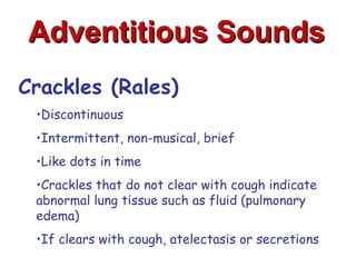
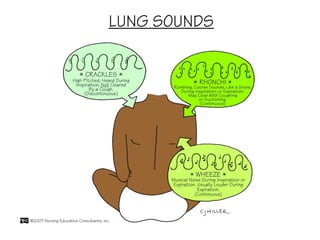
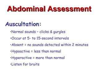
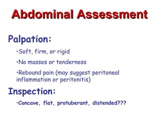
This document discusses the importance of comprehensive cardiac assessment beyond just listening to heart sounds. It emphasizes that nurses are responsible for around-the-clock surveillance of patients and should act as detectives to identify any abnormalities. The document provides guidance on key components of cardiac assessment including taking a thorough history, performing a focused physical exam, reviewing vital signs, and considering diagnostic testing based on findings.
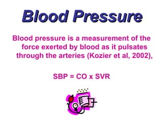
Ventricular tachycardia (VT) is a broad complex tachycardia originating from a ventricular ectopic focus. It is defined as three or more ventricular extrasystoles in succession at a rate of more than 120 beats per minute (bpm). Accelerated idioventricular rhythm refers to ventricular rhythms with rates of 100-120 bpm
Basics on heart murmurs, differentiate physiologic murmur from pathologic. Learn when to investigate further and when to monitor. Know the effect of different maneuvers on murmurs and physiology behind them. Listen to the heart sounds on the slides to appreciate the distinctive nature of each murmur.
Vital signs
The four main vital signs routinely monitored by medical professionals and health care providers include the following:
Body temperature.
Pulse rate.
Respiration rate (rate of breathing)
Blood pressure
It describes how to check blood pressure effectively. Indications to checking and contriandications. Role of continuous blood pressure measurement. Preparation before checking blood pressure and precautions.
These lecture slides, by Dr Sidra Arshad, offer a quick overview of the physiological basis of a normal electrocardiogram.
Learning objectives:
1. Define an electrocardiogram (ECG) and electrocardiography
2. Describe how dipoles generated by the heart produce the waveforms of the ECG
3. Describe the components of a normal electrocardiogram of a typical bipolar lead (limb II)
4. Differentiate between intervals and segments
5. Enlist some common indications for obtaining an ECG
6. Describe the flow of current around the heart during the cardiac cycle
7. Discuss the placement and polarity of the leads of electrocardiograph
8. Describe the normal electrocardiograms recorded from the limb leads and explain the physiological basis of the different records that are obtained
9. Define mean electrical vector (axis) of the heart and give the normal range
10. Define the mean QRS vector
11. Describe the axes of leads (hexagonal reference system)
12. Comprehend the vectorial analysis of the normal ECG
13. Determine the mean electrical axis of the ventricular QRS and appreciate the mean axis deviation
14. Explain the concepts of current of injury, J point, and their significance
Study Resources:
1. Chapter 11, Guyton and Hall Textbook of Medical Physiology, 14th edition
2. Chapter 9, Human Physiology - From Cells to Systems, Lauralee Sherwood, 9th edition
3. Chapter 29, Ganong’s Review of Medical Physiology, 26th edition
4. Electrocardiogram, StatPearls - https://www.ncbi.nlm.nih.gov/books/NBK549803/
5. ECG in Medical Practice by ABM Abdullah, 4th edition
6. Chapter 3, Cardiology Explained, https://www.ncbi.nlm.nih.gov/books/NBK2214/
7. ECG Basics, http://www.nataliescasebook.com/tag/e-c-g-basics
Muktapishti is a traditional Ayurvedic preparation made from Shoditha Mukta (Purified Pearl), is believed to help regulate thyroid function and reduce symptoms of hyperthyroidism due to its cooling and balancing properties. Clinical evidence on its efficacy remains limited, necessitating further research to validate its therapeutic benefits.
Integrating Ayurveda into Parkinson’s Management: A Holistic ApproachAyurveda ForAll
Explore the benefits of combining Ayurveda with conventional Parkinson's treatments. Learn how a holistic approach can manage symptoms, enhance well-being, and balance body energies. Discover the steps to safely integrate Ayurvedic practices into your Parkinson’s care plan, including expert guidance on diet, herbal remedies, and lifestyle modifications.
Title: Sense of Taste
Presenter: Dr. Faiza, Assistant Professor of Physiology
Qualifications:
MBBS (Best Graduate, AIMC Lahore)
FCPS Physiology
ICMT, CHPE, DHPE (STMU)
MPH (GC University, Faisalabad)
MBA (Virtual University of Pakistan)
Learning Objectives:
Describe the structure and function of taste buds.
Describe the relationship between the taste threshold and taste index of common substances.
Explain the chemical basis and signal transduction of taste perception for each type of primary taste sensation.
Recognize different abnormalities of taste perception and their causes.
Key Topics:
Significance of Taste Sensation:
Differentiation between pleasant and harmful food
Influence on behavior
Selection of food based on metabolic needs
Receptors of Taste:
Taste buds on the tongue
Influence of sense of smell, texture of food, and pain stimulation (e.g., by pepper)
Primary and Secondary Taste Sensations:
Primary taste sensations: Sweet, Sour, Salty, Bitter, Umami
Chemical basis and signal transduction mechanisms for each taste
Taste Threshold and Index:
Taste threshold values for Sweet (sucrose), Salty (NaCl), Sour (HCl), and Bitter (Quinine)
Taste index relationship: Inversely proportional to taste threshold
Taste Blindness:
Inability to taste certain substances, particularly thiourea compounds
Example: Phenylthiocarbamide
Structure and Function of Taste Buds:
Composition: Epithelial cells, Sustentacular/Supporting cells, Taste cells, Basal cells
Features: Taste pores, Taste hairs/microvilli, and Taste nerve fibers
Location of Taste Buds:
Found in papillae of the tongue (Fungiform, Circumvallate, Foliate)
Also present on the palate, tonsillar pillars, epiglottis, and proximal esophagus
Mechanism of Taste Stimulation:
Interaction of taste substances with receptors on microvilli
Signal transduction pathways for Umami, Sweet, Bitter, Sour, and Salty tastes
Taste Sensitivity and Adaptation:
Decrease in sensitivity with age
Rapid adaptation of taste sensation
Role of Saliva in Taste:
Dissolution of tastants to reach receptors
Washing away the stimulus
Taste Preferences and Aversions:
Mechanisms behind taste preference and aversion
Influence of receptors and neural pathways
Impact of Sensory Nerve Damage:
Degeneration of taste buds if the sensory nerve fiber is cut
Abnormalities of Taste Detection:
Conditions: Ageusia, Hypogeusia, Dysgeusia (parageusia)
Causes: Nerve damage, neurological disorders, infections, poor oral hygiene, adverse drug effects, deficiencies, aging, tobacco use, altered neurotransmitter levels
Neurotransmitters and Taste Threshold:
Effects of serotonin (5-HT) and norepinephrine (NE) on taste sensitivity
Supertasters:
25% of the population with heightened sensitivity to taste, especially bitterness
Increased number of fungiform papillae
Title: Sense of Smell
Presenter: Dr. Faiza, Assistant Professor of Physiology
Qualifications:
MBBS (Best Graduate, AIMC Lahore)
FCPS Physiology
ICMT, CHPE, DHPE (STMU)
MPH (GC University, Faisalabad)
MBA (Virtual University of Pakistan)
Learning Objectives:
Describe the primary categories of smells and the concept of odor blindness.
Explain the structure and location of the olfactory membrane and mucosa, including the types and roles of cells involved in olfaction.
Describe the pathway and mechanisms of olfactory signal transmission from the olfactory receptors to the brain.
Illustrate the biochemical cascade triggered by odorant binding to olfactory receptors, including the role of G-proteins and second messengers in generating an action potential.
Identify different types of olfactory disorders such as anosmia, hyposmia, hyperosmia, and dysosmia, including their potential causes.
Key Topics:
Olfactory Genes:
3% of the human genome accounts for olfactory genes.
400 genes for odorant receptors.
Olfactory Membrane:
Located in the superior part of the nasal cavity.
Medially: Folds downward along the superior septum.
Laterally: Folds over the superior turbinate and upper surface of the middle turbinate.
Total surface area: 5-10 square centimeters.
Olfactory Mucosa:
Olfactory Cells: Bipolar nerve cells derived from the CNS (100 million), with 4-25 olfactory cilia per cell.
Sustentacular Cells: Produce mucus and maintain ionic and molecular environment.
Basal Cells: Replace worn-out olfactory cells with an average lifespan of 1-2 months.
Bowman’s Gland: Secretes mucus.
Stimulation of Olfactory Cells:
Odorant dissolves in mucus and attaches to receptors on olfactory cilia.
Involves a cascade effect through G-proteins and second messengers, leading to depolarization and action potential generation in the olfactory nerve.
Quality of a Good Odorant:
Small (3-20 Carbon atoms), volatile, water-soluble, and lipid-soluble.
Facilitated by odorant-binding proteins in mucus.
Membrane Potential and Action Potential:
Resting membrane potential: -55mV.
Action potential frequency in the olfactory nerve increases with odorant strength.
Adaptation Towards the Sense of Smell:
Rapid adaptation within the first second, with further slow adaptation.
Psychological adaptation greater than receptor adaptation, involving feedback inhibition from the central nervous system.
Primary Sensations of Smell:
Camphoraceous, Musky, Floral, Pepperminty, Ethereal, Pungent, Putrid.
Odor Detection Threshold:
Examples: Hydrogen sulfide (0.0005 ppm), Methyl-mercaptan (0.002 ppm).
Some toxic substances are odorless at lethal concentrations.
Characteristics of Smell:
Odor blindness for single substances due to lack of appropriate receptor protein.
Behavioral and emotional influences of smell.
Transmission of Olfactory Signals:
From olfactory cells to glomeruli in the olfactory bulb, involving lateral inhibition.
Primitive, less old, and new olfactory systems with different path
Adv. biopharm. APPLICATION OF PHARMACOKINETICS : TARGETED DRUG DELIVERY SYSTEMSAkankshaAshtankar
MIP 201T & MPH 202T
ADVANCED BIOPHARMACEUTICS & PHARMACOKINETICS : UNIT 5
APPLICATION OF PHARMACOKINETICS : TARGETED DRUG DELIVERY SYSTEMS By - AKANKSHA ASHTANKAR
Recomendações da OMS sobre cuidados maternos e neonatais para uma experiência pós-natal positiva.
Em consonância com os ODS – Objetivos do Desenvolvimento Sustentável e a Estratégia Global para a Saúde das Mulheres, Crianças e Adolescentes, e aplicando uma abordagem baseada nos direitos humanos, os esforços de cuidados pós-natais devem expandir-se para além da cobertura e da simples sobrevivência, de modo a incluir cuidados de qualidade.
Estas diretrizes visam melhorar a qualidade dos cuidados pós-natais essenciais e de rotina prestados às mulheres e aos recém-nascidos, com o objetivo final de melhorar a saúde e o bem-estar materno e neonatal.
Uma “experiência pós-natal positiva” é um resultado importante para todas as mulheres que dão à luz e para os seus recém-nascidos, estabelecendo as bases para a melhoria da saúde e do bem-estar a curto e longo prazo. Uma experiência pós-natal positiva é definida como aquela em que as mulheres, pessoas que gestam, os recém-nascidos, os casais, os pais, os cuidadores e as famílias recebem informação consistente, garantia e apoio de profissionais de saúde motivados; e onde um sistema de saúde flexível e com recursos reconheça as necessidades das mulheres e dos bebês e respeite o seu contexto cultural.
Estas diretrizes consolidadas apresentam algumas recomendações novas e já bem fundamentadas sobre cuidados pós-natais de rotina para mulheres e neonatos que recebem cuidados no pós-parto em unidades de saúde ou na comunidade, independentemente dos recursos disponíveis.
É fornecido um conjunto abrangente de recomendações para cuidados durante o período puerperal, com ênfase nos cuidados essenciais que todas as mulheres e recém-nascidos devem receber, e com a devida atenção à qualidade dos cuidados; isto é, a entrega e a experiência do cuidado recebido. Estas diretrizes atualizam e ampliam as recomendações da OMS de 2014 sobre cuidados pós-natais da mãe e do recém-nascido e complementam as atuais diretrizes da OMS sobre a gestão de complicações pós-natais.
O estabelecimento da amamentação e o manejo das principais intercorrências é contemplada.
Recomendamos muito.
Vamos discutir essas recomendações no nosso curso de pós-graduação em Aleitamento no Instituto Ciclos.
Esta publicação só está disponível em inglês até o momento.
Prof. Marcus Renato de Carvalho
www.agostodourado.com
Ozempic: Preoperative Management of Patients on GLP-1 Receptor Agonists Saeid Safari
Preoperative Management of Patients on GLP-1 Receptor Agonists like Ozempic and Semiglutide
ASA GUIDELINE
NYSORA Guideline
2 Case Reports of Gastric Ultrasound
Here is the updated list of Top Best Ayurvedic medicine for Gas and Indigestion and those are Gas-O-Go Syp for Dyspepsia | Lavizyme Syrup for Acidity | Yumzyme Hepatoprotective Capsules etc
1. Cardiac Assessment:
More Than Just Heart Sounds
Telemetry
Course
Natalie Bermudez, RN, BSN, MS
Clinical Educator for Cardiac Telemetry
2. Importance of Assessment
• RNs are the 24/7 surveillance system
for the patient (Linda Aiken)
• RNs are rescue workers (Suzanne
Gordon)
• RNs are the integrators of all things
(Maggie McClure)
• RNs are the coordinators of care
3. Essentials of Assessment
• Empathic listening
• Ability to interview patients of different
ages, moods, and backgrounds
• Techniques for examining different
body systems
• Clinical Reasoning (I.e. critical
thinking)
– Putting it all together!
4. Key Points of Assessment
• Listen to the patient, they will often
help in leading to a diagnosis
• Focus on the patient, not the task; be
observant
• Be a detective; dig for clues
• Don’t take anything for GRANTED!
– Always check things out, especially “gut
feelings”
5. Key Points of Assessment
• Be proactive; anticipate your patients
needs
– Act before your patients gets into trouble
• When possible, round with the
physician
– Discuss any abnormal findings, especially
when you’re not sure of their significance
6. History
• Drives the physical assessment as well
as the diagnostic studies and treatment
• Lays the groundwork for the nurse-
patient relationship
• Provides key information
• Should not be bypassed
7. History of Present Illness
• Why is the patient seeking care?
• Have patient describe in his/her own words
Presenting Symptoms:
• Ask patient to describe symptoms
• Use a systematic approach to evaluating
symptoms
– OLDCARTS
– NOPQRST
8. OLDCARTS
• O = Onset
• L = Location
• D = Duration
• C = Character
• A = Aggravating/Alleviating factors
• R = Radiation
• T = Timing
• S = Site
9. NOPQRST
• N = Normal
• O = Onset
• P = Precipitating, Provoking, Palliative
• Q = Quality or Quantity
• R = Radiation or Region
• S = Severity or other Symptoms
• T = Time and Treatment
10. Cardiovascular Complaints
Chest Pain or Pressure
• Most common symptom in CV presentation
• Utilize the NOPQRST method of assessment
• N = Normal
• O = Onset
• P = Precipitation, Provoking, Palliation
• Q = Quality and Quantity
• R = Radiation and Region
• S = Severity and other Symptoms
• T = Time and Treatment
11. Chest Pain or Pressure
Onset
• Start suddenly or gradually – most angina starts at
low intensity and builds
• Time of day that discomfort started - some MI’s
occur in the morning after the patient rises and
begins activity
• When did the discomfort 1st begin – today or a few
days ago???
• MI may occur with activity or after a heavy meal
• Periods of increased myocardial demand
12. Chest Pain or Pressure
Precipitation, Provoking, Palliation
• Chest pain caused by CAD is often precipitated by
exertion
• Other precipitants are exposure to cold or heavy meals
• Associated factors – does the discomfort change
with inspiration or position change?
• What relieves the discomfort? –
• NTG, how many; if no relief, ask about storage of NTG
• Does the discomfort change with activity change,
such as rest?
13. Chest Pain or Pressure
Quality and Quantity
• Angina or ischemic discomfort is often described as
heaviness, pressure, tightness, or squeezing
• Stabbing, intermittent, knife-like descriptions are
not likely to be due to cardiac ischemia
• Remember – Ask the patient to describe the
discomfort
14. Chest Pain or Pressure
Radiation and Region
• Substernal region in the most common location for
discomfort with cardiac origin
• Anginal or ischemic discomfort is likely to radiate to
the jaw, either arm, or back
• However, discomfort is not always substernal even if
it is of cardiac origin
• Region of discomfort is usually larger than a fingertip
and often the size of a hand or closed fist
15. Chest Pain or Pressure
Severity and Other Symptoms
• Severity is subjective
• Ischemic pain can range from mild to severe
• Rate on a scale of 0 – 10
• Assess for other symptoms – nausea, vomiting,
dyspnea, diaphoresis, etc.
16. Chest Pain or Pressure
Time and Treatment
• Length of time since onset of symptoms
• How long do the symptoms last?
• Treated in the past for the same symptoms?
17. Dyspnea
• Can be due to pulmonary or cardiac problems
• Symptoms occur with activity or rest?
• If with activity, what level?
• Decreased activity tolerance demonstrated by DOE
might be anginal
• Onset gradual or sudden?
• Orthopnea - Difficulty breathing when flat
• PND – dyspnea that occurs 1-2 hours into sleep,
relieved by sitting
• How many pillows does the patient use?
18. Cough and Hemoptysis
• Heart Failure or Pulmonary Embolus
• Signs of Left-Sided HF
• Wet or dry cough
• Frequency – chronic or new onset
• Occurs only with activity?
• Sputum (amount, color, and consistency)
• Hemoptysis – blood-streaked, frothy pink, frank
• May be present with mitral valve stenosis, pulmonary embolus,
pulmonary hypertension, or tuberculosis
19. Palpitations
• Awareness of heartbeat
• May occur with fast or normal heart rate
• May be regular or irregular
• May occur with aortic or mitral regurgitation,
pregnancy
• Tachydysrhythmias may result in palpitations
• A-Fib or A-Flutter with RVR, SVT, VT
20. Syncope
• Distinguish between dizziness, fainting and
syncope
• Room spinning or whirling indicates a
vestibular disorder
• Fading off or blacking out is usually caused by
insufficient blood supply to the brain
• Hypotension or marked bradycardia or tachycardia
• Usually occurs when systolic BP < 70 mmHg
• Suspect orthostatic hypotension if occurs with position
changes
• Vasovagal stimulation
21. Physical Assessment
• Find a systematic approach that works for you
• Always begin your shift with a thorough
physical assessment (baseline)
• Always complete assessment with respect for
patient’s privacy
• Room should be quiet
• Perform assessment from patients right side
22. Physical Assessment
• Find a systematic approach that works for you
• Always begin your shift with a thorough
physical assessment (baseline)
• Always complete assessment with respect for
patient’s privacy
• Room should be quiet
• Perform assessment from patients right side
23. Vital Signs
Blood Pressure
Hypotensive or Hypertensive
Heart Rate
Bradycardia or Tachycardia
Respiratory Rate
Bradypneic or Tachypneic
O2 Saturation
Hypoxia/Hypoxemia
24. Blood Pressure
Blood pressure is a measurement of the
force exerted by blood as it pulsates
through the arteries (Kozier et al, 2002),
SBP = CO x SVR
25. Blood Pressure
Systolic blood pressure (SBP) is the
pressure of the blood as a result of
contraction of the ventricles
Diastolic blood pressure (DBP) is the
pressure when the ventricles are at rest
DBP is the lower pressure that is present at
all times within the arteries
(Kozier et al, 2002, p. 33)
26. Blood Pressure
Blood pressure is affected by factors such as
CO [preload, contractility, afterload]; distension
of the arteries; and the volume, velocity, and
viscosity of the blood (Smeltzer et al, 2008, p. 799)
Blood pressure is an indicator of adequate or
inadequate perfusion
Inadequate perfusion may be a result of high or
low blood pressures
28. Blood Pressure
• Technique for measuring blood pressure
is important
– Sitting up
– Arm at the level of the heart with support
– Place cuff over brachial artery
– Use appropriate cuff size
• Too small – falsely elevated BP
• Too big – falsely decreased BP
29. Orthostatic Blood Pressure
• Technique for measuring orthostatic BP
– Use the same arm
– Wait at least 5 minutes between measurements
– Lying, sitting, standing
• Orthostatic Hypotension if:
– Fall of SBP > 20 mmHg
– Fall of DBP > 10 mmHg
30. Mean Arterial Pressure
Mean Arterial Pressure (MAP)
Range = 70 – 110 mmHg
The average pressure of the arteries
MAP = (2 x DBP) + SBP
3
MAP is multiplied by 2 because diastolic phase
lasts longer than the systolic phase
If B/P 120/75, then MAP = ______
31. Mean Arterial Pressure
MAP is the average arterial pressure during a
cardiac cycle
MAP is considered to be the perfusion
pressure seen by organs in the body
MAP that is > 60 mmHg is enough to sustain
the organs of the average person
If MAP is < 60 mmHg, then the organs are not
being adequately perfused and they will
become ischemic
32. Noninvasive BP
Measurement
Two Common Noninvasive Indirect
Methods of B/P Measurement
Ausculatory & Palpatory
33. Ausculatory BP Measurement
External pressure is applied to a superficial
artery (most commonly the brachial).
The stethoscope, or a Doppler device,is
placed over the artery and the pressure is
assessed by listening for the 5 phases of
sounds
a.k.a. Korotkoff’s sounds
35. Palpatory BP Measurement
Used when Korotkoff’s sounds cannot be
heard and electronic equipment to
amplify the sound (i.e. doppler) is not
available
The pulses are palpated, instead of
auscultated
The first palpation is the SBP
DBP is not able to be assessed
36. Invasive BP Measurement
Common Invasive Methods of B/P
Measurement:
• Arterial B/P Monitoring
• Pulmonary Artery Pressure Monitoring
• Cardiac Output Monitoring
• Cardiac Catheterization
• Central Venous Pressure Monitoring
(Donofrio et al, 2005)
Cardiac Telemetry Patients are not monitored
invasively!!!!
37. Factors Affecting BP:
• Age: Increased r/t arterial wall rigidity
• Sex: Male BP > Female B/P
• Exercise: Increases B/P
• Medications: Some Increase, some decrease
• Stress: Increases B/P
• Race: African American males –
increased after age 35
38. Factors Affecting BP:
• Obesity: Predisposed to hypertension
• Diurnal Variations: lowest in AM, peaks
in late afternoon/early evening
• Fever/Heat/Cold: Increased with fever
(increased metabolic rate), decreased
w/ external heat (vasodilation), and
increased with cold (vasoconstriction)
39. Heart Rate
Pulse is the term used to describe rate,
rhythm, and volume of the heartbeat
A pulse is produced by ventricular
contraction which creates a wave of
blood through the arteries
The pulse reflects the heartbeat
(Kozier et al, 2002, p. 23)
40. Characteristics of a Pulse
Pulse should be characterized as:
• Thready, weak, strong, or bounding
• Equal bilaterally or not
• Rhythm regular or irregular
41. Heart Rate & Blood
Pressure
Blood pressure is directly affected by the
heart rate
Heart rate is directly affected by blood
pressure
What does this mean…?
42. Heart Rate & Blood
Pressure
• HR is Within Defined Parameters if 60–
100
– Bradycardia if HR < 60
– Tachycardia if HR > 100
• Blood pressure affects HR and HR affects
BP
– If HR > 100, then BP decreases
– If HR < 60, then BP decreases
– If BP decreases then HR increases
44. Respiratory Rate
Respiratory rate is calculated by counting
the number of inspirations/respirations
per minute
Normal range is 15 – 20 bpm
Depth & Rhythm (pattern)
(Kozier et al, 2002)
45. Breathing Rates
Eupnea – normal RR that is quiet, rhythmic, and
effortless
Tachypnea – rapid respirations, marked by
shallow breaths (> 20 per minute)
Bradypnea – abnormally slow breathing
(< 8 per minute)
Apnea – cessation of breathing
(Kozier et al, 2002, p. 31)
46. Breathing Rates
Cheyne-Stoke – Fast, deep respirations of 30 –
170 seconds punctuated by periods of apnea
lasting 20 – 60 seconds
Kussmaul’s – fast (over 20 per minute), deep
(resembling sighs), labored respirations without
a pause
(Goldberg et al, 1997, p. 764)
47. Factors Affecting RR
• Age: rate & depth decrease with age
• Exercise: Increased rate & depth
• Fever: Increased
• Medications: Narcotics cause respiratory
depression
• Stress: Increased rate & depth
• Homeostasis (acidosis/alkalosis): Increased
or decreased rate
(Kozier et al, 2002)
48. Oxygen Saturation
Normal = 95% - 100%
Below 70% is life threatening
Pulse oximeter - measures arterial blood
oxygen saturation
Can detect hypoxemia before clinical signs
& symptoms are apparent
(Kozier et al, 2002)
49. Pulse Oximeter
2-Part Sensor
2. Two light-emitting diodes (LEDs) – one
red and one infrared
Transmit light through nails, tissue, venous blood, &
arterial blood
2. Photodetector (opposite side of LEDs)
Measures the amount of red and infrared light absorbed
by oxygenated & deoxygenated hemoglobin in arterial
blood and reports it as SaO2.
(Kozier et al, 2002)
50. Factors Affecting 02 Sat:
• Hemoglobin: regardless of low Hemoglobin
levels, if the hemoglobin is fully saturated
the SaO2 will still be “normal”
• Circulation: Will be inaccurate if the area
under the sensor has impaired circulation
• Activity: Shivering or excessive movement
of the sensor site may interfere with
accurate readings
(Kozier et al, 2002, p. 39)
54. Assessment of Cardiac Perfusion and
Pulmonary Congestion
WARM and DRY WARM and WET
No Congestion Congestion
Normal Perfusion Normal Perfusion
COLD and DRY COLD and WET
No Congestion Congestion
Low Perfusion Low Perfusion
62. Edema 4-Point Scale
Grade Description Depth of Indentation
0 None N/A
1+ Trace Up to ¼-inch
2+ Mild ¼- to ½-inch
3+ Moderate ½- to 1-inch
4+ Severe Greater than 1-inch
64. Pulse 4-Point Scale
Grade Description
0 Absent
1+ Palpable, but thready and
weak; easily obliterated
2+ Normal, easily identified; not
easily obliterated
3+ Increased pulse; moderate
pressure for obliteration
4+ Full, bounding; cannot
obliterate
65. Terminology of Pulse
Variations
•Pulsus Magnus – strong and bounding
•Pulsus Parvus – thready
•Pulsus Alternans – large amplitude followed by low
amplitude (with a regular rhythm)
•Pulsus Bisferiens – double-peaked systolic impulse
(cardiomyopathy)
•Water-Hammer pulse – rapid rising and collapsing
(aortic regurgitation)
66.
67. General Points
When assessing heart sounds:
•Need a quiet room
•Stand to the right of the patient
•Having patient roll slightly to the left accentuates S3, S4
and mitral murmurs, especially mitral stenosis
•Having patient lean forward accentuates aortic
regurgitation
•Right-sided heart sounds are better heard on inspiration
•Left-sided heart sounds are better heard during
expiration
70. First Heart Sounds
S1 = Lub
•Closure of the mitral and tricuspid valves
•Beginning of ventricular systole and atrial
diastole
•Palpate the carotid pulse to assist with ID
•Occurs just before carotid pulse
•Best heard in mitral area
71. Second Heart Sounds
S2 = Dub
•Closure of the aortic and pulmonic valves
•End of ventricular systole
•Beginning of ventricular diastole
•Best heard at pulmonic area and Erb’s
point
72. Third Heart Sounds
S3 = Lub DubDa
•Ventricular gallop
•Caused by increased atrial or ventriuclar filling
•May be normal in children and pregnancy
•Best heard in left lateral decub position
•Associated with R or L ventricular failure,
ischemia, aortic regurg, mitral regurg, or systolic
dysfunction
73. Fourth Heart Sounds
S4 = DaLub Dub
•Atrial gallop
•Occurs during late ventricular diastole
•Caused by atrial contraction and propulsion of
blood into a noncompliant, stiff ventricle
•Best heard in left lateral decub position
•Associated with restrictive cardiomyopathy,
ischemia, and aortic stenosis
78. Grading Murmurs
Gradation of Description
Murmurs
Very faint, heard only after listener has "tuned in"; may not be heard
Grade 1 in all positions
Quiet, but heard immediately after placing the stethoscope on the
Grade 2 chest
Moderately loud
Grade 3
Murmur is very loud, with palpable thrill
Grade 4
Murmur is extremely loud, with palpable thrill, and can be heard if
Grade 5 only the edge of the stethoscope is in contact with the skin, but
cannot be heard if the stethoscope is removed from the skin
Murmur is exceptionally loud, with palpable thrill, and can be heard
Grade 6 with the stethoscope just removed from contact with the chest.
80. Normal Breath Sounds
Bronchial (upper)
•Expiratory longer than inspiratory
•Loud and higher in pitch
Bronchovesicular (middle)
•Equal inspiratory and expiratory
Vesicular (lower)
•Soft or low pitched
•Heard through inspiration and 1/3 expiration
81. Adventitious Sounds
Crackles (Rales)
•Discontinuous
•Intermittent, non-musical, brief
•Like dots in time
•Crackles that do not clear with cough indicate
abnormal lung tissue such as fluid (pulmonary
edema)
•If clears with cough, atelectasis or secretions
85. Abdominal Assessment
Auscultation:
•Normal sounds – clicks & gurgles
•Occur at 5- to 15-second intervals
•Absent = no sounds detected within 2 minutes
•Hypoactive = less than normal
•Hyperactive = more than normal
•Listen for bruits
86. Abdominal Assessment
Palpation:
•Soft, firm, or rigid
•No masses or tenderness
•Rebound pain (may suggest peritoneal
inflammation or peritonitis)
Inspection:
•Concave, flat, protuberant, distended???
87. Genitourinary Assessment
•Intake and output
•Indicates both renal and cardiac function
•Foley catheter
•Check for orders and insertion date
88. References
Bickley, L. S. (2007). Bates’ pocket guide to physical examination and history
taking, (5th ed.). Philadelphia, PA: Lippincott, Wilkins, and Williams.
Davis, L. (2004). Cardiovascular nursing secrets: Your cardiovascular
questions answered by exoerts you trust. St. Louis, MO: Elsevier Mosby.
Donofrio, J., Haworth, K., Schaeffer, L., & Thompson, G. (Eds.). (2005).
Cardiovascular care made incredibly easy. Ambler, PA: Lippincott, Williams,
and Wilkins.
Goldberg, K., Johnson, P., & Lear-Olimpi, M. (1997). Handbook of clinical skills.
Springhouse, PA: Springhouse Corporation.
Kozier, B., Erb, G., Berman, A., & Snyder, S. (2002). Kozier’s and erb’s
techniques in clinical nursing: Basic to intermediate skills, (5th ed.). Upper
Saddle, NJ: Prentice Hall.
Moser, D. K., & Riegel, B. (2008). Cardiac nursing: A companion to braunwald’s
heart disease. St. Louis, MO: Saunders Elsevier.
Smeltzer et al. (2008). Brunner and suddarth’s textbook of medical-surgical
nursing, (11th ed.). Philadelphia, PA: Lippincott Williams and Wilkins.
Editor's Notes
Thorough cardiac assessment Head – to – Toe Fashion
Blood pressure tells us about adequate perfusion B/P that is too low can cause poor perfusion SBP < 90 is considered unstable if symptomatic Elevated B/P can cause problems such as stroke Different factors cause elevations in B/P Pulse Too low or too high can cause poor perfusion Different factors cause elevations in pulse rates RR -elevated RR’s can indicate compensation for a cardiac problem 02 sat -low O2 sat’s may reflect poor perfusion or other cardiac disorders
Brief synopsis of a head – to – toe cardiac assessment tool Identifies key assessment findings that could indicate cardiac problems