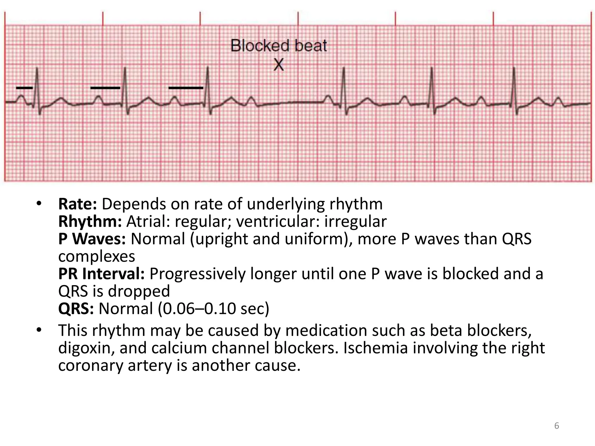
Atrio-ventricular (AV) Blocks
Post Basic Specialization in Nursing
1
Atrioventricular (AV) Blocks
• AV blocks are divided into three categories:
first, second, and third degree.
• First-Degree AV Block
• Second Degree AV Block
• Third Degree AV Block
2
First-degree AV block
• It is a type of AV block in which every impulse is
conducted to the ventricles but the duration of AV
conduction is prolonged.
– First-degree AV block is associated with MI, CAD,
rheumatic fever, hyperthyroidism, vagal stimulation, and
drugs such as digoxin, -adrenergic blockers, calcium
channel blockers, and flecainide.
– First-degree AV block is usually not serious but can be a
precursor of higher degrees of AV block. Patients with first-
degree AV block are asymptomatic.
– There is no treatment for first-degree AV block. Patients
should continue to be monitored for any new changes in
heart rhythm.
3
Rate: Depends on rate of underlying rhythm
Rhythm: Regular
P Waves: Normal (upright and uniform)
PR Interval: Prolonged (>0.20 sec)
QRS: Normal (0.06–0.10 sec)
• Usually a first-degree AV block is benign, but if associated with an acute
MI it may lead to further AV defects.
• Often AV block is caused by medications that prolong AV conduction;
these include digoxin, calcium channel blockers, and beta blockers.
4
Second-degree AV block
(Mobitz I or Wenckebach)
• Second-degree AV block, Type I (Mobitz I or
Wenckebach heart block) is a gradual
lengthening of the PR interval. It occurs
because of a prolonged AV conduction time
until an atrial impulse is nonconducted and a
QRS complex is blocked (missing).
5
• Rate: Depends on rate of underlying rhythm
Rhythm: Atrial: regular; ventricular: irregular
P Waves: Normal (upright and uniform), more P waves than QRS
complexes
PR Interval: Progressively longer until one P wave is blocked and a
QRS is dropped
QRS: Normal (0.06–0.10 sec)
• This rhythm may be caused by medication such as beta blockers,
digoxin, and calcium channel blockers. Ischemia involving the right
coronary artery is another cause.
6
Second-degree AV block
• Type I AV block may result from use of drugs such as
digoxin or -adrenergic blockers. It may also be
associated with CAD and other diseases that can slow
AV conduction.
• Type I AV block is usually a result of myocardial
ischemia or infarction. It is almost always transient and
is usually well tolerated. However, it may be a warning
signal of a more serious AV conduction disturbance.
• If the patient is symptomatic, atropine is used to
increase HR, or a temporary pacemaker may be
needed.
7
Second-degree AV block, Type II
(Mobitz II)
• Second-degree AV block, Type II (Mobitz II heart
block), involves a P wave that is nonconducted
without progressive antecedent PR lengthening.
This almost always occurs when a block in one of
the bundle branches is present.
• Conduction ratio (P waves to QRS complexes) is
commonly 2:1, 3:1, or 4:1, or variable.
■ QRS complexes are usually wide because this
block usually involves both bundle branches.
8
9
Rate: Atrial: usually 60–100 bpm; ventricular: slower than atrial rate
Rhythm: Atrial: regular; ventricular: regular or irregular
P Waves: Normal (upright and uniform); more P waves than QRS complexes
PR Interval: Normal or prolonged but constant
QRS: May be normal, but usually wide (>0.10 sec) if the bundle branches are
involved
Resulting bradycardia can compromise cardiac output and lead to complete AV
block.
This rhythm often occurs with cardiac ischemia or an MI.
Second-degree AV block, Type II
(Mobitz II)
• Type II second-degree AV block is a more serious type of block in
which a certain number of impulses from the SA node are not
conducted to the ventricles.
• Type II AV block is associated with rheumatic heart disease, CAD,
anterior MI, and digitalis toxicity.
• Type II AV block often progresses to third-degree AV block and is
associated with a poor prognosis. The reduced HR often results in
decreased CO with subsequent hypotension and myocardial
ischemia.
• Temporary treatment before the insertion of a permanent
pacemaker may be necessary if the patient becomes symptomatic
(e.g., hypotension, angina) and involves the use of a temporary
transvenous or transcutaneous pacemaker.
10
Third-degree AV block
• Third-degree AV block, or complete heart block, constitutes one
form of AV dissociation in which no impulses from the atria are
conducted to the ventricles.
– Third-degree AV block is associated with severe heart disease,
including CAD, MI, myocarditis, cardiomyopathy, and some systemic
diseases such as amyloidosis and progressive systemic sclerosis
(scleroderma).
– Third-degree AV block almost always results in reduced CO with
subsequent ischemia, HF, and shock. Syncope from third-degree AV
block may result from severe bradycardia or even periods of asystole.
– Conduction between atria and ventricles is totally absent because of
complete electrical block at or
below the AV node. This is known as AV dissociation.
– Treatment. For symptomatic patients, a transcutaneous pacemaker is
used until a temporary transvenous pacemaker can be inserted.
11
• Rate: Atrial: 60–100 bpm; ventricular: 40–60 bpm if escape focus is
junctional, <40 bpm if escape focus
is ventricular
Rhythm: Usually regular, but atria and ventricles act independently
P Waves: Normal (upright and uniform); may be superimposed on
QRS complexes or T waves
PR Interval: Varies greatly
QRS: Normal if ventricles are activated by junctional escape focus;
wide if escape focus is ventricular
• Third-degree AV block may be associated with ischemia involving
the left coronary arteries.
12
SUDDEN CARDIAC DEATH
• Sudden cardiac death (SCD) refers to death from a
cardiac cause.The majority of SCDs result from
ventricular dysrhythmias, specifically ventricular
tachycardia or fibrillation.
PRODYSRHYTHMIA
• Antidysrhythmia drugs may cause life-threatening
dysrhythmias similar to those for which they are
administered. This concept is termed prodysrhythmia.
– The patient who has severe left ventricular dysfunction is
the most susceptible to prodysrhythmias.
– Digoxin and some antidysrhythmia drugs can cause a
prodysrhythmic response.
13
DEFIBRILLATION
• Defibrillation is the most effective method of terminating
VF and pulseless VT.
• Defibrillation is accomplished by the passage of a DC
electrical shock through the heart to depolarize the cells of
the myocardium. The intent is that subsequent
repolarization of myocardial cells will allow the SA node to
resume the role of pacemaker.
• Rapid defibrillation can be performed using a manual or
automatic device.
– Manual defibrillators require health care providers to interpret
cardiac rhythms, determine the need for a shock, and deliver a
shock.
– Automatic external defibrillators (AEDs) are defibrillators that
have rhythm detection capability and the ability to advise the
operator to deliver a shock using hands-free defibrillator pads.
14
SYNCHRONIZED CARDIOVERSION
• Synchronized cardioversion is the therapy of
choice for the patient with hemodynamically
unstable ventricular or supraventricular
tachydysrhythmias.
– A synchronized circuit in the defibrillator is used to
deliver a countershock that is programmed to occur
on the R wave of the QRS complex of the ECG.
– The synchronizer switch must be turned on when
cardioversion is planned.
• The procedure for synchronized cardioversion is
the same as for defibrillation, with some
exceptions
15
THANKS
16

Heart blocks slide for pharmacy and study

  • 1.
    Atrio-ventricular (AV) Blocks PostBasic Specialization in Nursing 1
  • 2.
    Atrioventricular (AV) Blocks •AV blocks are divided into three categories: first, second, and third degree. • First-Degree AV Block • Second Degree AV Block • Third Degree AV Block 2
  • 3.
    First-degree AV block •It is a type of AV block in which every impulse is conducted to the ventricles but the duration of AV conduction is prolonged. – First-degree AV block is associated with MI, CAD, rheumatic fever, hyperthyroidism, vagal stimulation, and drugs such as digoxin, -adrenergic blockers, calcium channel blockers, and flecainide. – First-degree AV block is usually not serious but can be a precursor of higher degrees of AV block. Patients with first- degree AV block are asymptomatic. – There is no treatment for first-degree AV block. Patients should continue to be monitored for any new changes in heart rhythm. 3
  • 4.
    Rate: Depends onrate of underlying rhythm Rhythm: Regular P Waves: Normal (upright and uniform) PR Interval: Prolonged (>0.20 sec) QRS: Normal (0.06–0.10 sec) • Usually a first-degree AV block is benign, but if associated with an acute MI it may lead to further AV defects. • Often AV block is caused by medications that prolong AV conduction; these include digoxin, calcium channel blockers, and beta blockers. 4
  • 5.
    Second-degree AV block (MobitzI or Wenckebach) • Second-degree AV block, Type I (Mobitz I or Wenckebach heart block) is a gradual lengthening of the PR interval. It occurs because of a prolonged AV conduction time until an atrial impulse is nonconducted and a QRS complex is blocked (missing). 5
  • 6.
    • Rate: Dependson rate of underlying rhythm Rhythm: Atrial: regular; ventricular: irregular P Waves: Normal (upright and uniform), more P waves than QRS complexes PR Interval: Progressively longer until one P wave is blocked and a QRS is dropped QRS: Normal (0.06–0.10 sec) • This rhythm may be caused by medication such as beta blockers, digoxin, and calcium channel blockers. Ischemia involving the right coronary artery is another cause. 6
  • 7.
    Second-degree AV block •Type I AV block may result from use of drugs such as digoxin or -adrenergic blockers. It may also be associated with CAD and other diseases that can slow AV conduction. • Type I AV block is usually a result of myocardial ischemia or infarction. It is almost always transient and is usually well tolerated. However, it may be a warning signal of a more serious AV conduction disturbance. • If the patient is symptomatic, atropine is used to increase HR, or a temporary pacemaker may be needed. 7
  • 8.
    Second-degree AV block,Type II (Mobitz II) • Second-degree AV block, Type II (Mobitz II heart block), involves a P wave that is nonconducted without progressive antecedent PR lengthening. This almost always occurs when a block in one of the bundle branches is present. • Conduction ratio (P waves to QRS complexes) is commonly 2:1, 3:1, or 4:1, or variable. ■ QRS complexes are usually wide because this block usually involves both bundle branches. 8
  • 9.
    9 Rate: Atrial: usually60–100 bpm; ventricular: slower than atrial rate Rhythm: Atrial: regular; ventricular: regular or irregular P Waves: Normal (upright and uniform); more P waves than QRS complexes PR Interval: Normal or prolonged but constant QRS: May be normal, but usually wide (>0.10 sec) if the bundle branches are involved Resulting bradycardia can compromise cardiac output and lead to complete AV block. This rhythm often occurs with cardiac ischemia or an MI.
  • 10.
    Second-degree AV block,Type II (Mobitz II) • Type II second-degree AV block is a more serious type of block in which a certain number of impulses from the SA node are not conducted to the ventricles. • Type II AV block is associated with rheumatic heart disease, CAD, anterior MI, and digitalis toxicity. • Type II AV block often progresses to third-degree AV block and is associated with a poor prognosis. The reduced HR often results in decreased CO with subsequent hypotension and myocardial ischemia. • Temporary treatment before the insertion of a permanent pacemaker may be necessary if the patient becomes symptomatic (e.g., hypotension, angina) and involves the use of a temporary transvenous or transcutaneous pacemaker. 10
  • 11.
    Third-degree AV block •Third-degree AV block, or complete heart block, constitutes one form of AV dissociation in which no impulses from the atria are conducted to the ventricles. – Third-degree AV block is associated with severe heart disease, including CAD, MI, myocarditis, cardiomyopathy, and some systemic diseases such as amyloidosis and progressive systemic sclerosis (scleroderma). – Third-degree AV block almost always results in reduced CO with subsequent ischemia, HF, and shock. Syncope from third-degree AV block may result from severe bradycardia or even periods of asystole. – Conduction between atria and ventricles is totally absent because of complete electrical block at or below the AV node. This is known as AV dissociation. – Treatment. For symptomatic patients, a transcutaneous pacemaker is used until a temporary transvenous pacemaker can be inserted. 11
  • 12.
    • Rate: Atrial:60–100 bpm; ventricular: 40–60 bpm if escape focus is junctional, <40 bpm if escape focus is ventricular Rhythm: Usually regular, but atria and ventricles act independently P Waves: Normal (upright and uniform); may be superimposed on QRS complexes or T waves PR Interval: Varies greatly QRS: Normal if ventricles are activated by junctional escape focus; wide if escape focus is ventricular • Third-degree AV block may be associated with ischemia involving the left coronary arteries. 12
  • 13.
    SUDDEN CARDIAC DEATH •Sudden cardiac death (SCD) refers to death from a cardiac cause.The majority of SCDs result from ventricular dysrhythmias, specifically ventricular tachycardia or fibrillation. PRODYSRHYTHMIA • Antidysrhythmia drugs may cause life-threatening dysrhythmias similar to those for which they are administered. This concept is termed prodysrhythmia. – The patient who has severe left ventricular dysfunction is the most susceptible to prodysrhythmias. – Digoxin and some antidysrhythmia drugs can cause a prodysrhythmic response. 13
  • 14.
    DEFIBRILLATION • Defibrillation isthe most effective method of terminating VF and pulseless VT. • Defibrillation is accomplished by the passage of a DC electrical shock through the heart to depolarize the cells of the myocardium. The intent is that subsequent repolarization of myocardial cells will allow the SA node to resume the role of pacemaker. • Rapid defibrillation can be performed using a manual or automatic device. – Manual defibrillators require health care providers to interpret cardiac rhythms, determine the need for a shock, and deliver a shock. – Automatic external defibrillators (AEDs) are defibrillators that have rhythm detection capability and the ability to advise the operator to deliver a shock using hands-free defibrillator pads. 14
  • 15.
    SYNCHRONIZED CARDIOVERSION • Synchronizedcardioversion is the therapy of choice for the patient with hemodynamically unstable ventricular or supraventricular tachydysrhythmias. – A synchronized circuit in the defibrillator is used to deliver a countershock that is programmed to occur on the R wave of the QRS complex of the ECG. – The synchronizer switch must be turned on when cardioversion is planned. • The procedure for synchronized cardioversion is the same as for defibrillation, with some exceptions 15
  • 16.