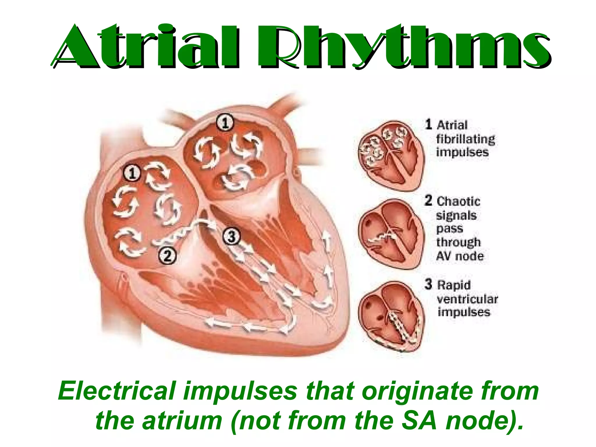
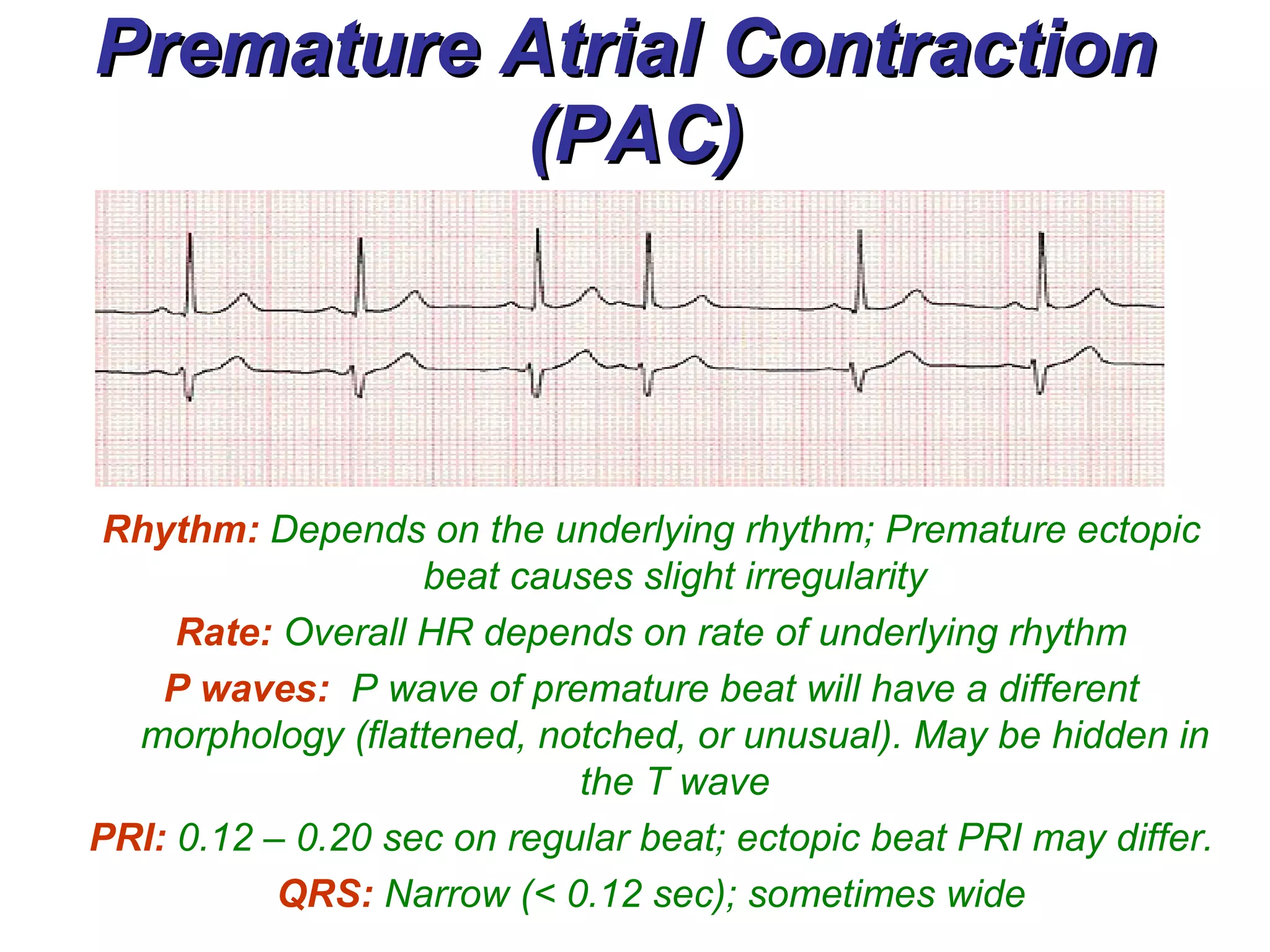
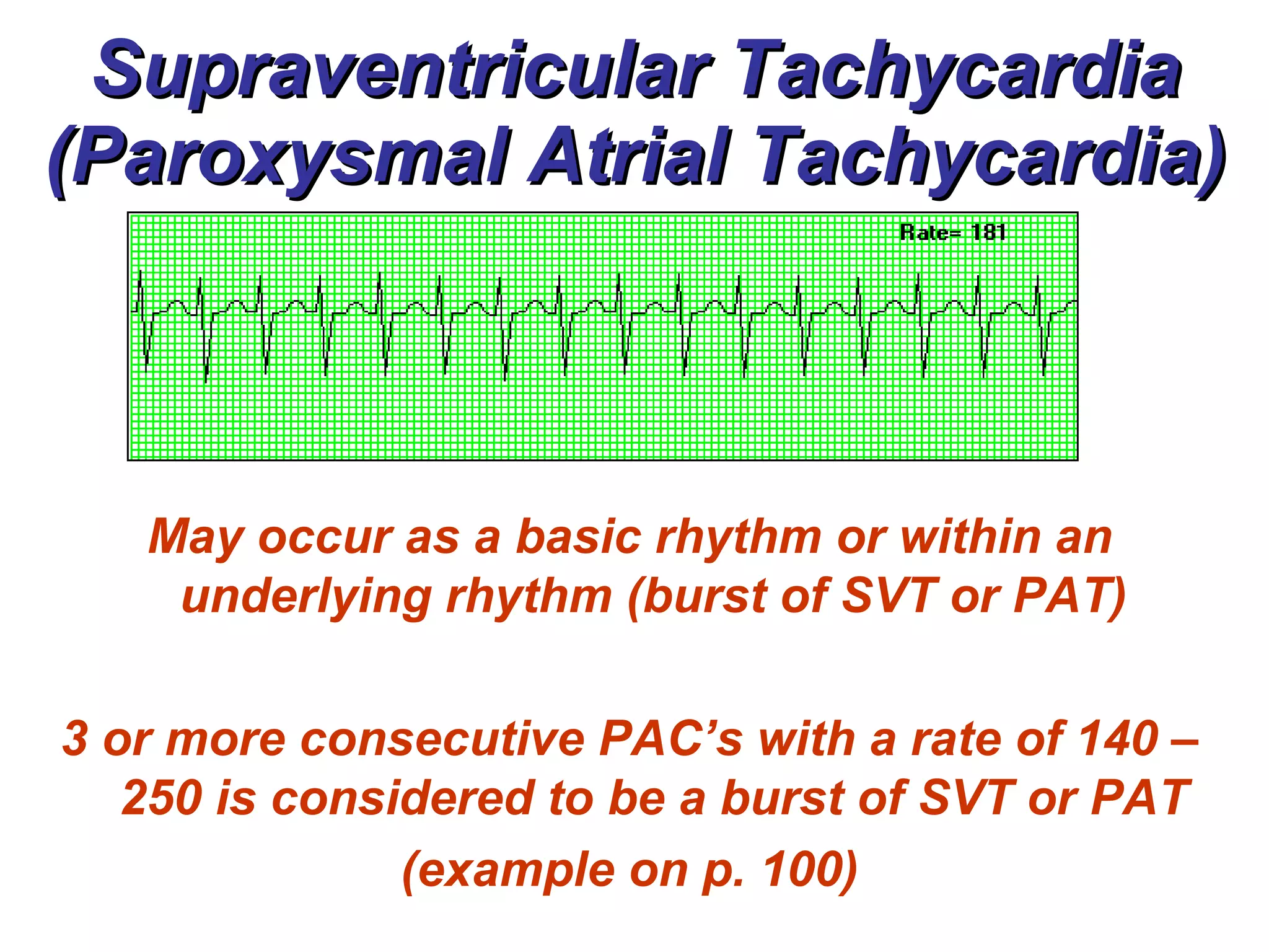
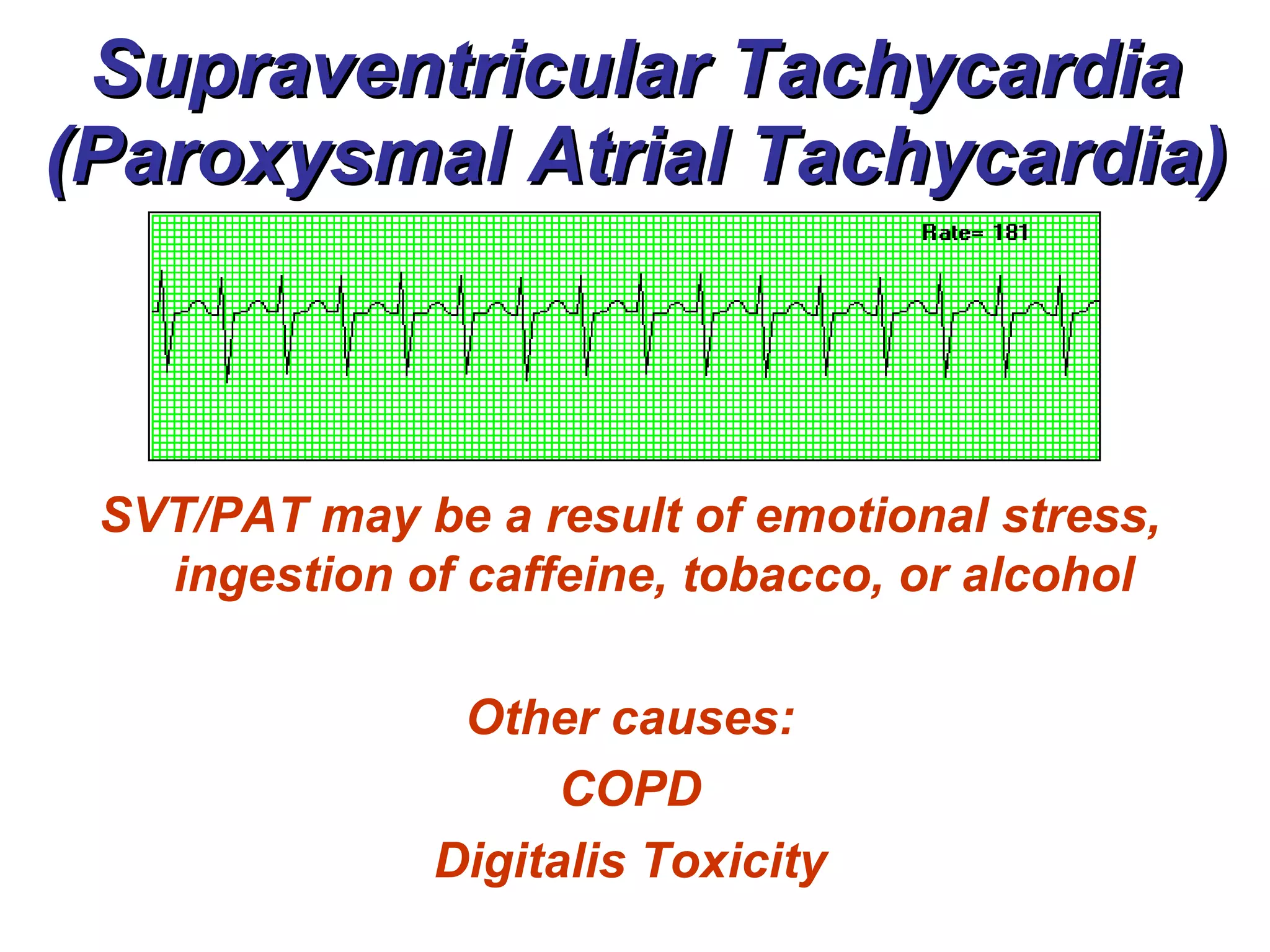
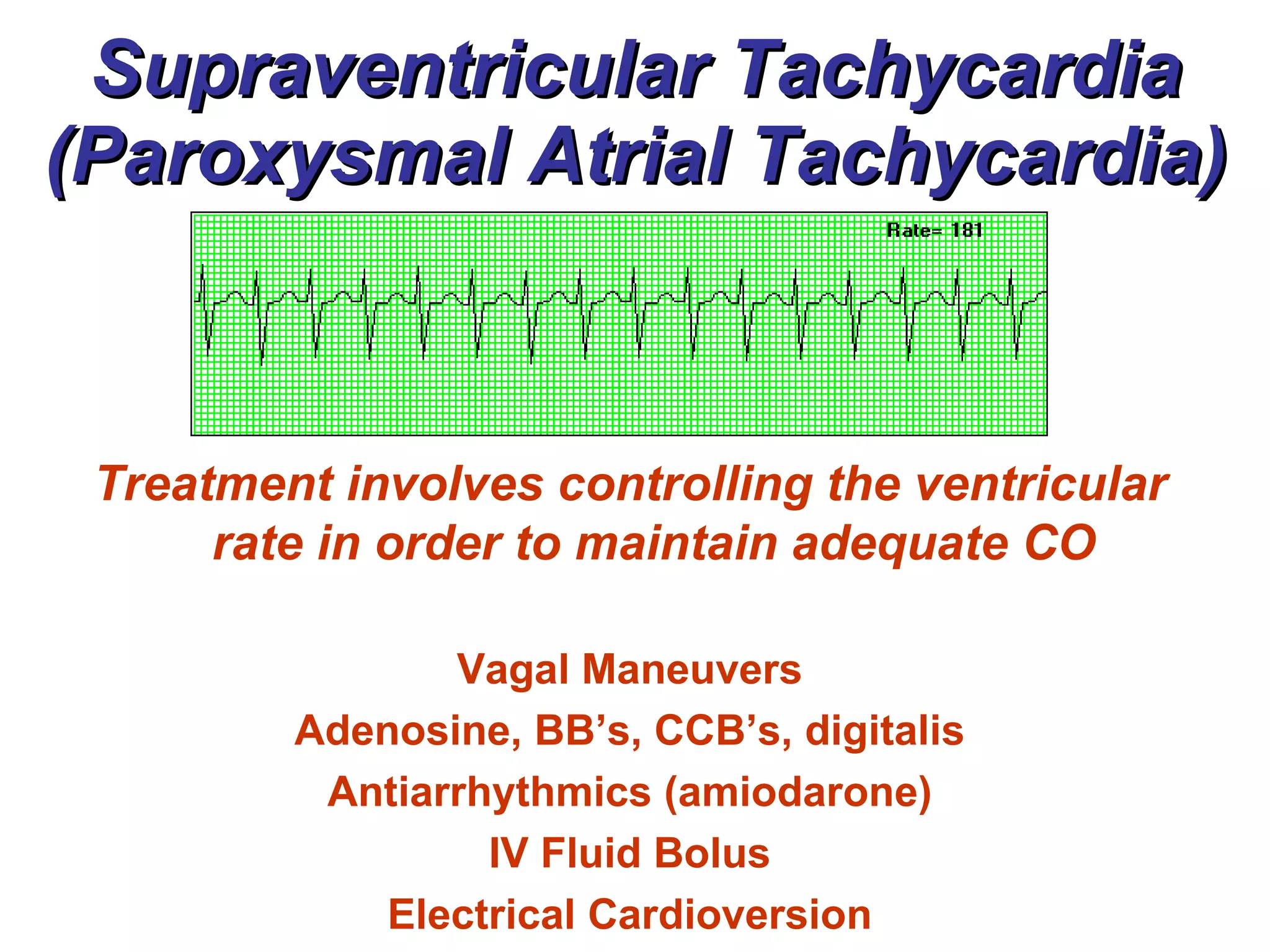
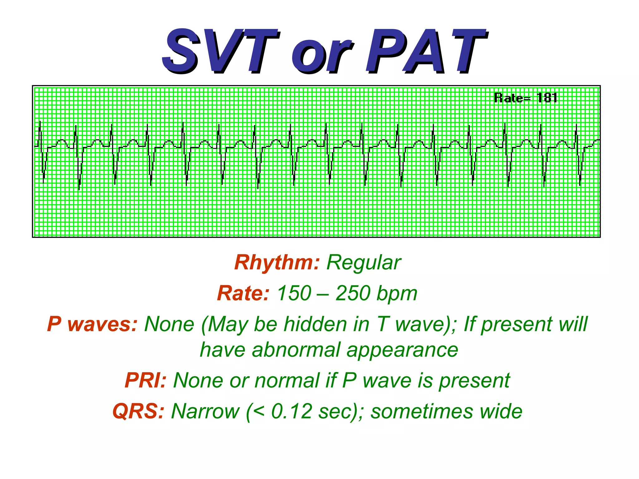
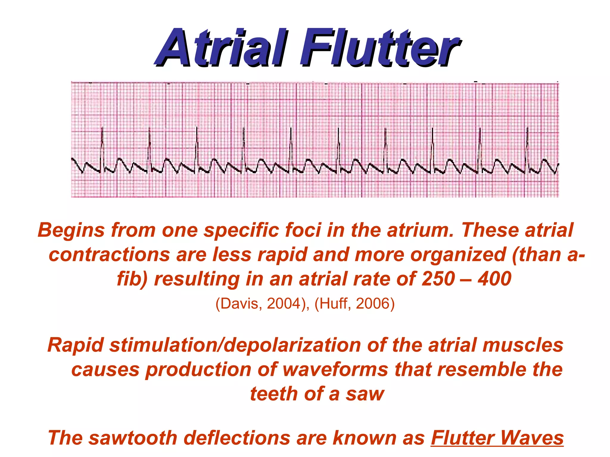
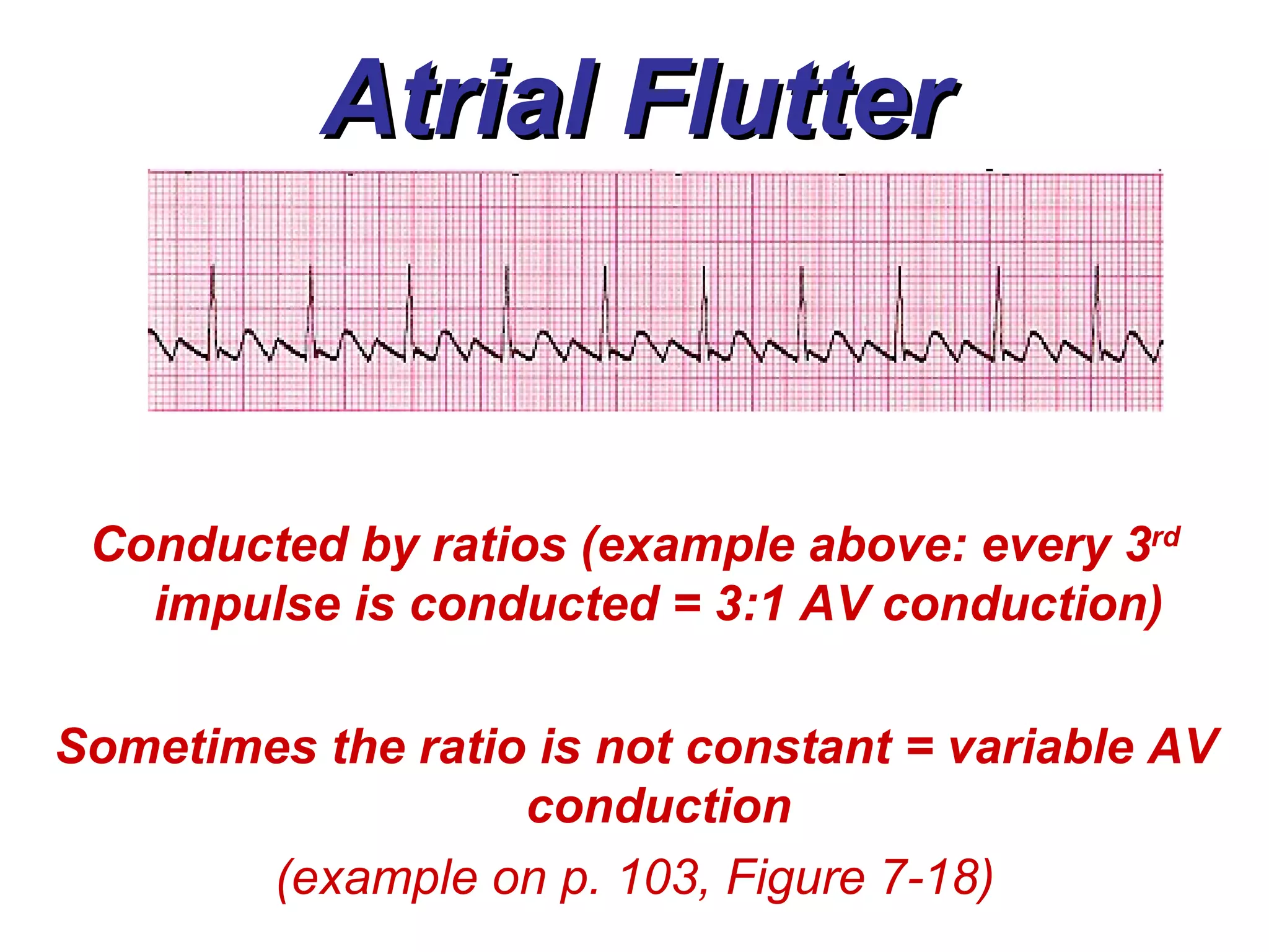
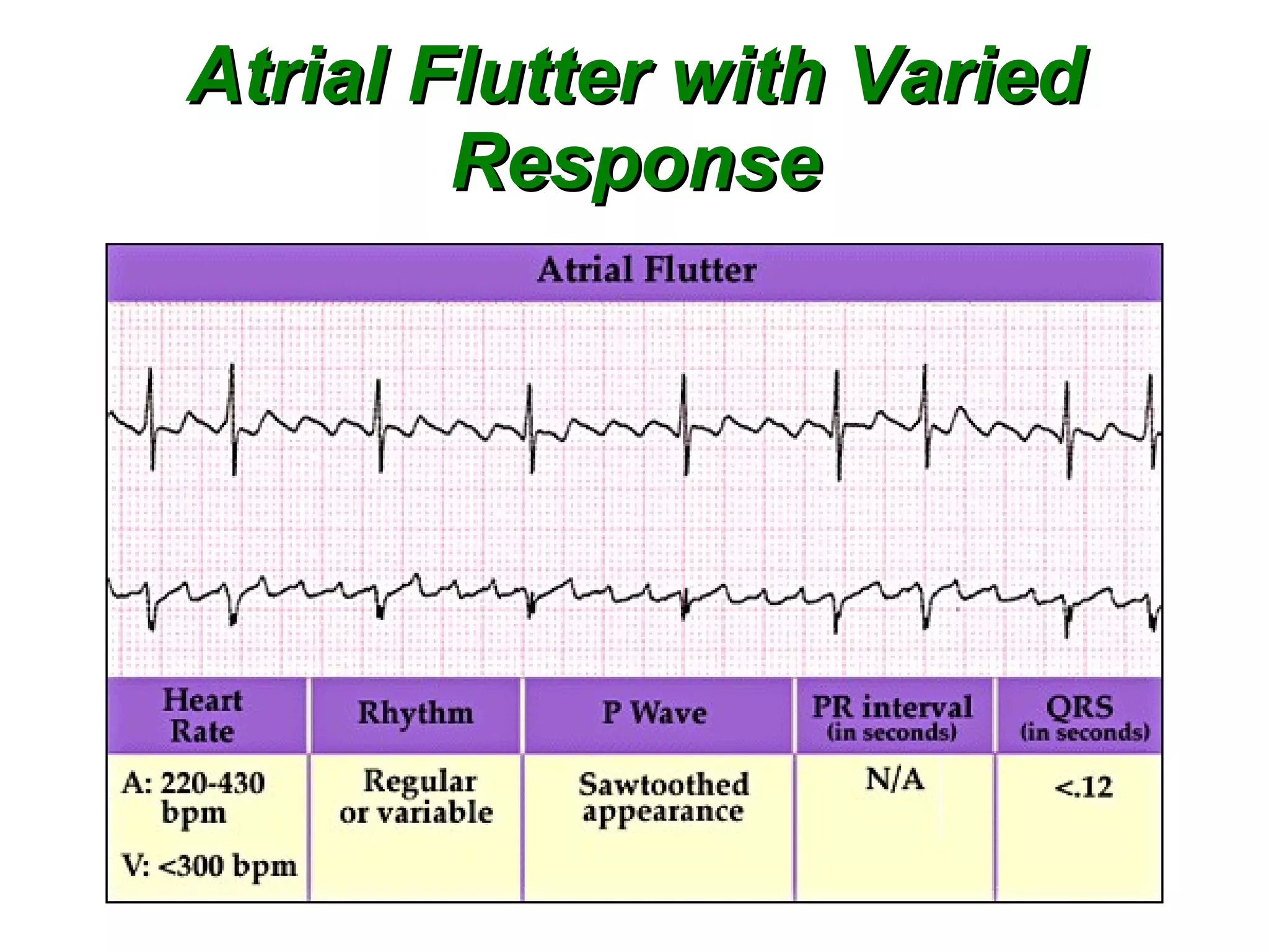
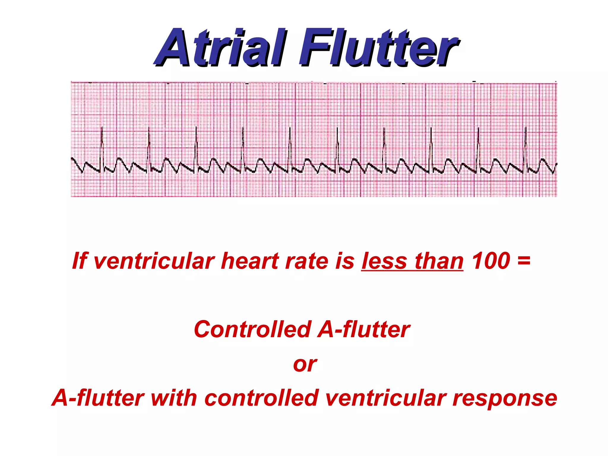
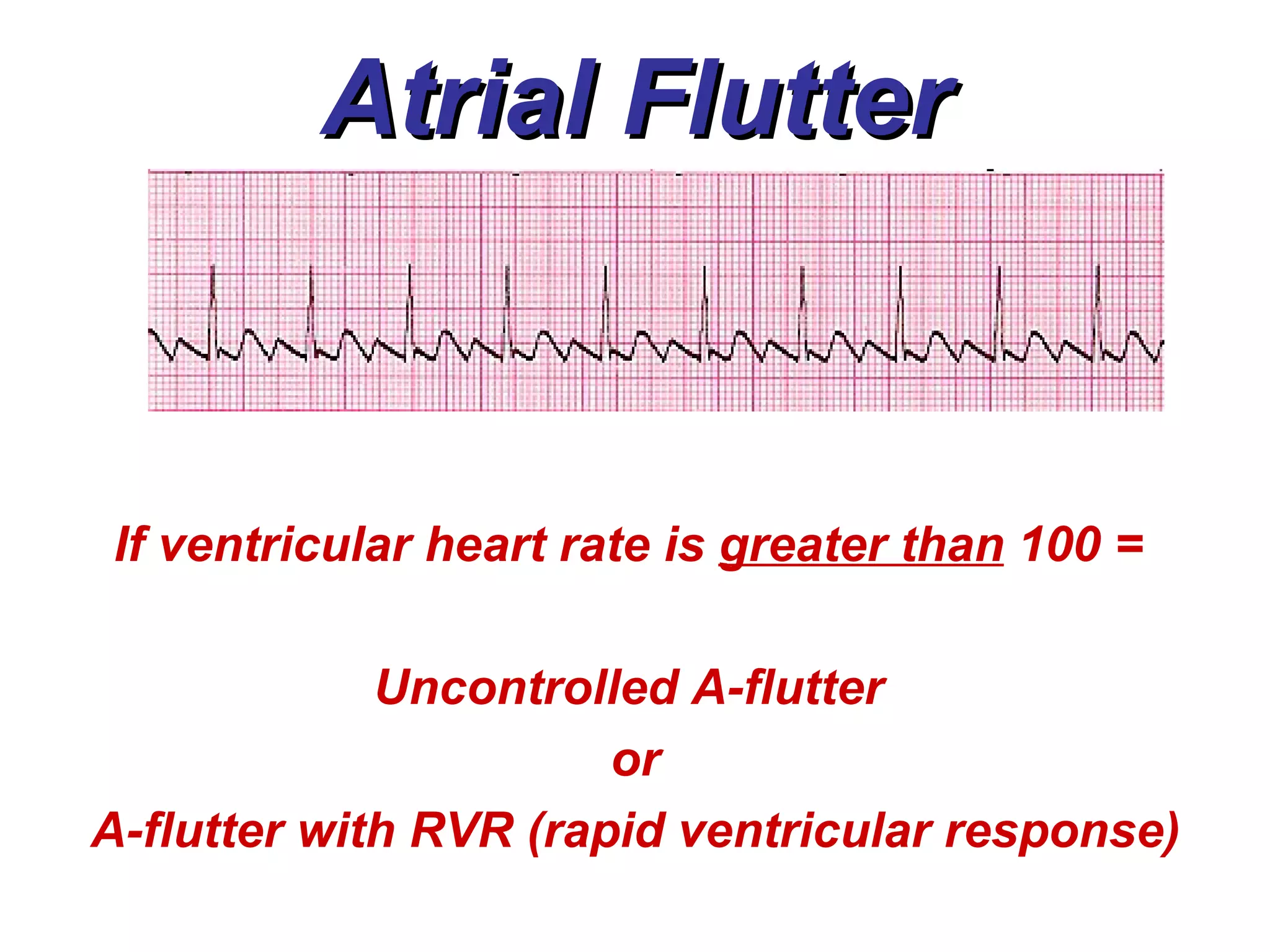
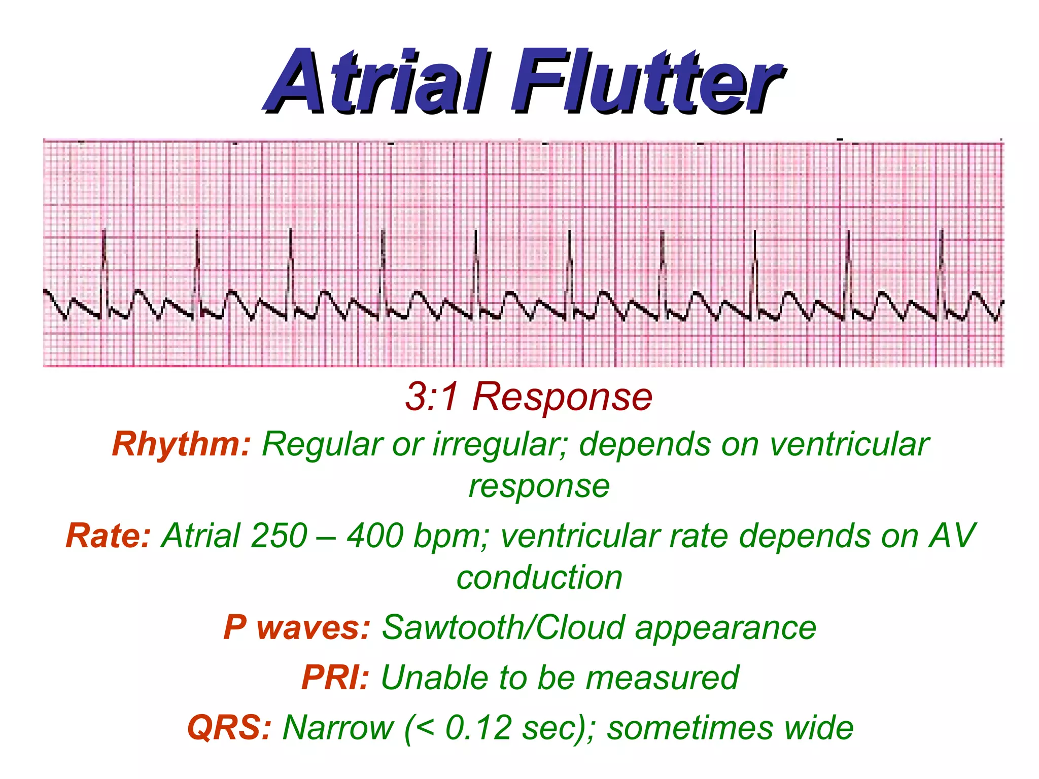
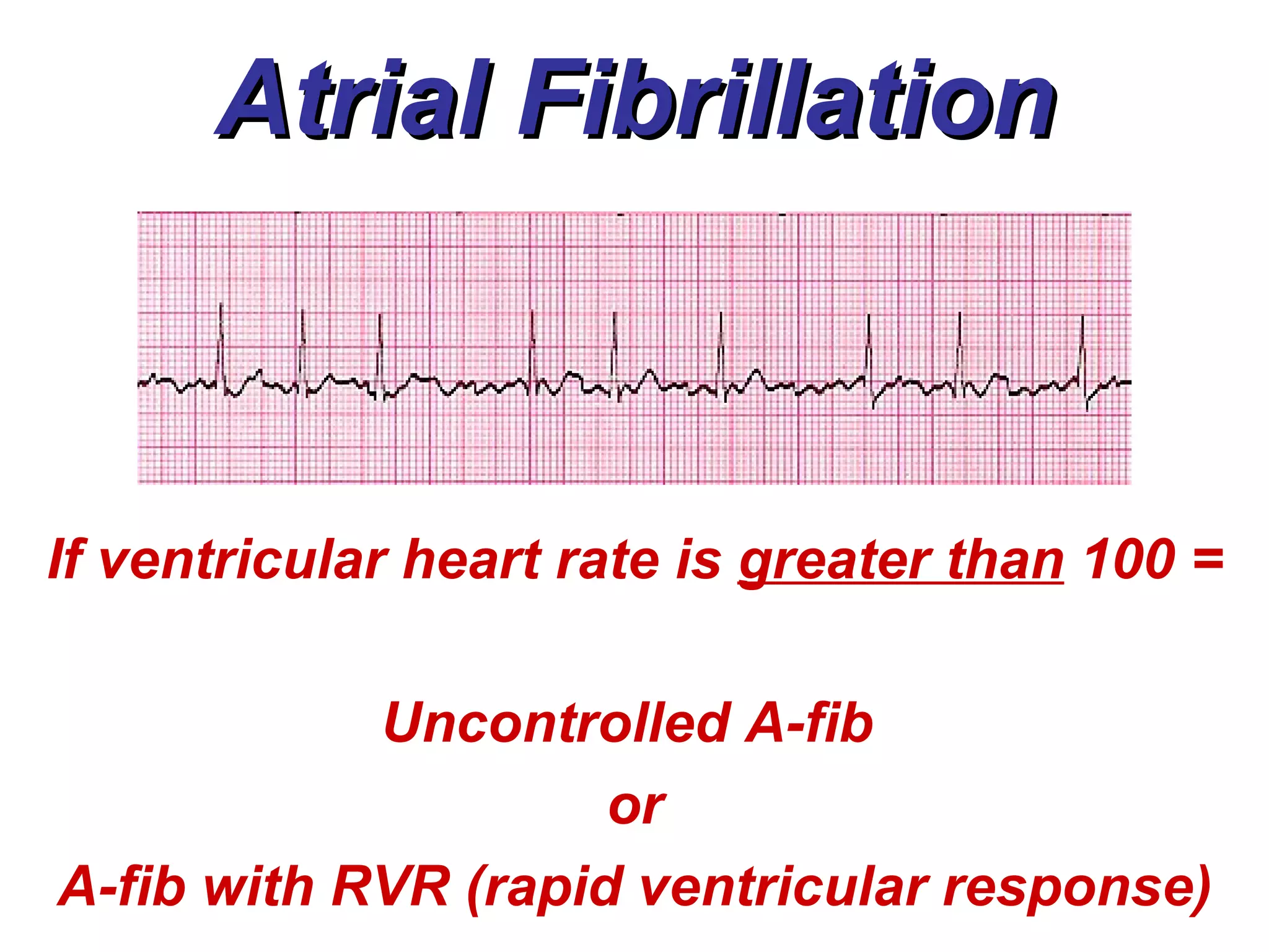
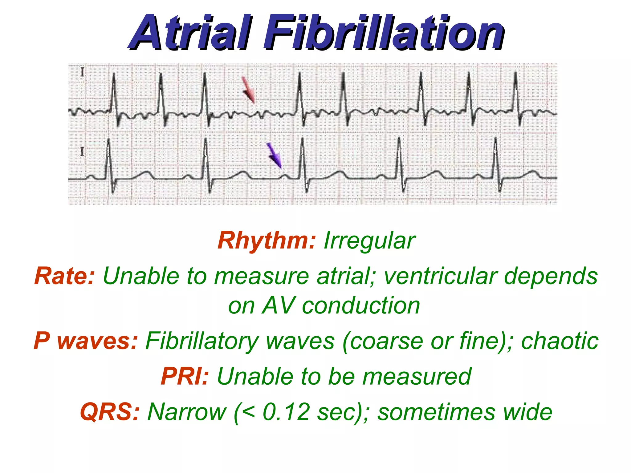
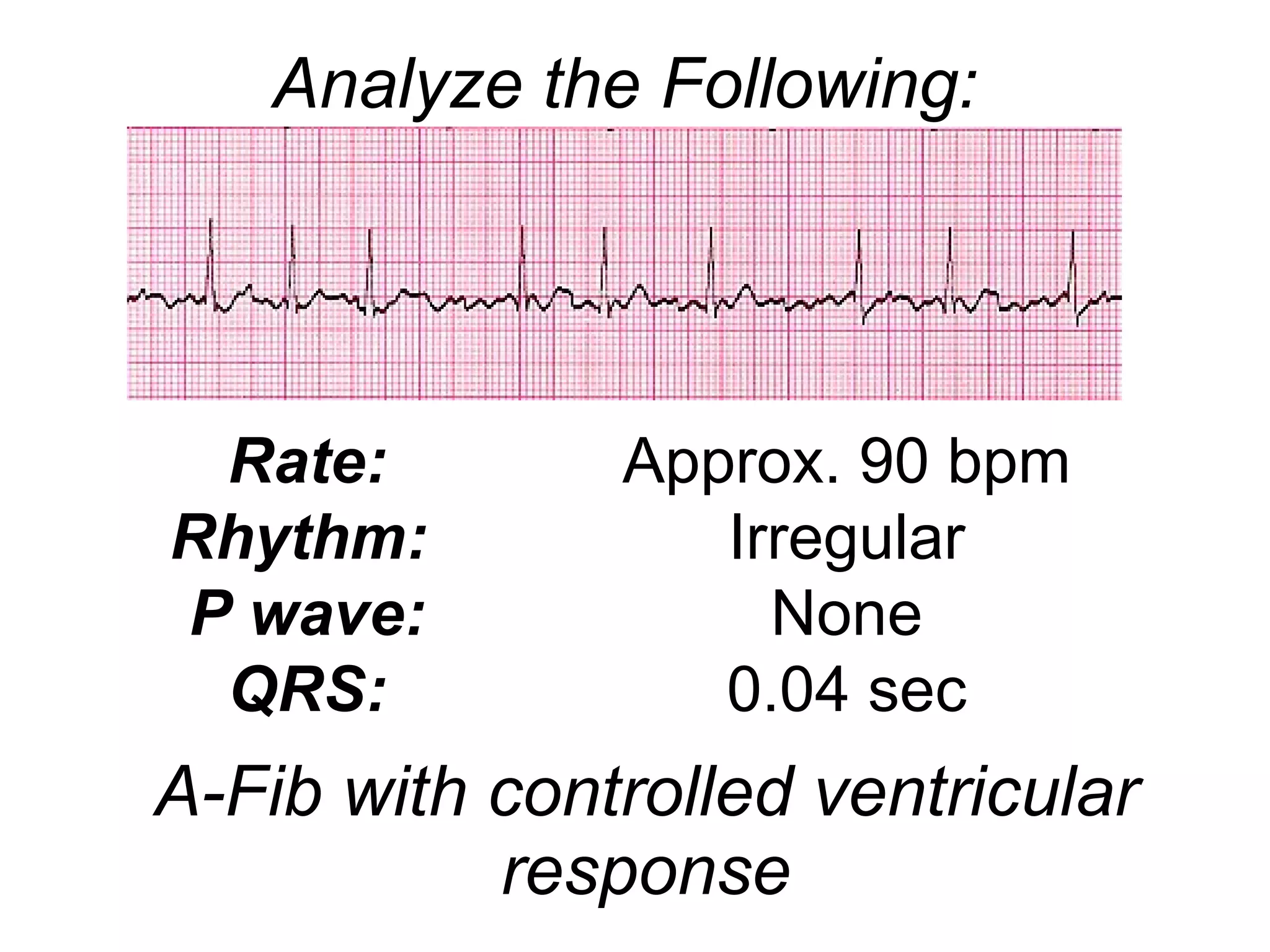
Atrial Rhythms Electrical impulses that originate from the atrium (not from the SA node).
Wandering Atrial Pacemaker (WAP) Rhythm:   Slightly Irregular Rate:  Usually 60 – 100 bpm; sometimes slower P waves:  Morphology of each P wave differs PRI:  0.12 – 0.20 sec; inconsistent QRS:  Narrow (< 0.12 sec); sometimes wide
Wandering Atrial Pacemaker (WAP) Causes: Increased vagal effect on the sinoatrial node, slowing the sinus rate and allowing other pacemaker sites an opportunity to compete for control of the heart rate It is a normal phenomenon in young healthy hearts (especially those of athletes) Coughing may decrease vagal tone and encourage reappearance of a sinus rhythm
Premature Atrial Contraction (PAC)  A premature atrial contraction (PAC) is an early beat that occurs when an ectopic site within the atria discharges an impulse before the sinus node impulse is discharged.  Ectopic site is speeding up in an attempt to take over as the pacemaker – irritability; (or the SA node has slowed down)  Can originate from one ectopic site or from multiple sites in the atria.
PAC  If the ectopic site is near the SA node, the appearance of the P wave may closely resemble the sinus P wave Most often the P wave morphology of the PAC is significantly different from that of the sinus P wave Sometimes with very premature PAC’s, the P wave is hidden within the T wave of the previous complex
PAC with Aberrant Ventricular Conduction PAC with Aberrant Ventricular Conduction An early beat from an atrial site (other than the SA node) that is abnormally conducted through the ventricles Causes a change in the morphology of the QRS complex (Example on p. 96)
Non-Conducted PAC P wave of PAC is not followed by a QRS complex AV node is refractory and is unable to respond to the electrical stimulus P wave is followed by a pause P wave may be hidden in the T wave (changes morphology of the T wave) - May be confused with a block/arrest (example on p. 99)
Compensatory vs. Noncompensatory PAC’s are followed by a pause What makes it compensatory versus noncompensatory??? (Overhead slide 3-1)
Premature Atrial Contraction  (PAC) Rhythm:   Depends on the underlying rhythm; Premature ectopic beat causes slight irregularity Rate:  Overall HR depends on rate of underlying rhythm P waves:  P wave of premature beat will have a different morphology (flattened, notched, or unusual). May be hidden in the T wave PRI:  0.12 – 0.20 sec on regular beat; ectopic beat PRI may differ. QRS:  Narrow (< 0.12 sec); sometimes wide
Supraventricular Tachycardia (Paroxysmal Atrial Tachycardia) May occur as a basic rhythm or within an underlying rhythm (burst of SVT or PAT) 3 or more consecutive PAC’s with a rate of 140 – 250 is considered to be a burst of SVT or PAT (example on p. 100)
Supraventricular Tachycardia (Paroxysmal Atrial Tachycardia) SVT/PAT may be a result of emotional stress, ingestion of caffeine, tobacco, or alcohol Other causes: COPD Digitalis Toxicity
Supraventricular Tachycardia (Paroxysmal Atrial Tachycardia) During episodes of SVT/PAT, patient may experience the following s/s: Hypotension Decreased LOC  Palpitations Chest Pain S.O.B.
Supraventricular Tachycardia (Paroxysmal Atrial Tachycardia) Treatment involves controlling the ventricular rate in order to maintain adequate CO Vagal Maneuvers Adenosine, BB’s, CCB’s, digitalis Antiarrhythmics (amiodarone) IV Fluid Bolus Electrical Cardioversion
SVT or PAT Rhythm:   Regular Rate:   150 – 250 bpm P waves:   None (May be hidden in T wave); If present will have abnormal appearance PRI:   None or normal if P wave is present QRS:   Narrow (< 0.12 sec); sometimes wide
Atrial Flutter Begins from one specific foci in the atrium. These atrial contractions are less rapid and more organized (than a-fib) resulting in an atrial rate of 250 – 400  (Davis, 2004), (Huff, 2006) Rapid stimulation/depolarization of the atrial muscles causes production of waveforms that resemble the teeth of a saw The sawtooth deflections are known as  Flutter Waves
Atrial Flutter Sometimes the flutter waves have a softer appearance and appear as “clouds” (Example on p. 112, Strip 7-16) Atrial firing is continuous; does not stop while impulses are traveling through the ventricles   Ventricular rate is controlled by AV node (the gatekeeper)
Atrial Flutter Conducted by ratios (example above: every 3 rd  impulse is conducted = 3:1 AV conduction) Sometimes the ratio is not constant = variable AV conduction (example on p. 103, Figure 7-18)
Atrial Flutter with Varied Response
Atrial Flutter If ventricular heart rate is  less than  100 =  Controlled A-flutter  or A-flutter with controlled ventricular response
Atrial Flutter If ventricular heart rate is  greater than  100 =  Uncontrolled A-flutter  or A-flutter with RVR (rapid ventricular response)
Atrial Flutter 3:1 Response Rhythm:   Regular or irregular; depends on ventricular response Rate:  Atrial 250 – 400 bpm; ventricular rate depends on AV conduction P waves:   Sawtooth/Cloud appearance PRI:   Unable to be measured QRS:   Narrow (< 0.12 sec); sometimes wide
Atrial Fibrillation It is the most common rhythm next to sinus rhythm Is a result of various ectopic sites within the atria that are firing at a rate of 400 – 600 bpm
Atrial Fibrillation Rapid depolarization of various sites causes the atria to quiver = fibrillatory waveforms Some of the atrial firing does not go to the AV node; remains in the same place and only depolarizes that small area
 
Atrial Fibrillation Fib-Flutter waves Flutter waves mixed with fibrillatory waves between the QRS complexes (Example on p. 111, Strip 7-14)
Atrial Fibrillation May produce fine fibrillatory waves or coarse fibrillatory waves Coarse fibrillatory waves are “very wavy” Fine waves are so small that they may appear as flat lines between the QRS’s (example on p. 105, Figure 7-22)
Atrial Fibrillation Atrial firing is continuous; does not stop while impulses are traveling through the ventricles   Ventricular rate is controlled by AV node (the gatekeeper) Goal is to control the AV conduction to the ventricles
Atrial Fibrillation If ventricular heart rate is  less than  100 =  Controlled A-fib  or A-fib with controlled ventricular response)
Atrial Fibrillation If ventricular heart rate is  greater than  100 =  Uncontrolled A-fib  or A-fib with RVR (rapid ventricular response)
Atrial Fibrillation Rhythm:   Irregular Rate:  Unable to measure atrial; ventricular depends on AV conduction  P waves:   Fibrillatory waves (coarse or fine); chaotic PRI:   Unable to be measured QRS:   Narrow (< 0.12 sec); sometimes wide
A-Fib & A-Flutter Common Causes: Valvular Heart Disease (especially with mitral valve) Hypertensive Heart Disease CAD Pulmonary Emboli Hyperthyroidism S/P Cardiac Surgery
A-Flutter & A-Fib Treatment involves converting the rhythm and/or controlling the ventricular rate in order to maintain adequate CO, in addition to prevention of blood clots BB’s, CCB’s, digitalis Antiarrhythmics (amiodarone) IV Fluid Bolus Anticoagulants Electrical Cardioversion
A-Fib with controlled ventricular response Rate: Rhythm:   P wave: QRS: Approx. 90 bpm Irregular None 0.04 sec Analyze the Following:
TIME TO WORKOUT!!!
References Chernecky, C., et al. (2002).  Real world nursing survival guide: ECG’s & the heart.  United States of America: W. B. Saunders Company. Huff, J. (2006).  ECG workout: Exercises in arrhythmia interpretation  (5 th  ed.). United States of America: Lippincott, Williams & Wilkins. Walraven, G. (1999).  Basic arrhythmias  (5 th  ed.). United States of America: Prentice-Hall, Inc. www.madsci.com/manu/ekg_rhy.htm

3a atrial rhythms

  • 1.
    Atrial Rhythms Electricalimpulses that originate from the atrium (not from the SA node).
  • 2.
    Wandering Atrial Pacemaker(WAP) Rhythm: Slightly Irregular Rate: Usually 60 – 100 bpm; sometimes slower P waves: Morphology of each P wave differs PRI: 0.12 – 0.20 sec; inconsistent QRS: Narrow (< 0.12 sec); sometimes wide
  • 3.
    Wandering Atrial Pacemaker(WAP) Causes: Increased vagal effect on the sinoatrial node, slowing the sinus rate and allowing other pacemaker sites an opportunity to compete for control of the heart rate It is a normal phenomenon in young healthy hearts (especially those of athletes) Coughing may decrease vagal tone and encourage reappearance of a sinus rhythm
  • 4.
    Premature Atrial Contraction(PAC) A premature atrial contraction (PAC) is an early beat that occurs when an ectopic site within the atria discharges an impulse before the sinus node impulse is discharged. Ectopic site is speeding up in an attempt to take over as the pacemaker – irritability; (or the SA node has slowed down) Can originate from one ectopic site or from multiple sites in the atria.
  • 5.
    PAC Ifthe ectopic site is near the SA node, the appearance of the P wave may closely resemble the sinus P wave Most often the P wave morphology of the PAC is significantly different from that of the sinus P wave Sometimes with very premature PAC’s, the P wave is hidden within the T wave of the previous complex
  • 6.
    PAC with AberrantVentricular Conduction PAC with Aberrant Ventricular Conduction An early beat from an atrial site (other than the SA node) that is abnormally conducted through the ventricles Causes a change in the morphology of the QRS complex (Example on p. 96)
  • 7.
    Non-Conducted PAC Pwave of PAC is not followed by a QRS complex AV node is refractory and is unable to respond to the electrical stimulus P wave is followed by a pause P wave may be hidden in the T wave (changes morphology of the T wave) - May be confused with a block/arrest (example on p. 99)
  • 8.
    Compensatory vs. NoncompensatoryPAC’s are followed by a pause What makes it compensatory versus noncompensatory??? (Overhead slide 3-1)
  • 9.
    Premature Atrial Contraction (PAC) Rhythm: Depends on the underlying rhythm; Premature ectopic beat causes slight irregularity Rate: Overall HR depends on rate of underlying rhythm P waves: P wave of premature beat will have a different morphology (flattened, notched, or unusual). May be hidden in the T wave PRI: 0.12 – 0.20 sec on regular beat; ectopic beat PRI may differ. QRS: Narrow (< 0.12 sec); sometimes wide
  • 10.
    Supraventricular Tachycardia (ParoxysmalAtrial Tachycardia) May occur as a basic rhythm or within an underlying rhythm (burst of SVT or PAT) 3 or more consecutive PAC’s with a rate of 140 – 250 is considered to be a burst of SVT or PAT (example on p. 100)
  • 11.
    Supraventricular Tachycardia (ParoxysmalAtrial Tachycardia) SVT/PAT may be a result of emotional stress, ingestion of caffeine, tobacco, or alcohol Other causes: COPD Digitalis Toxicity
  • 12.
    Supraventricular Tachycardia (ParoxysmalAtrial Tachycardia) During episodes of SVT/PAT, patient may experience the following s/s: Hypotension Decreased LOC Palpitations Chest Pain S.O.B.
  • 13.
    Supraventricular Tachycardia (ParoxysmalAtrial Tachycardia) Treatment involves controlling the ventricular rate in order to maintain adequate CO Vagal Maneuvers Adenosine, BB’s, CCB’s, digitalis Antiarrhythmics (amiodarone) IV Fluid Bolus Electrical Cardioversion
  • 14.
    SVT or PATRhythm: Regular Rate: 150 – 250 bpm P waves: None (May be hidden in T wave); If present will have abnormal appearance PRI: None or normal if P wave is present QRS: Narrow (< 0.12 sec); sometimes wide
  • 15.
    Atrial Flutter Beginsfrom one specific foci in the atrium. These atrial contractions are less rapid and more organized (than a-fib) resulting in an atrial rate of 250 – 400 (Davis, 2004), (Huff, 2006) Rapid stimulation/depolarization of the atrial muscles causes production of waveforms that resemble the teeth of a saw The sawtooth deflections are known as Flutter Waves
  • 16.
    Atrial Flutter Sometimesthe flutter waves have a softer appearance and appear as “clouds” (Example on p. 112, Strip 7-16) Atrial firing is continuous; does not stop while impulses are traveling through the ventricles Ventricular rate is controlled by AV node (the gatekeeper)
  • 17.
    Atrial Flutter Conductedby ratios (example above: every 3 rd impulse is conducted = 3:1 AV conduction) Sometimes the ratio is not constant = variable AV conduction (example on p. 103, Figure 7-18)
  • 18.
    Atrial Flutter withVaried Response
  • 19.
    Atrial Flutter Ifventricular heart rate is less than 100 = Controlled A-flutter or A-flutter with controlled ventricular response
  • 20.
    Atrial Flutter Ifventricular heart rate is greater than 100 = Uncontrolled A-flutter or A-flutter with RVR (rapid ventricular response)
  • 21.
    Atrial Flutter 3:1Response Rhythm: Regular or irregular; depends on ventricular response Rate: Atrial 250 – 400 bpm; ventricular rate depends on AV conduction P waves: Sawtooth/Cloud appearance PRI: Unable to be measured QRS: Narrow (< 0.12 sec); sometimes wide
  • 22.
    Atrial Fibrillation Itis the most common rhythm next to sinus rhythm Is a result of various ectopic sites within the atria that are firing at a rate of 400 – 600 bpm
  • 23.
    Atrial Fibrillation Rapiddepolarization of various sites causes the atria to quiver = fibrillatory waveforms Some of the atrial firing does not go to the AV node; remains in the same place and only depolarizes that small area
  • 24.
  • 25.
    Atrial Fibrillation Fib-Flutterwaves Flutter waves mixed with fibrillatory waves between the QRS complexes (Example on p. 111, Strip 7-14)
  • 26.
    Atrial Fibrillation Mayproduce fine fibrillatory waves or coarse fibrillatory waves Coarse fibrillatory waves are “very wavy” Fine waves are so small that they may appear as flat lines between the QRS’s (example on p. 105, Figure 7-22)
  • 27.
    Atrial Fibrillation Atrialfiring is continuous; does not stop while impulses are traveling through the ventricles Ventricular rate is controlled by AV node (the gatekeeper) Goal is to control the AV conduction to the ventricles
  • 28.
    Atrial Fibrillation Ifventricular heart rate is less than 100 = Controlled A-fib or A-fib with controlled ventricular response)
  • 29.
    Atrial Fibrillation Ifventricular heart rate is greater than 100 = Uncontrolled A-fib or A-fib with RVR (rapid ventricular response)
  • 30.
    Atrial Fibrillation Rhythm: Irregular Rate: Unable to measure atrial; ventricular depends on AV conduction P waves: Fibrillatory waves (coarse or fine); chaotic PRI: Unable to be measured QRS: Narrow (< 0.12 sec); sometimes wide
  • 31.
    A-Fib & A-FlutterCommon Causes: Valvular Heart Disease (especially with mitral valve) Hypertensive Heart Disease CAD Pulmonary Emboli Hyperthyroidism S/P Cardiac Surgery
  • 32.
    A-Flutter & A-FibTreatment involves converting the rhythm and/or controlling the ventricular rate in order to maintain adequate CO, in addition to prevention of blood clots BB’s, CCB’s, digitalis Antiarrhythmics (amiodarone) IV Fluid Bolus Anticoagulants Electrical Cardioversion
  • 33.
    A-Fib with controlledventricular response Rate: Rhythm: P wave: QRS: Approx. 90 bpm Irregular None 0.04 sec Analyze the Following:
  • 34.
  • 35.
    References Chernecky, C.,et al. (2002). Real world nursing survival guide: ECG’s & the heart. United States of America: W. B. Saunders Company. Huff, J. (2006). ECG workout: Exercises in arrhythmia interpretation (5 th ed.). United States of America: Lippincott, Williams & Wilkins. Walraven, G. (1999). Basic arrhythmias (5 th ed.). United States of America: Prentice-Hall, Inc. www.madsci.com/manu/ekg_rhy.htm