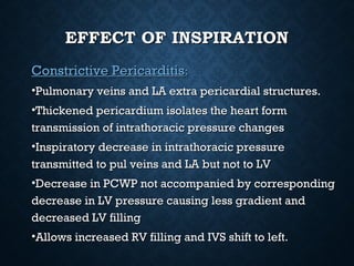
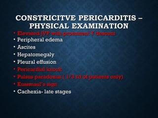
CONSTRICTIVECONSTRICTIVE
PERICARDITISPERICARDITIS
MAJ WAQAS KHALIDMAJ WAQAS KHALID
•CONSTRICTIVE PERICARDITISCONSTRICTIVE PERICARDITIS —result of scarring and—result of scarring and
consequent loss of elasticity of the pericardial sac.consequent loss of elasticity of the pericardial sac.
Typically chronic. The pathological changes areTypically chronic. The pathological changes are
inflammation, sometimes calcification. Grossly,inflammation, sometimes calcification. Grossly,
pericardium thicker than normal -80% of time.pericardium thicker than normal -80% of time.
•EFFUSIVE-CONSTRICTIVE PERICARDITISEFFUSIVE-CONSTRICTIVE PERICARDITIS ——
characterized by constrictive physiology with acharacterized by constrictive physiology with a
coexisting pericardial effusion, usually with tamponade.coexisting pericardial effusion, usually with tamponade.
PERICARDIUMPERICARDIUM
Upto 50 mL fluid
Parietal and visceral layers
ANATOMYANATOMY
• Lt. AtriumLt. Atrium is notis not
completelycompletely
intrapericardialintrapericardial
• All other cardiacAll other cardiac
chamberschambers areare
completelycompletely
intrapericardialintrapericardial
• Pulmonary Veins arePulmonary Veins are
completelycompletely
intrathoracic (extraintrathoracic (extra
pericardial)pericardial)
PHYSIOLOGY OF CONSTRICTIONPHYSIOLOGY OF CONSTRICTION
• In the pericardial compressive syndromes, theIn the pericardial compressive syndromes, the
pericardium is inelastic andpericardium is inelastic and total cardiac volumetotal cardiac volume
cannot changecannot change
• The result is enhanced ventricular interaction or…The result is enhanced ventricular interaction or…
ventricular interdependenceventricular interdependence
PHYSIOLOGY OF CONSTRICTIONPHYSIOLOGY OF CONSTRICTION
• Pericardial constriction leads to impairment ofPericardial constriction leads to impairment of
ventricular filling, usually affecting all four cardiacventricular filling, usually affecting all four cardiac
chambers, preventing ventricular filling in mid andchambers, preventing ventricular filling in mid and
late diastole.late diastole.
• As a result, the majority of ventricular filling occursAs a result, the majority of ventricular filling occurs
rapidly in early diastole and the ventricular volumerapidly in early diastole and the ventricular volume
does not increase after the end of the early fillingdoes not increase after the end of the early filling
period.period.
EFFECT OF INSPIRATIONEFFECT OF INSPIRATION
• Normal PericardiumNormal Pericardium::
• Inspiratory decrease in intrathoracic pressure isInspiratory decrease in intrathoracic pressure is
uniformly transmitted to the lungs, PVs, and alluniformly transmitted to the lungs, PVs, and all
cardiac chamberscardiac chambers..
• Decrease in PCWP accompanied byDecrease in PCWP accompanied by
corresponding decrease in LV Pressurecorresponding decrease in LV Pressure
• Normal pericardium contributes to diastolicNormal pericardium contributes to diastolic
interaction ( transmission of intracavitary fillinginteraction ( transmission of intracavitary filling
pressures to adjoining chambers)pressures to adjoining chambers)
EFFECT OF INSPIRATIONEFFECT OF INSPIRATION
Constrictive PericarditisConstrictive Pericarditis::
•Pulmonary veins and LA extra pericardial structures.Pulmonary veins and LA extra pericardial structures.
•Thickened pericardium isolates the heart formThickened pericardium isolates the heart form
transmission of intrathoracic pressure changestransmission of intrathoracic pressure changes
•Inspiratory decrease in intrathoracic pressureInspiratory decrease in intrathoracic pressure
transmitted to pul veins and LA but not to LVtransmitted to pul veins and LA but not to LV
•Decrease in PCWP not accompanied by correspondingDecrease in PCWP not accompanied by corresponding
decrease in LV pressure causing less gradient anddecrease in LV pressure causing less gradient and
decreased LV fillingdecreased LV filling
•Allows increased RV filling and IVS shift to left.Allows increased RV filling and IVS shift to left.
CONSTRICTIVECONSTRICTIVE
PERICARDITIS - ETIOLOGYPERICARDITIS - ETIOLOGY
• Idiopathic or viral — 42 to 49 %Idiopathic or viral — 42 to 49 %
• Post cardiac surgery — 11 to 37 %Post cardiac surgery — 11 to 37 %
• Post radiation therapy — 9 to 31 %Post radiation therapy — 9 to 31 %
• Connective tissue disorder — 3 to 7 %Connective tissue disorder — 3 to 7 %
• Post infectious (Post infectious (tuberculoustuberculous or purulent pericarditis)or purulent pericarditis)
— 3 to 6 % (— 3 to 6 % ( Most common in our setup)Most common in our setup)
• Miscellaneous causes (malignancy, trauma, drug-Miscellaneous causes (malignancy, trauma, drug-
induced, asbestosis, sarcoidosis, uremicinduced, asbestosis, sarcoidosis, uremic
pericarditis) — 1 to 10 %pericarditis) — 1 to 10 %
CONSTRICTIVE PERICARDITIS –CONSTRICTIVE PERICARDITIS –
CLINICAL PRESENTATIONCLINICAL PRESENTATION
• 67 % presented with symptoms of Right heart failure (HF)67 % presented with symptoms of Right heart failure (HF)
• 8 % with chest pain8 % with chest pain
• 6 % with abdominal symptoms6 % with abdominal symptoms
• 4 % with atrial arrhythmia4 % with atrial arrhythmia
• 5 % with symptoms of cardiac tamponade5 % with symptoms of cardiac tamponade
• Constrictive pericarditis can be mistaken for any cause ofConstrictive pericarditis can be mistaken for any cause of
right heart failure as well as end stage liver diseaseright heart failure as well as end stage liver disease
• Most patients have thickened pericardiumMost patients have thickened pericardium
• Normal thickness on direct histopathologicalNormal thickness on direct histopathological
examination (18%) and on CT scanning ( 28%)examination (18%) and on CT scanning ( 28%)
• Transient constrictive pericarditis ( Post cardiacTransient constrictive pericarditis ( Post cardiac
surgery)surgery)
CONSTRICTIVE PERICARDITISCONSTRICTIVE PERICARDITIS
CONSTRICITVE PERICARDITIS –CONSTRICITVE PERICARDITIS –
PHYSICAL EXAMINATIONPHYSICAL EXAMINATION
• Elevated JVP with prominent Y descentElevated JVP with prominent Y descent
• Peripheral edemaPeripheral edema
• AscitesAscites
• HepatomegalyHepatomegaly
• Pleural effusionPleural effusion
• Pericardial knockPericardial knock
• Pulsus paradoxus ( 1/3 rd of patients only)Pulsus paradoxus ( 1/3 rd of patients only)
• Kussmaul’s signKussmaul’s sign
• Cachexia- late stagesCachexia- late stages
• Inspiratory increase in mean venous pressureInspiratory increase in mean venous pressure
or the pressure may simply fail to decrease onor the pressure may simply fail to decrease on
inspirationinspiration
• Reflects loss of normal increase in right heartReflects loss of normal increase in right heart
venous return on inspiration, even thoughvenous return on inspiration, even though
tricuspid flow increasestricuspid flow increases
• Absent in tamponadeAbsent in tamponade
Kussmaul’s sign
KUSSMAUL’S SIGNKUSSMAUL’S SIGN
PERICARDIAL THICKENINGPERICARDIAL THICKENING
• This can be visualized by transesophageal echoThis can be visualized by transesophageal echo
(often requiring multiple views), however, this is(often requiring multiple views), however, this is
best seen using other imaging modalities such asbest seen using other imaging modalities such as
CT or MRI.CT or MRI.
CALCIFIED PERICARDIUMCALCIFIED PERICARDIUM
PERICARDIAL CALCIFICATIONSPERICARDIAL CALCIFICATIONS
CTCT
PERICARDIAL CALCIFICATIONPERICARDIAL CALCIFICATION
ON ECHOON ECHO
• Normal pericardium is highlyNormal pericardium is highly
reflectivereflective
• Bright pericardial echoBright pericardial echo
cannot alone diagnosecannot alone diagnose
constrictive pericarditisconstrictive pericarditis
SPECIFIC ECHO EXAM FORSPECIFIC ECHO EXAM FOR
CONSTRICTIONCONSTRICTION
• Neither sensitive nor specificNeither sensitive nor specific
• Must diagnose via a combination of physicalMust diagnose via a combination of physical
exam/ history findings and echo findingsexam/ history findings and echo findings
M-MODE FINDINGS INM-MODE FINDINGS IN
CONSTRICTIONCONSTRICTION
DILATION AND LACK OFDILATION AND LACK OF
RESPIRATORY VARIATION IN IVCRESPIRATORY VARIATION IN IVC
DIASTOLIC SEPTAL BOUNCE:DIASTOLIC SEPTAL BOUNCE:
• Thought to be due to the rapid filling during earlyThought to be due to the rapid filling during early
diastole leading to asymmetrical filling of the rightdiastole leading to asymmetrical filling of the right
and left ventricals which creates a fluctuatingand left ventricals which creates a fluctuating
pressure gradient that manifests as an abrupt shift ofpressure gradient that manifests as an abrupt shift of
the septum.the septum.
SEPTAL BOUNCE ON ECHO ANDSEPTAL BOUNCE ON ECHO AND
MRIMRI
ATRIAL DILATIONATRIAL DILATION
• MildMild
• Secondary to elevated atrial pressuresSecondary to elevated atrial pressures
• More severe atrial dilatation seen inMore severe atrial dilatation seen in
restrictive cardiomyopathy.restrictive cardiomyopathy.
DOPPLER ECHO FINDINGS INDOPPLER ECHO FINDINGS IN
CONSTRICTIONCONSTRICTION
UN MASKING RESP VARIATIONSUN MASKING RESP VARIATIONS
TISSUE DOPPLER TOTISSUE DOPPLER TO
DISTINGUISH ENTITIESDISTINGUISH ENTITIES
Dimunitive E’
<8 cm/s
E’ similar to E
>12cm/s
DOPPLER ECHO AND RESPDOPPLER ECHO AND RESP
VARIATIONSVARIATIONS
VENTRICULARVENTRICULAR
INTERDEPENDENCEINTERDEPENDENCE
Insp Expir
Hatle LK, et. al.
Circ. 1989;79357-370
Ventricular Pressures
Are DISCORDANT
CONSTRICTIVE PERICARDITIS –CONSTRICTIVE PERICARDITIS –
OTHER TESTS?OTHER TESTS?
• ECG sync CT – helps in detecting minute amount ofECG sync CT – helps in detecting minute amount of
calcification. Important adjunt to Doppler echocalcification. Important adjunt to Doppler echo
examinationexamination
• Cardiac MRI – Role in diagnosing transient post opCardiac MRI – Role in diagnosing transient post op
constrictive pericarditis( late gadolinium enhancementconstrictive pericarditis( late gadolinium enhancement
and pericardial thickness > 3mm)and pericardial thickness > 3mm)
• BNP – usually only a mild elevation due to limited wallBNP – usually only a mild elevation due to limited wall
stretchstretch
EFFUSIVE CONSTRICTIVEEFFUSIVE CONSTRICTIVE
PERICARDITISPERICARDITIS
• Combination of tamponade and constrictionCombination of tamponade and constriction
• Common etiologies: malignancy and radiation therapyCommon etiologies: malignancy and radiation therapy
• Pericardial thickening may prevent RA collapsePericardial thickening may prevent RA collapse
• Hemodynamic compromise and JVD persist even after tapHemodynamic compromise and JVD persist even after tap
TREATMENTTREATMENT
• Surgical pericardiectomy is definitive treatment except transientSurgical pericardiectomy is definitive treatment except transient
constrictive pericarditisconstrictive pericarditis
• Patients with major comorbidities or severe debilitation and radiationPatients with major comorbidities or severe debilitation and radiation
induced disease are relatively contraindicated for pericardiectomyinduced disease are relatively contraindicated for pericardiectomy
• Medical management with diuretics and salt restrictionMedical management with diuretics and salt restriction
• Because sinus tachycardia is a compensatory mechanism beta blockersBecause sinus tachycardia is a compensatory mechanism beta blockers
and Ca antagonists should be avoidedand Ca antagonists should be avoided
• In pattients with AF and rapid vent response, digoxin is recommendedIn pattients with AF and rapid vent response, digoxin is recommended
• Patients with late gadolinium enhancement on MRI and pericardialPatients with late gadolinium enhancement on MRI and pericardial
thickness of more than 3 mm should be considered for a trial of antithickness of more than 3 mm should be considered for a trial of anti
inflammatory therapy for 2-3 months.inflammatory therapy for 2-3 months.
ThanksThanks
ForFor
Listening!!Listening!!

Constrictive pericarditis

  • 1.
  • 2.
    •CONSTRICTIVE PERICARDITISCONSTRICTIVE PERICARDITIS—result of scarring and—result of scarring and consequent loss of elasticity of the pericardial sac.consequent loss of elasticity of the pericardial sac. Typically chronic. The pathological changes areTypically chronic. The pathological changes are inflammation, sometimes calcification. Grossly,inflammation, sometimes calcification. Grossly, pericardium thicker than normal -80% of time.pericardium thicker than normal -80% of time. •EFFUSIVE-CONSTRICTIVE PERICARDITISEFFUSIVE-CONSTRICTIVE PERICARDITIS —— characterized by constrictive physiology with acharacterized by constrictive physiology with a coexisting pericardial effusion, usually with tamponade.coexisting pericardial effusion, usually with tamponade.
  • 3.
    PERICARDIUMPERICARDIUM Upto 50 mLfluid Parietal and visceral layers
  • 4.
    ANATOMYANATOMY • Lt. AtriumLt.Atrium is notis not completelycompletely intrapericardialintrapericardial • All other cardiacAll other cardiac chamberschambers areare completelycompletely intrapericardialintrapericardial • Pulmonary Veins arePulmonary Veins are completelycompletely intrathoracic (extraintrathoracic (extra pericardial)pericardial)
  • 5.
    PHYSIOLOGY OF CONSTRICTIONPHYSIOLOGYOF CONSTRICTION • In the pericardial compressive syndromes, theIn the pericardial compressive syndromes, the pericardium is inelastic andpericardium is inelastic and total cardiac volumetotal cardiac volume cannot changecannot change • The result is enhanced ventricular interaction or…The result is enhanced ventricular interaction or… ventricular interdependenceventricular interdependence
  • 6.
    PHYSIOLOGY OF CONSTRICTIONPHYSIOLOGYOF CONSTRICTION • Pericardial constriction leads to impairment ofPericardial constriction leads to impairment of ventricular filling, usually affecting all four cardiacventricular filling, usually affecting all four cardiac chambers, preventing ventricular filling in mid andchambers, preventing ventricular filling in mid and late diastole.late diastole. • As a result, the majority of ventricular filling occursAs a result, the majority of ventricular filling occurs rapidly in early diastole and the ventricular volumerapidly in early diastole and the ventricular volume does not increase after the end of the early fillingdoes not increase after the end of the early filling period.period.
  • 7.
    EFFECT OF INSPIRATIONEFFECTOF INSPIRATION • Normal PericardiumNormal Pericardium:: • Inspiratory decrease in intrathoracic pressure isInspiratory decrease in intrathoracic pressure is uniformly transmitted to the lungs, PVs, and alluniformly transmitted to the lungs, PVs, and all cardiac chamberscardiac chambers.. • Decrease in PCWP accompanied byDecrease in PCWP accompanied by corresponding decrease in LV Pressurecorresponding decrease in LV Pressure • Normal pericardium contributes to diastolicNormal pericardium contributes to diastolic interaction ( transmission of intracavitary fillinginteraction ( transmission of intracavitary filling pressures to adjoining chambers)pressures to adjoining chambers)
  • 8.
    EFFECT OF INSPIRATIONEFFECTOF INSPIRATION Constrictive PericarditisConstrictive Pericarditis:: •Pulmonary veins and LA extra pericardial structures.Pulmonary veins and LA extra pericardial structures. •Thickened pericardium isolates the heart formThickened pericardium isolates the heart form transmission of intrathoracic pressure changestransmission of intrathoracic pressure changes •Inspiratory decrease in intrathoracic pressureInspiratory decrease in intrathoracic pressure transmitted to pul veins and LA but not to LVtransmitted to pul veins and LA but not to LV •Decrease in PCWP not accompanied by correspondingDecrease in PCWP not accompanied by corresponding decrease in LV pressure causing less gradient anddecrease in LV pressure causing less gradient and decreased LV fillingdecreased LV filling •Allows increased RV filling and IVS shift to left.Allows increased RV filling and IVS shift to left.
  • 10.
    CONSTRICTIVECONSTRICTIVE PERICARDITIS - ETIOLOGYPERICARDITIS- ETIOLOGY • Idiopathic or viral — 42 to 49 %Idiopathic or viral — 42 to 49 % • Post cardiac surgery — 11 to 37 %Post cardiac surgery — 11 to 37 % • Post radiation therapy — 9 to 31 %Post radiation therapy — 9 to 31 % • Connective tissue disorder — 3 to 7 %Connective tissue disorder — 3 to 7 % • Post infectious (Post infectious (tuberculoustuberculous or purulent pericarditis)or purulent pericarditis) — 3 to 6 % (— 3 to 6 % ( Most common in our setup)Most common in our setup) • Miscellaneous causes (malignancy, trauma, drug-Miscellaneous causes (malignancy, trauma, drug- induced, asbestosis, sarcoidosis, uremicinduced, asbestosis, sarcoidosis, uremic pericarditis) — 1 to 10 %pericarditis) — 1 to 10 %
  • 11.
    CONSTRICTIVE PERICARDITIS –CONSTRICTIVEPERICARDITIS – CLINICAL PRESENTATIONCLINICAL PRESENTATION • 67 % presented with symptoms of Right heart failure (HF)67 % presented with symptoms of Right heart failure (HF) • 8 % with chest pain8 % with chest pain • 6 % with abdominal symptoms6 % with abdominal symptoms • 4 % with atrial arrhythmia4 % with atrial arrhythmia • 5 % with symptoms of cardiac tamponade5 % with symptoms of cardiac tamponade • Constrictive pericarditis can be mistaken for any cause ofConstrictive pericarditis can be mistaken for any cause of right heart failure as well as end stage liver diseaseright heart failure as well as end stage liver disease
  • 12.
    • Most patientshave thickened pericardiumMost patients have thickened pericardium • Normal thickness on direct histopathologicalNormal thickness on direct histopathological examination (18%) and on CT scanning ( 28%)examination (18%) and on CT scanning ( 28%) • Transient constrictive pericarditis ( Post cardiacTransient constrictive pericarditis ( Post cardiac surgery)surgery) CONSTRICTIVE PERICARDITISCONSTRICTIVE PERICARDITIS
  • 14.
    CONSTRICITVE PERICARDITIS –CONSTRICITVEPERICARDITIS – PHYSICAL EXAMINATIONPHYSICAL EXAMINATION • Elevated JVP with prominent Y descentElevated JVP with prominent Y descent • Peripheral edemaPeripheral edema • AscitesAscites • HepatomegalyHepatomegaly • Pleural effusionPleural effusion • Pericardial knockPericardial knock • Pulsus paradoxus ( 1/3 rd of patients only)Pulsus paradoxus ( 1/3 rd of patients only) • Kussmaul’s signKussmaul’s sign • Cachexia- late stagesCachexia- late stages
  • 15.
    • Inspiratory increasein mean venous pressureInspiratory increase in mean venous pressure or the pressure may simply fail to decrease onor the pressure may simply fail to decrease on inspirationinspiration • Reflects loss of normal increase in right heartReflects loss of normal increase in right heart venous return on inspiration, even thoughvenous return on inspiration, even though tricuspid flow increasestricuspid flow increases • Absent in tamponadeAbsent in tamponade Kussmaul’s sign
  • 16.
  • 17.
    PERICARDIAL THICKENINGPERICARDIAL THICKENING •This can be visualized by transesophageal echoThis can be visualized by transesophageal echo (often requiring multiple views), however, this is(often requiring multiple views), however, this is best seen using other imaging modalities such asbest seen using other imaging modalities such as CT or MRI.CT or MRI.
  • 18.
  • 19.
  • 20.
    PERICARDIAL CALCIFICATIONPERICARDIAL CALCIFICATION ONECHOON ECHO • Normal pericardium is highlyNormal pericardium is highly reflectivereflective • Bright pericardial echoBright pericardial echo cannot alone diagnosecannot alone diagnose constrictive pericarditisconstrictive pericarditis
  • 21.
    SPECIFIC ECHO EXAMFORSPECIFIC ECHO EXAM FOR CONSTRICTIONCONSTRICTION • Neither sensitive nor specificNeither sensitive nor specific • Must diagnose via a combination of physicalMust diagnose via a combination of physical exam/ history findings and echo findingsexam/ history findings and echo findings
  • 22.
    M-MODE FINDINGS INM-MODEFINDINGS IN CONSTRICTIONCONSTRICTION
  • 23.
    DILATION AND LACKOFDILATION AND LACK OF RESPIRATORY VARIATION IN IVCRESPIRATORY VARIATION IN IVC
  • 24.
    DIASTOLIC SEPTAL BOUNCE:DIASTOLICSEPTAL BOUNCE: • Thought to be due to the rapid filling during earlyThought to be due to the rapid filling during early diastole leading to asymmetrical filling of the rightdiastole leading to asymmetrical filling of the right and left ventricals which creates a fluctuatingand left ventricals which creates a fluctuating pressure gradient that manifests as an abrupt shift ofpressure gradient that manifests as an abrupt shift of the septum.the septum.
  • 26.
    SEPTAL BOUNCE ONECHO ANDSEPTAL BOUNCE ON ECHO AND MRIMRI
  • 27.
    ATRIAL DILATIONATRIAL DILATION •MildMild • Secondary to elevated atrial pressuresSecondary to elevated atrial pressures • More severe atrial dilatation seen inMore severe atrial dilatation seen in restrictive cardiomyopathy.restrictive cardiomyopathy.
  • 30.
    DOPPLER ECHO FINDINGSINDOPPLER ECHO FINDINGS IN CONSTRICTIONCONSTRICTION
  • 33.
    UN MASKING RESPVARIATIONSUN MASKING RESP VARIATIONS
  • 35.
    TISSUE DOPPLER TOTISSUEDOPPLER TO DISTINGUISH ENTITIESDISTINGUISH ENTITIES Dimunitive E’ <8 cm/s E’ similar to E >12cm/s
  • 38.
    DOPPLER ECHO ANDRESPDOPPLER ECHO AND RESP VARIATIONSVARIATIONS
  • 44.
    VENTRICULARVENTRICULAR INTERDEPENDENCEINTERDEPENDENCE Insp Expir Hatle LK,et. al. Circ. 1989;79357-370 Ventricular Pressures Are DISCORDANT
  • 45.
    CONSTRICTIVE PERICARDITIS –CONSTRICTIVEPERICARDITIS – OTHER TESTS?OTHER TESTS? • ECG sync CT – helps in detecting minute amount ofECG sync CT – helps in detecting minute amount of calcification. Important adjunt to Doppler echocalcification. Important adjunt to Doppler echo examinationexamination • Cardiac MRI – Role in diagnosing transient post opCardiac MRI – Role in diagnosing transient post op constrictive pericarditis( late gadolinium enhancementconstrictive pericarditis( late gadolinium enhancement and pericardial thickness > 3mm)and pericardial thickness > 3mm) • BNP – usually only a mild elevation due to limited wallBNP – usually only a mild elevation due to limited wall stretchstretch
  • 46.
    EFFUSIVE CONSTRICTIVEEFFUSIVE CONSTRICTIVE PERICARDITISPERICARDITIS •Combination of tamponade and constrictionCombination of tamponade and constriction • Common etiologies: malignancy and radiation therapyCommon etiologies: malignancy and radiation therapy • Pericardial thickening may prevent RA collapsePericardial thickening may prevent RA collapse • Hemodynamic compromise and JVD persist even after tapHemodynamic compromise and JVD persist even after tap
  • 47.
    TREATMENTTREATMENT • Surgical pericardiectomyis definitive treatment except transientSurgical pericardiectomy is definitive treatment except transient constrictive pericarditisconstrictive pericarditis • Patients with major comorbidities or severe debilitation and radiationPatients with major comorbidities or severe debilitation and radiation induced disease are relatively contraindicated for pericardiectomyinduced disease are relatively contraindicated for pericardiectomy • Medical management with diuretics and salt restrictionMedical management with diuretics and salt restriction • Because sinus tachycardia is a compensatory mechanism beta blockersBecause sinus tachycardia is a compensatory mechanism beta blockers and Ca antagonists should be avoidedand Ca antagonists should be avoided • In pattients with AF and rapid vent response, digoxin is recommendedIn pattients with AF and rapid vent response, digoxin is recommended • Patients with late gadolinium enhancement on MRI and pericardialPatients with late gadolinium enhancement on MRI and pericardial thickness of more than 3 mm should be considered for a trial of antithickness of more than 3 mm should be considered for a trial of anti inflammatory therapy for 2-3 months.inflammatory therapy for 2-3 months.
  • 50.

Editor's Notes

  • #4 The pericardium is composed of two layers, the visceral pericardium, a monolayer of mesothelial cells and collagen and elastin fibers adherent to the epicardial surface of the heart, and the fibrous parietal layer, which is normally about 2 mm thick and surrounds most of the heart. The parietal pericardium is largely acellular and contains collagen and elastin fibers.
  • #7 There is impairment of ventricular filling and majority of vent filling occurs in early diastole
  • #14 Y desent signifies early diastolic filling which is augmented in constrictive pericarditis.
  • #15 Rapid y desent signifies early rapid filling in initial diastole. Normal X desent and rapid Y desent results in W or M shaped venous pressure contour. Y desent is absent in cardiac tamponade. Pericardial knock is an early diastolic sound best heard at left sternal border and higher frequency content than 3rd heart sound. It corresponds to abrupt cessation of ventricular filling. Kussmauls sign is an inspiratory increase in mean venous pressure or may fail to decrease in inspiration. It reflects loss of normal increase in venous return to right heart on inspiration, even though tricuspid flow increases.
  • #16 Kussmauls sign is an inspiratory increase in mean venous pressure or may fail to decrease in inspiration. It reflects loss of normal increase in venous return to right heart on inspiration, even though tricuspid flow increases.
  • #34 Examination after manuvers that decrease the preload ( upright position, head up tilt ) can unmask characteristic resp variation. This maneuver should be performed if constrictive pericarditis is suspected but not evident in basal state.
  • #38 Annulus reversus. The lateral mitral annular e prime is lower than medial annular e prime termed annulus reversus. In restrictive cardiomyopathy the characterstic tall and narrow transmitral E is present but e prime is reduced.
  • #39 Hepatic vein diastolic flow reversal exaggerated in expiration.
  • #42 The RA and RV diastolic pressure, PCWP, and pre a wave LV diastolic pressures are elevated and equal at around 20 mm Hg. Differences of more than 5 mmHg between left and right filling pressures are rare and favours restriction.
  • #43 RV and LV pressures reveal an early marked diastolic dip followed by a plateau suggestive of dip and plateau or square root sign.