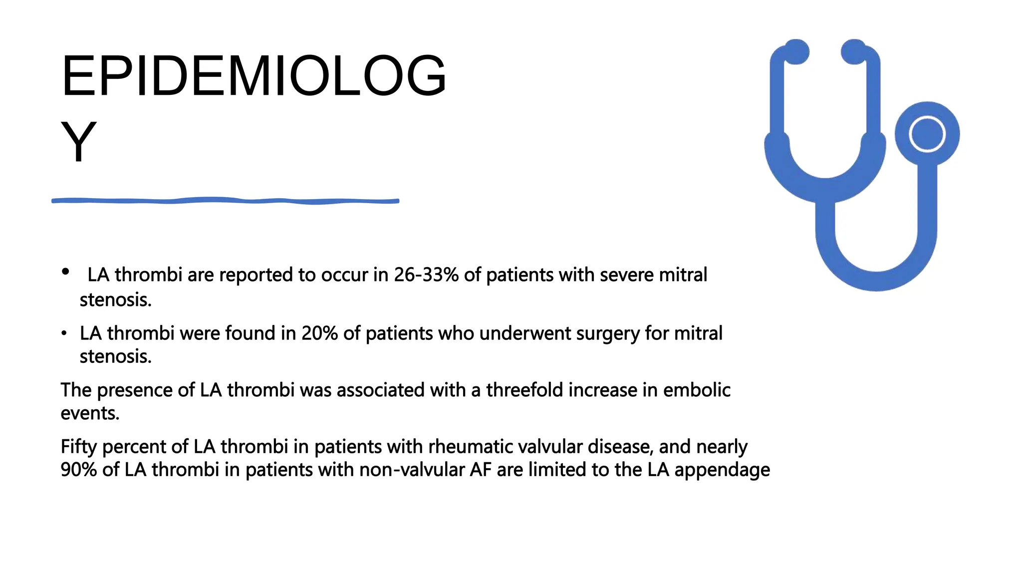
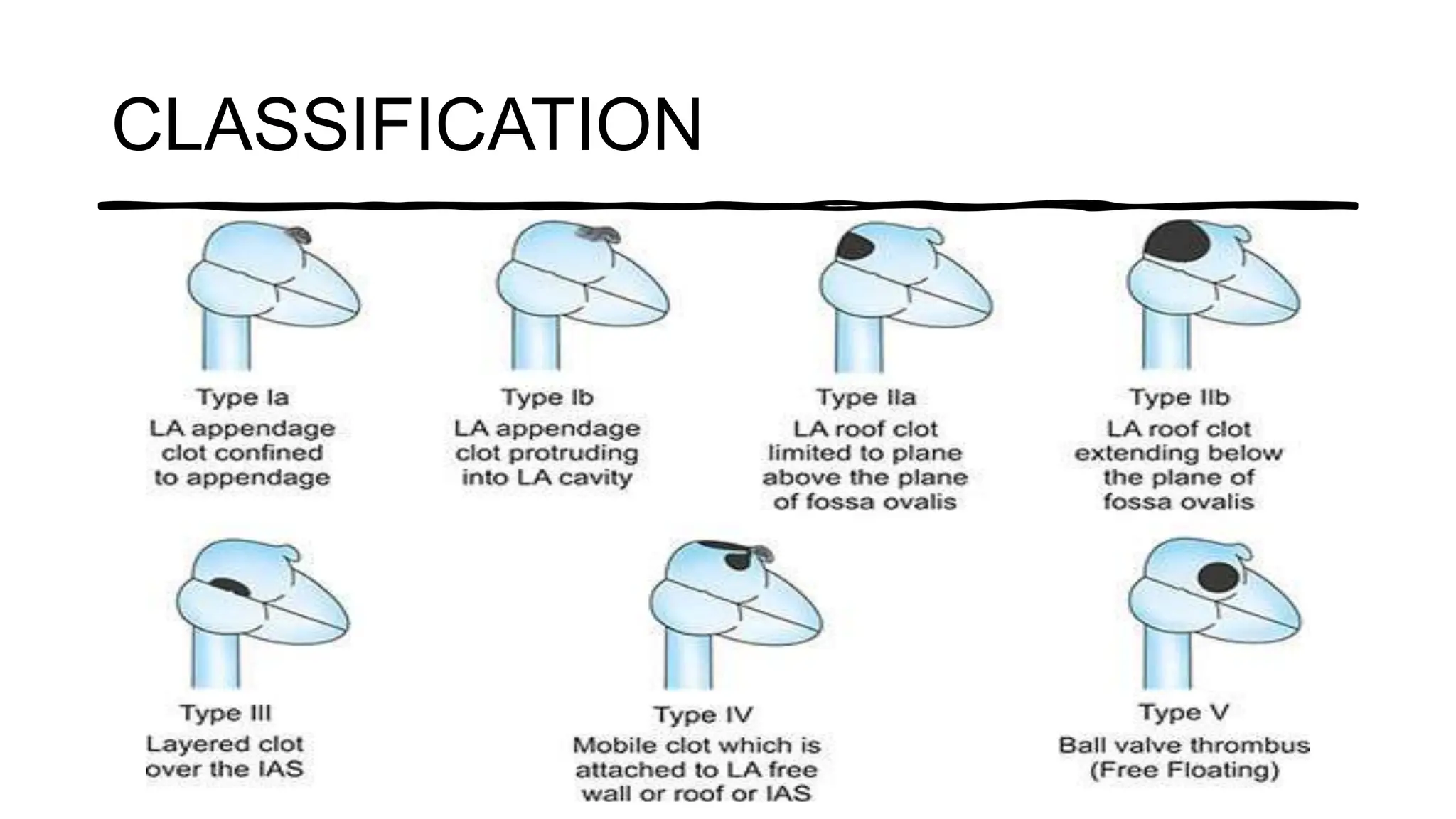
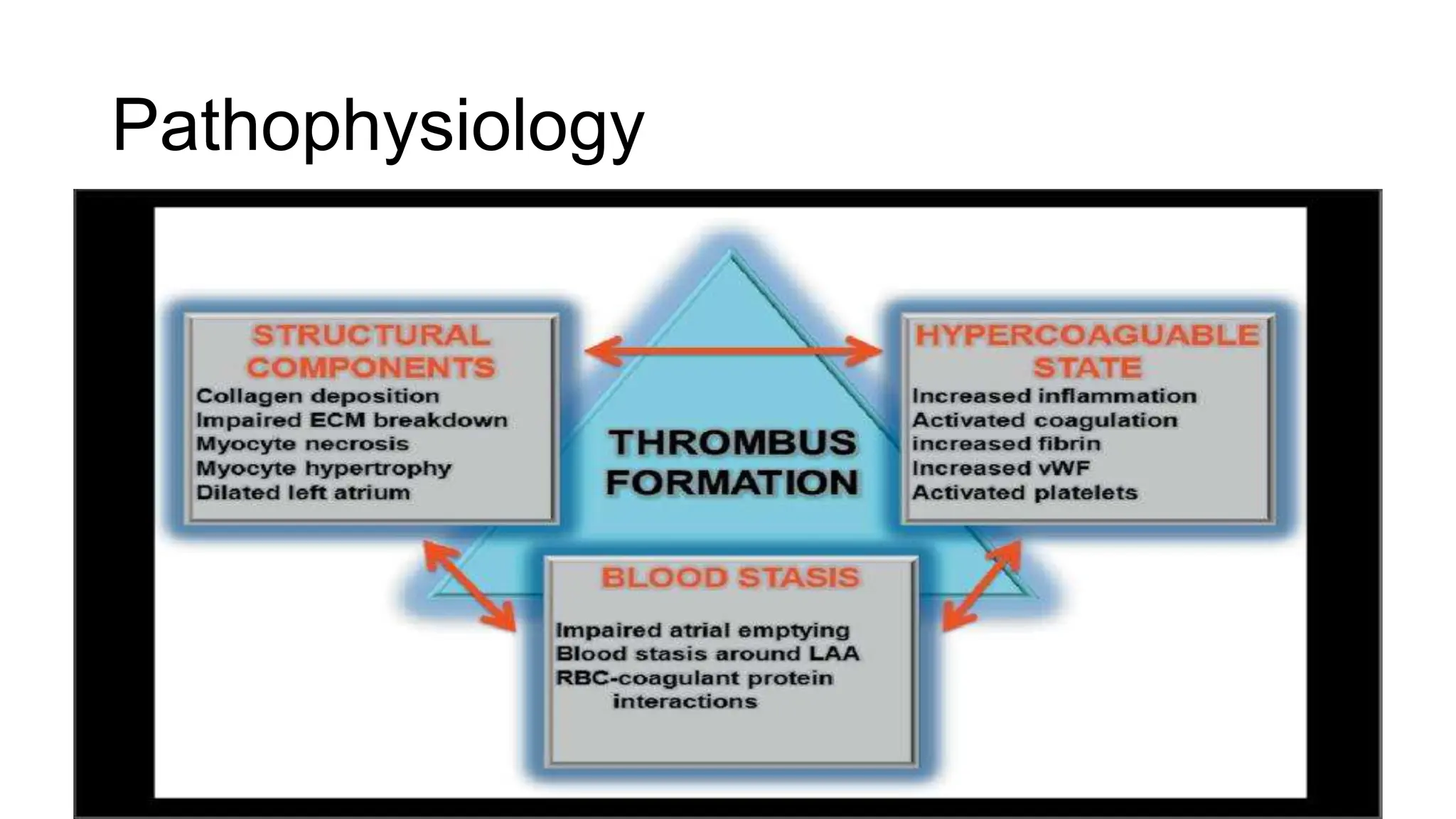
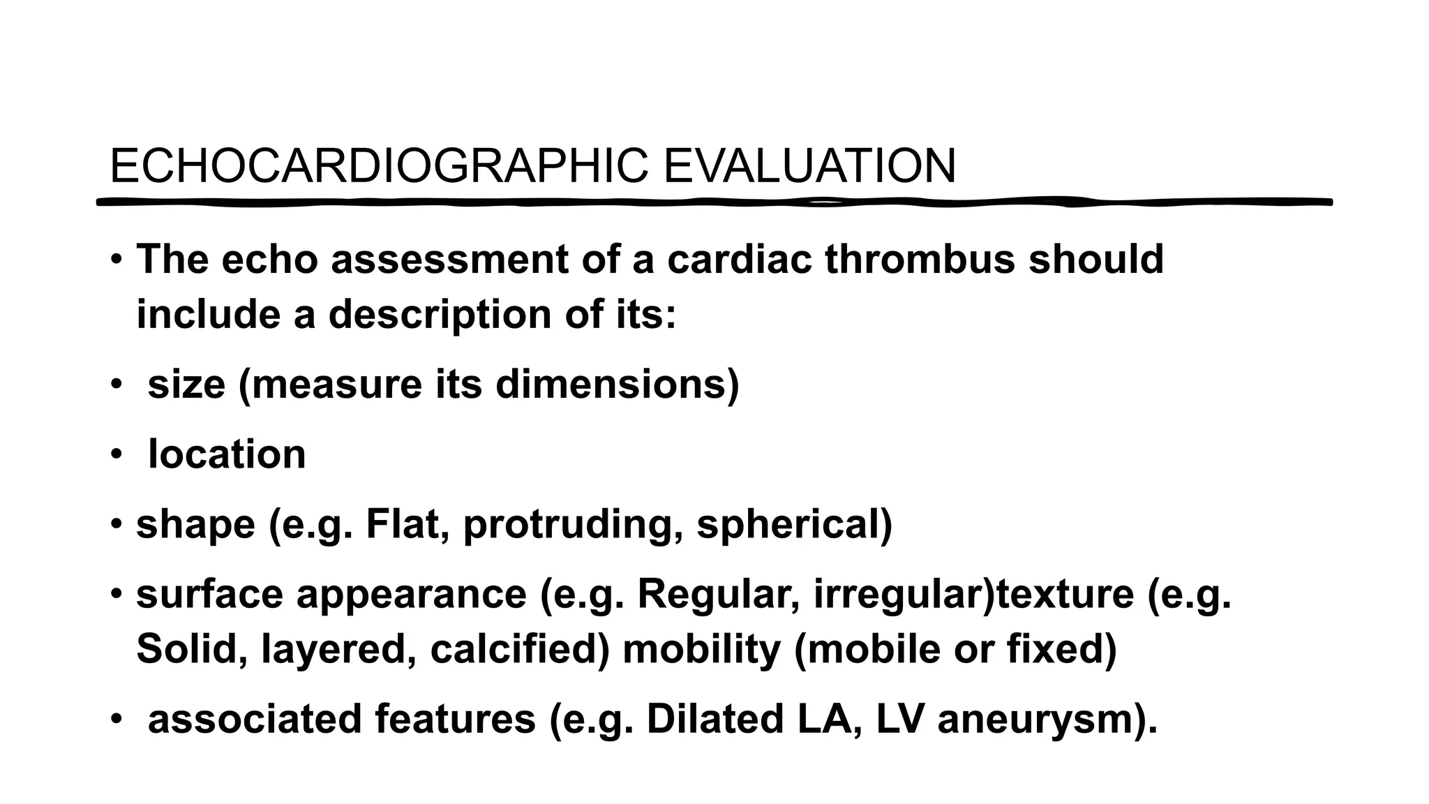
Ok
LA THROMBUS
Ms.Afrin , department of
Cct
Definition
Thrombus is a blood clot that forms inside a circulatory system
that impedes the blood flow
Cardiac thrombus is more likely to form
when there is Stasis or slow
flow of blood
there is abnormal endocardium
allowing thrombus to attach
the blood is hypercoagulable
making it more likely to Clot
DETERMINANT
S
Thrombus formation is therefore more likely in the
LEFT ATRIUM in patients with ATRIAL FIBRILLATION
when there is a loss of normal atrial contraction
left ventricle following myocardial infarction when
reduced Contractility predisposes to thrombus
formation particularly if there is anneurysm formation
the presence of an intra cardiac device such as
prosthetic valve or a pacing wire can also act as a
focus for thrombosis
When there is a high risk of thrombosis anti
coagulation with the drug such as war farin is used
DETERMINANT
S
Various factors determine the individual risk for the development of left
atrial clot and thromboembolizationin patients with rheumaticmitral
valve disease which include Atrial fibrillation
Left atrial size
Duration of symptoms
Older age
Severity of mitral stenosis
However the presence of moderate to severe mitral
regurgitation has a negative predictive value for the
development of clot in the left atrium.
EPIDEMIOLOG
Y
• Patients with severe symptomatic MS, 50% or more
have chronic AF.
• The incidence of thromboembolic complications is
higher in patients with rheumatic mitral stenosis and
is a major cause of morbidity and mortality.
• The left atrium and left atrial appendage are well
recognised sites of clot formation in these patients.
• Patients with a clot in the left atrium and left atrial
appendage had a higher incidence of systemic
embolization.
EPIDEMIOLOG
Y
• LA thrombi are reported to occur in 26-33% of patients with severe mitral
stenosis.
• LA thrombi were found in 20% of patients who underwent surgery for mitral
stenosis.
The presence of LA thrombi was associated with a threefold increase in embolic
events.
Fifty percent of LA thrombi in patients with rheumatic valvular disease, and nearly
90% of LA thrombi in patients with non-valvular AF are limited to the LA appendage
CLASSIFICATIO
N
The thrombus in the left atrium can be
classified as
la (thrombus confined to LAA) lb (thrombus
in LAA and protruding into LA cavity)Ila
(attached to LA roof but above the plane of
fossa ovalis)
IIb (reaching below plane of fossa ovalis) III
(attached to interatrial septum)IV (mobile
with attachment to roof or lateral wall) V (ball
valve thrombus)
CLASSIFICATION
TYPES( Based on morphology )
Pathophysiology
• Rudolf Virchow postulated that
the pathophysiology of thrombus
formation forms a triad which
includes abnormalities of the
vessel wall, blood stasis, and a
hypercoaguable state. This triad
can be applied to thrombus
formation in atrial fibrillation,
Pathophysiology
ECHOCARDIOGRAPHIC EVALUATION
• Transesophageal
Echocardiography and
Intracardiac Echocardiography.
TOE is considered the gold
standard modality for diagnosis of
LAA thrombi with a sensitivity and
specificity of 95%–100%
• The LAA is a small, pyramidal structure, which is situated
on the lateral aspect of the left atrium (LA), extending
between the pulmonary artery above and the left ventricle
(LV) below. It is usually a mul- tilobed structure..
• Although the LAA can be visualized on transthoracic
echocardiography also, in most patients a detailed
assessment is not possible due to the posterior location of
the LAA. In contrast, TEE, with the close proximity of the
transducer to the LAA, allows excellent imaging of the LAA
and is therefore mandatory whenever an as- sessment of
ECHOCARDIOGRAPHIC EVALUATION
TEE VIEWS
• On TEE, the LAA is best visualized in the mid- esophageal
two-chamber view (80-100°) and the mid-esophageal aortic
valve short-axis view (30-60°). In most pa- tients, these two
views allow satisfactory imaging of the LAA and are
therefore the recommended views for this purpose
• The echo assessment of a cardiac thrombus should
include a description of its:
• size (measure its dimensions)
• location
• shape (e.g. Flat, protruding, spherical)
• surface appearance (e.g. Regular, irregular)texture (e.g.
Solid, layered, calcified) mobility (mobile or fixed)
• associated features (e.g. Dilated LA, LV aneurysm).
ECHOCARDIOGRAPHIC EVALUATION
LEFT ATRIUM DIMENSION
• LA diameter is measured at end-systole in the parasternal
long axis view, using either 2D or M-mode imaging
LEFT ATRIUM DIMENSION
ECHOCARDIOGRAPHIC
EVALUATION
• Compared with a myxoma, a thrombus usually has a more
irregular shape.
• Thrombus usually attaches to the endocardium via a broad
base rather than a pedicle, and is consequently less
mobile.
• A large proportion of LA thrombi are within the LA
appendage, which can be difficult to inspect fully on TTE.
• The appendage is, however, clearly seen on TOE. It is
important to try to distinguish between thrombus and the
• Stasis of blood within the heart (and sometimes even the
aorta) can be evident as ‘spontaneous echo contrast.
• This has the appearance of a swirling ‘cloud’ of tiny
particles, hence it is sometimes referred to as ‘smoke’.
• Although it is most often (and mostly clearly) seen during
TOE studies, it can also be observed during TTE.
• Spontaneous contrast is caused by echo reflections from
aggregations of red blood cells moving at low velocity, and
it is most often observed in the LA in patients in AF,
SPONTANEOUS ECHO CONTRAST
THROMBUS IN OTHER CHAMBERS
• Thrombus in the LV normally occurs in association with an
area of wall motion abnormality and/or aneurysm
formation, commonly the apex
• The echo texture of the thrombus is usually distinct from
the adjacent myocardium.
• With the passage of time, the thrombus may become
organized and layered, and there may be associated
calcification.
• TTE is better than TOE for the detection of ventricular
• Thrombus in the right heart is less commonly found, and
may represent a thromboembolism that has arisen in the
peripheral veins and is ‘in transit’ to becoming a pulmonary
embolism.
• Another cause of right heart thrombi is the presence of
devices such as pacing/defibrillator leads or intravascular
catheters, which can act as a focus for thrombus
formation.
THROMBUS IN OTHER CHAMBERS

Left atrial thrombus in Echocardiography

  • 1.
    Ok LA THROMBUS Ms.Afrin ,department of Cct
  • 2.
    Definition Thrombus is ablood clot that forms inside a circulatory system that impedes the blood flow Cardiac thrombus is more likely to form when there is Stasis or slow flow of blood there is abnormal endocardium allowing thrombus to attach the blood is hypercoagulable making it more likely to Clot
  • 3.
    DETERMINANT S Thrombus formation istherefore more likely in the LEFT ATRIUM in patients with ATRIAL FIBRILLATION when there is a loss of normal atrial contraction left ventricle following myocardial infarction when reduced Contractility predisposes to thrombus formation particularly if there is anneurysm formation the presence of an intra cardiac device such as prosthetic valve or a pacing wire can also act as a focus for thrombosis When there is a high risk of thrombosis anti coagulation with the drug such as war farin is used
  • 4.
    DETERMINANT S Various factors determinethe individual risk for the development of left atrial clot and thromboembolizationin patients with rheumaticmitral valve disease which include Atrial fibrillation Left atrial size Duration of symptoms Older age Severity of mitral stenosis However the presence of moderate to severe mitral regurgitation has a negative predictive value for the development of clot in the left atrium.
  • 5.
    EPIDEMIOLOG Y • Patients withsevere symptomatic MS, 50% or more have chronic AF. • The incidence of thromboembolic complications is higher in patients with rheumatic mitral stenosis and is a major cause of morbidity and mortality. • The left atrium and left atrial appendage are well recognised sites of clot formation in these patients. • Patients with a clot in the left atrium and left atrial appendage had a higher incidence of systemic embolization.
  • 6.
    EPIDEMIOLOG Y • LA thrombiare reported to occur in 26-33% of patients with severe mitral stenosis. • LA thrombi were found in 20% of patients who underwent surgery for mitral stenosis. The presence of LA thrombi was associated with a threefold increase in embolic events. Fifty percent of LA thrombi in patients with rheumatic valvular disease, and nearly 90% of LA thrombi in patients with non-valvular AF are limited to the LA appendage
  • 7.
    CLASSIFICATIO N The thrombus inthe left atrium can be classified as la (thrombus confined to LAA) lb (thrombus in LAA and protruding into LA cavity)Ila (attached to LA roof but above the plane of fossa ovalis) IIb (reaching below plane of fossa ovalis) III (attached to interatrial septum)IV (mobile with attachment to roof or lateral wall) V (ball valve thrombus)
  • 8.
  • 9.
    TYPES( Based onmorphology )
  • 10.
    Pathophysiology • Rudolf Virchowpostulated that the pathophysiology of thrombus formation forms a triad which includes abnormalities of the vessel wall, blood stasis, and a hypercoaguable state. This triad can be applied to thrombus formation in atrial fibrillation,
  • 11.
  • 12.
    ECHOCARDIOGRAPHIC EVALUATION • Transesophageal Echocardiographyand Intracardiac Echocardiography. TOE is considered the gold standard modality for diagnosis of LAA thrombi with a sensitivity and specificity of 95%–100%
  • 13.
    • The LAAis a small, pyramidal structure, which is situated on the lateral aspect of the left atrium (LA), extending between the pulmonary artery above and the left ventricle (LV) below. It is usually a mul- tilobed structure.. • Although the LAA can be visualized on transthoracic echocardiography also, in most patients a detailed assessment is not possible due to the posterior location of the LAA. In contrast, TEE, with the close proximity of the transducer to the LAA, allows excellent imaging of the LAA and is therefore mandatory whenever an as- sessment of ECHOCARDIOGRAPHIC EVALUATION
  • 14.
    TEE VIEWS • OnTEE, the LAA is best visualized in the mid- esophageal two-chamber view (80-100°) and the mid-esophageal aortic valve short-axis view (30-60°). In most pa- tients, these two views allow satisfactory imaging of the LAA and are therefore the recommended views for this purpose
  • 15.
    • The echoassessment of a cardiac thrombus should include a description of its: • size (measure its dimensions) • location • shape (e.g. Flat, protruding, spherical) • surface appearance (e.g. Regular, irregular)texture (e.g. Solid, layered, calcified) mobility (mobile or fixed) • associated features (e.g. Dilated LA, LV aneurysm). ECHOCARDIOGRAPHIC EVALUATION
  • 16.
    LEFT ATRIUM DIMENSION •LA diameter is measured at end-systole in the parasternal long axis view, using either 2D or M-mode imaging
  • 17.
  • 18.
    ECHOCARDIOGRAPHIC EVALUATION • Compared witha myxoma, a thrombus usually has a more irregular shape. • Thrombus usually attaches to the endocardium via a broad base rather than a pedicle, and is consequently less mobile. • A large proportion of LA thrombi are within the LA appendage, which can be difficult to inspect fully on TTE. • The appendage is, however, clearly seen on TOE. It is important to try to distinguish between thrombus and the
  • 19.
    • Stasis ofblood within the heart (and sometimes even the aorta) can be evident as ‘spontaneous echo contrast. • This has the appearance of a swirling ‘cloud’ of tiny particles, hence it is sometimes referred to as ‘smoke’. • Although it is most often (and mostly clearly) seen during TOE studies, it can also be observed during TTE. • Spontaneous contrast is caused by echo reflections from aggregations of red blood cells moving at low velocity, and it is most often observed in the LA in patients in AF, SPONTANEOUS ECHO CONTRAST
  • 21.
    THROMBUS IN OTHERCHAMBERS • Thrombus in the LV normally occurs in association with an area of wall motion abnormality and/or aneurysm formation, commonly the apex • The echo texture of the thrombus is usually distinct from the adjacent myocardium. • With the passage of time, the thrombus may become organized and layered, and there may be associated calcification. • TTE is better than TOE for the detection of ventricular
  • 22.
    • Thrombus inthe right heart is less commonly found, and may represent a thromboembolism that has arisen in the peripheral veins and is ‘in transit’ to becoming a pulmonary embolism. • Another cause of right heart thrombi is the presence of devices such as pacing/defibrillator leads or intravascular catheters, which can act as a focus for thrombus formation. THROMBUS IN OTHER CHAMBERS