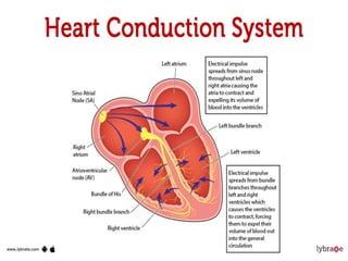
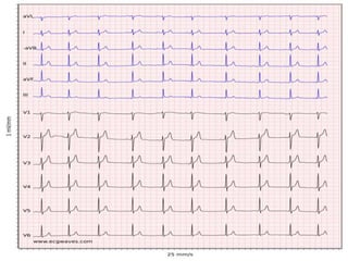
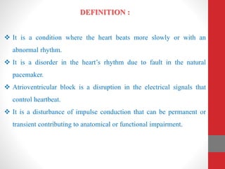
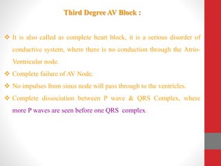
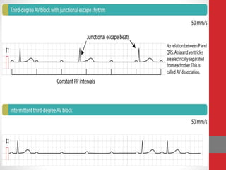
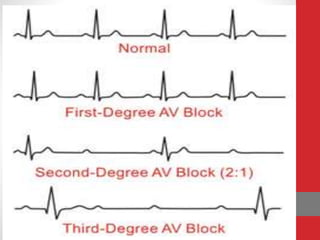
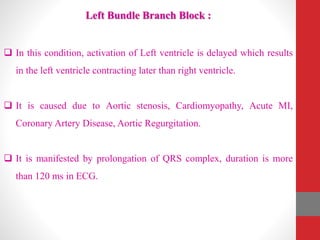
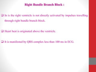
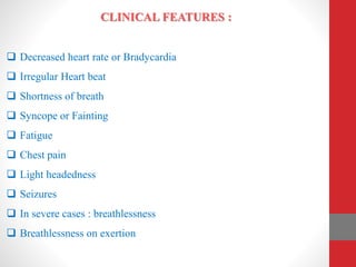
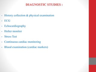
HEART BLOCK
Presented By:
Mr. Nandish.S
Asso. Professor
Mandya Institute of Nursing Sciences
DEFINITION :
 It is a condition where the heart beats more slowly or with an
abnormal rhythm.
 It is a disorder in the heart’s rhythm due to fault in the natural
pacemaker.
 Atrioventricular block is a disruption in the electrical signals that
control heartbeat.
 It is a disturbance of impulse conduction that can be permanent or
transient contributing to anatomical or functional impairment.
ETIOLOGY & RISK FACTORS :
 Medications like digitalis, calcium channel blockers, beta blockers ….
 Lyme disease
 Myocardial Infarction & Ischemia
 Valvular disorders
 Cardiomyopathy
 Endocarditis & Myocarditis
 Increased Vagal Tone.
 Rheumatic Heart Disease
 Hyperthyroidism
TYPES OF HEART BLOCK :
1. First degree AV Block
2. Second Degree AV Block
- Mobitz type I or Wenckebach phenomena
- Mobitz type II AV Block
3. Third Degree AV Block
FIRST DEGREE AV BLOCK :
 It occurs when all the impulses are conducted through AV Node into
the ventricles at a rate slower than normal.
 It is a disease of electrical conduct system in which PR interval is
lengthened than 0.20 seconds.
Causes for First Degree Heart Block :
- Intrinsic AVN Disease
- Acute Myocardial Infarction
- Myocarditis
- Hypokalemia
- Medications (that increase refractory time of AVN)
SECOND DEGREE HEART / AV BLOCK
Type I or Mobitz Type I or Wenckebach phenomena
 It is characterized by disturbance, delay or interruption of atrial
impulse conduction through the AV Node to ventricles.
 PR interval prolongs with each beat until a dropped beat is seen. After
each dropped beat, new cycle begins which is normal.
Causes for Mobitz type I AV Block :
 Cardiomyopathy
 Rheumatic Fever
 Varicella zoster infection
 Hyperkalemia
 Hypothyroidism
 Hypoxia
 Inferior wall MI
 Medications like Beta blockers, calcium channel blockers,
Amiodarone…
Type II or Mobitz type II AV Block :
 It occurs when only some of the atrial impulses are conducted
through AV Node into ventricles.
 PR interval is usually regular & constant, but may be irregular.
 Occasionally a dropped beat is seen.
 It is more significant disease.
Third Degree AV Block :
 It is also called as complete heart block, it is a serious disorder of
conductive system, where there is no conduction through the Atrio-
Ventricular node.
 Complete failure of AV Node.
 No impulses from sinus node will pass through to the ventricles.
 Complete dissociation between P wave & QRS Complex, where
more P waves are seen before one QRS complex.
Types of Infra – Hisian Block :
- Left Bundle Branch Block
a. Left Anterior Fascicular Block
b. Left Posterior Fascicular Block
- Right Bundle Branch Block
Left Bundle Branch Block :
 In this condition, activation of Left ventricle is delayed which results
in the left ventricle contracting later than right ventricle.
 It is caused due to Aortic stenosis, Cardiomyopathy, Acute MI,
Coronary Artery Disease, Aortic Regurgitation.
 It is manifested by prolongation of QRS complex, duration is more
than 120 ms in ECG.
Right Bundle Branch Block :
 In is the right ventricle is not directly activated by impulses travelling
through right bundle branch block.
 Heart beat is originated above the ventricle.
 It is manifested by QRS complex less than 100 ms in ECG.
CLINICAL FEATURES :
 Decreased heart rate or Bradycardia
 Irregular Heart beat
 Shortness of breath
 Syncope or Fainting
 Fatigue
 Chest pain
 Light headedness
 Seizures
 In severe cases : breathlessness
 Breathlessness on exertion
DIAGNOSTIC STUDIES :
- History collection & physical examination
- ECG
- Echocardiography
- Holter monitor
- Stress Test
- Continuous cardiac monitoring
- Blood examination (cardiac markers)
Management :
- For asymptomatic patients, decreasing or eliminating the cause is
planned by holding the medications.
- For symptomatic patients, medications such as Atropine, Epinephrine,
Isoproterenol, Dopamine are given to increase BP and Heart rate.
- Temporary Pacing can be initiated for advanced AV Block, acute MI.
- Permanent Pacemaker is planned if the block persists.
PACE MAKER THERAPY
Definition :
o It is an electric instrument that provides electrical stimuli to the heart
muscle.
o They are usually used when a patient has a slower than normal
impulse formation or a conduction disturbances that cause symptoms.
Types :
 Temporary
 Permanent
Temporary Pacemaker :
It is used until long term therapy can be initiated.
Indications :
• Cardiac catheterization
• Coronary Angioplasty
• Before implantation of permanent Pace maker
• After open heart surgery
• Acute Anterior MI with 2nd or 3rd Degree block
• Acute Inferior MI
• Ventricular Tachycardia
• Atrial Flutter
• Brady or Tachy Arrhythmias
Types of Temporary Pacemaker
- Transcutaneous pacing (External)
- Transvenous pacing
- Transthoracic pacing
- Transesophageal pacing
Transcutaneous Cardiac Pacing :
• It is a non invasive & delivers electricity from the external power
source.
• This causes the depolarization of excitable myocardial tissue by
pulsed electrical current conducted through the chest wall, between
electrodes adherent to the skin.
Transvenous Pacing :
An intravenous catheter electrode is positioned endocardially through
subclavian or external Jugular venous route and this is then connected to
an external Generator by a lead connector.
Types :
 Bipolar Electrodes – both anode & cathode are intra thoracic.
 Unipolar – anode is extra thoracic.
Insertion Site :
- Right External Jugular vein - Left Subclavian vein
- Femoral vein - Brachial vein.
Transthoracic Cardiac Pacing :
 It is a technique of pacing the heart with an electrode introduced
percutaneously into the ventricular cavity using a needle Trocar
introducer.
 It was used in patients with acute unstable dysrhythmia until
transcutaneous pacing is introduced.
Trans esophageal pacing :
Trans esophageal pacing & recording is done by using specialized or
simple catheters.
There are 2 types.
- The pill electrode, connected to flexible wire that patient swallows
with water, which needs patient's collaboration.
- A flexible catheter that can be used in unconscious or intubated
patients.
Bipolar flexible catheter is introduced into esophagus through nose after
local anaesthesia / throat anaesthesia. Lead is introduced with guide
wires. It is positioned into esophagus in order to record posterior
paraseptal atrial electro gram.
Complications :
 Haemo or Pneumothorax
 Rupture of major blood vessels
 Perforation of right ventricle
 VT or VF
 Cardiac Arrest
 Heart Failure
Heart Block.pptx

Heart Block.pptx

  • 1.
    HEART BLOCK Presented By: Mr.Nandish.S Asso. Professor Mandya Institute of Nursing Sciences
  • 6.
    DEFINITION :  Itis a condition where the heart beats more slowly or with an abnormal rhythm.  It is a disorder in the heart’s rhythm due to fault in the natural pacemaker.  Atrioventricular block is a disruption in the electrical signals that control heartbeat.  It is a disturbance of impulse conduction that can be permanent or transient contributing to anatomical or functional impairment.
  • 7.
    ETIOLOGY & RISKFACTORS :  Medications like digitalis, calcium channel blockers, beta blockers ….  Lyme disease  Myocardial Infarction & Ischemia  Valvular disorders  Cardiomyopathy  Endocarditis & Myocarditis  Increased Vagal Tone.  Rheumatic Heart Disease  Hyperthyroidism
  • 8.
    TYPES OF HEARTBLOCK : 1. First degree AV Block 2. Second Degree AV Block - Mobitz type I or Wenckebach phenomena - Mobitz type II AV Block 3. Third Degree AV Block
  • 9.
    FIRST DEGREE AVBLOCK :  It occurs when all the impulses are conducted through AV Node into the ventricles at a rate slower than normal.  It is a disease of electrical conduct system in which PR interval is lengthened than 0.20 seconds.
  • 10.
    Causes for FirstDegree Heart Block : - Intrinsic AVN Disease - Acute Myocardial Infarction - Myocarditis - Hypokalemia - Medications (that increase refractory time of AVN)
  • 11.
    SECOND DEGREE HEART/ AV BLOCK Type I or Mobitz Type I or Wenckebach phenomena  It is characterized by disturbance, delay or interruption of atrial impulse conduction through the AV Node to ventricles.  PR interval prolongs with each beat until a dropped beat is seen. After each dropped beat, new cycle begins which is normal.
  • 12.
    Causes for Mobitztype I AV Block :  Cardiomyopathy  Rheumatic Fever  Varicella zoster infection  Hyperkalemia  Hypothyroidism  Hypoxia  Inferior wall MI  Medications like Beta blockers, calcium channel blockers, Amiodarone…
  • 13.
    Type II orMobitz type II AV Block :  It occurs when only some of the atrial impulses are conducted through AV Node into ventricles.  PR interval is usually regular & constant, but may be irregular.  Occasionally a dropped beat is seen.  It is more significant disease.
  • 14.
    Third Degree AVBlock :  It is also called as complete heart block, it is a serious disorder of conductive system, where there is no conduction through the Atrio- Ventricular node.  Complete failure of AV Node.  No impulses from sinus node will pass through to the ventricles.  Complete dissociation between P wave & QRS Complex, where more P waves are seen before one QRS complex.
  • 17.
    Types of Infra– Hisian Block : - Left Bundle Branch Block a. Left Anterior Fascicular Block b. Left Posterior Fascicular Block - Right Bundle Branch Block
  • 18.
    Left Bundle BranchBlock :  In this condition, activation of Left ventricle is delayed which results in the left ventricle contracting later than right ventricle.  It is caused due to Aortic stenosis, Cardiomyopathy, Acute MI, Coronary Artery Disease, Aortic Regurgitation.  It is manifested by prolongation of QRS complex, duration is more than 120 ms in ECG.
  • 19.
    Right Bundle BranchBlock :  In is the right ventricle is not directly activated by impulses travelling through right bundle branch block.  Heart beat is originated above the ventricle.  It is manifested by QRS complex less than 100 ms in ECG.
  • 20.
    CLINICAL FEATURES : Decreased heart rate or Bradycardia  Irregular Heart beat  Shortness of breath  Syncope or Fainting  Fatigue  Chest pain  Light headedness  Seizures  In severe cases : breathlessness  Breathlessness on exertion
  • 21.
    DIAGNOSTIC STUDIES : -History collection & physical examination - ECG - Echocardiography - Holter monitor - Stress Test - Continuous cardiac monitoring - Blood examination (cardiac markers)
  • 22.
    Management : - Forasymptomatic patients, decreasing or eliminating the cause is planned by holding the medications. - For symptomatic patients, medications such as Atropine, Epinephrine, Isoproterenol, Dopamine are given to increase BP and Heart rate. - Temporary Pacing can be initiated for advanced AV Block, acute MI. - Permanent Pacemaker is planned if the block persists.
  • 23.
    PACE MAKER THERAPY Definition: o It is an electric instrument that provides electrical stimuli to the heart muscle. o They are usually used when a patient has a slower than normal impulse formation or a conduction disturbances that cause symptoms. Types :  Temporary  Permanent
  • 24.
    Temporary Pacemaker : Itis used until long term therapy can be initiated. Indications : • Cardiac catheterization • Coronary Angioplasty • Before implantation of permanent Pace maker • After open heart surgery • Acute Anterior MI with 2nd or 3rd Degree block • Acute Inferior MI • Ventricular Tachycardia • Atrial Flutter • Brady or Tachy Arrhythmias
  • 25.
    Types of TemporaryPacemaker - Transcutaneous pacing (External) - Transvenous pacing - Transthoracic pacing - Transesophageal pacing
  • 26.
    Transcutaneous Cardiac Pacing: • It is a non invasive & delivers electricity from the external power source. • This causes the depolarization of excitable myocardial tissue by pulsed electrical current conducted through the chest wall, between electrodes adherent to the skin.
  • 27.
    Transvenous Pacing : Anintravenous catheter electrode is positioned endocardially through subclavian or external Jugular venous route and this is then connected to an external Generator by a lead connector. Types :  Bipolar Electrodes – both anode & cathode are intra thoracic.  Unipolar – anode is extra thoracic. Insertion Site : - Right External Jugular vein - Left Subclavian vein - Femoral vein - Brachial vein.
  • 30.
    Transthoracic Cardiac Pacing:  It is a technique of pacing the heart with an electrode introduced percutaneously into the ventricular cavity using a needle Trocar introducer.  It was used in patients with acute unstable dysrhythmia until transcutaneous pacing is introduced.
  • 31.
    Trans esophageal pacing: Trans esophageal pacing & recording is done by using specialized or simple catheters. There are 2 types. - The pill electrode, connected to flexible wire that patient swallows with water, which needs patient's collaboration. - A flexible catheter that can be used in unconscious or intubated patients. Bipolar flexible catheter is introduced into esophagus through nose after local anaesthesia / throat anaesthesia. Lead is introduced with guide wires. It is positioned into esophagus in order to record posterior paraseptal atrial electro gram.
  • 32.
    Complications :  Haemoor Pneumothorax  Rupture of major blood vessels  Perforation of right ventricle  VT or VF  Cardiac Arrest  Heart Failure