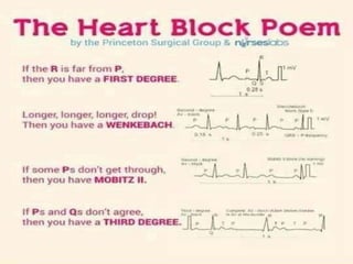
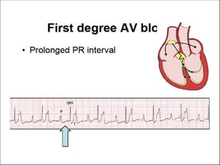
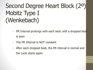
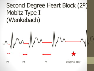
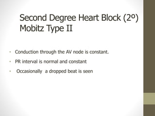
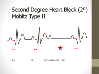
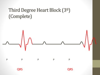
HEART BLOCK
RATHEESH R.L
Normal conduction pathway:
SA node -> atrial muscle -> AV node -> bundle of His ->
Left and Right Bundle Branches -> Ventricular muscle
DEFINITION
Heart block is an abnormal heart rhythm where
the heart beats too slowly (bradycardia).
In this condition, the electrical signals that tell the heart to
contract are partially or totally blocked between the upper
chambers (atria) and the lower chambers (ventricles).
TYPES
• They are further classified as,
First degree heart block ( first degree AV
block)
 second degree heart block (second degree AV
block)
 third degree heart block (third degree AV
block)
FIRST DEGREE HEART BLOCK
• First-degree atrio-ventricular block (AV block), or PR
prolongation, is a disease of the electrical conduction
system of the heart in which the PR interval is
lengthened beyond 0.20 seconds.
The following are the most common causes of first-
degree AV block:
• Intrinsic AVN disease
• Acute myocardial infarction (MI), particularly acute
inferior wall MI
• Myocarditis
• Electrolyte disturbances (eg, hypokalemia,
hypomagnesemia)
• Drugs (especially those drugs that increase the refractory
time of the AVN, thereby slowing conduction)
First Degree Heart Block (1º)
• SA Node – normal
• Normal P wave
• AV Node conducts more slowly than normal
• Prolonged PR Interval
• Rest of conduction is normal
• Normal QRS
Significance
• Clinical significance
• None
• Treatment
• None
• Note – this can progress to 2º or 3º heart block
Second Degree Heart Block (2º)
• Mobitz Type I (Wenkebach)
• Mobitz Type II
Second Degree Heart Block (2º)
Mobitz Type I
(Wenkebach)
• Conduction through the AV Node – progressively delayed until
a drop beat is seen
Second-degree atrio-ventricular (AV) block, or
second-degree heart block, is characterized by
disturbance, delay, or interruption of atrial impulse
conduction through the AV node to the ventricles.
CAUSES
• Drugs (beta-blockers, calcium channel blockers,
amiodarone)
• Cardiomyopathy
• rheumatic fever, myocarditis
• varicella-zoster virus infection
• Rheumatic diseases
• Hypoxia
• Hyperkalemia
• Hypothyroidism
• inferior wall myocardial infarction
Second Degree Heart Block (2º)
Mobitz Type I
(Wenkebach)
• PR Interval prolongs with each beat until a dropped beat
is seen
• The PR Interval is NOT constant
• After each dropped beat, the PR interval is normal and
the cycle starts again
Second Degree Heart Block (2º)
Mobitz Type I
(Wenkebach)
PR PR PR DROPPED BEAT
Significance
• Clinical Significance
• Slight symptoms e.g.. Lethargy,
Confusion
• Treatment
• Pacemaker if during day &/or
symptoms
• No treatment if at night
• Note – this can progress to 3º Heart Block
Second Degree Heart Block (2º)
Mobitz Type II
• Conduction through the AV node is constant.
• PR interval is normal and constant
• Occasionally a dropped beat is seen
Second Degree Heart Block (2º)
Mobitz Type II
PR PR DROPPED BEAT PR
Significance
• Clinical significance – this is more significant disease
• Treatment – pacemaker
• Note – this can progress to 3º Heart Block
Third Degree Heart Block (3º)
(Complete)
• Complete failure of the AV Node
• No impulses from Sinus Node will pass through to the
ventricles
• Some part if the conducting system will take over as
pacemaker of the heart (even a myocardial cell 10-15
bpm)
Third-degree atrioventricular (AV) block, also referred to
as third-degree heart block or complete heart block, is a
disorder of the cardiac conduction system where there is
no conduction through the atrioventricular node.
Third Degree Heart Block (3º)
(Complete)
• P wave rate – normal
• Ventricular rate – slow
• Ventricular complex may be broad
• Idioventricular rhythm
• Complete dissociation between P waves & QRS
Third Degree Heart Block (3º)
(Complete)
P P P P P
QRS QRS
Significance
• clinical significance
• Symptoms LOC, Confusion,
Dizziness, Low BP
• Can lead to standstill, VT or VF
(stokes Adams)
• Treatment - pacemaker
NURSING MANAGEMENT
1. Assess the high risk patients
2. Monitor ECG of the patient
3. Assess the family history of heart disease
4. Assess the history of smoking and alcoholism
5. Monitor lab values frequently especially serum
cholesterol levels.
6. Assess for CAD
7. Monitor vital signs
8. Instruct to avoid high fat and oil rich diet
Heart block

Heart block

  • 1.
  • 4.
    Normal conduction pathway: SAnode -> atrial muscle -> AV node -> bundle of His -> Left and Right Bundle Branches -> Ventricular muscle
  • 9.
    DEFINITION Heart block isan abnormal heart rhythm where the heart beats too slowly (bradycardia). In this condition, the electrical signals that tell the heart to contract are partially or totally blocked between the upper chambers (atria) and the lower chambers (ventricles).
  • 10.
    TYPES • They arefurther classified as, First degree heart block ( first degree AV block)  second degree heart block (second degree AV block)  third degree heart block (third degree AV block)
  • 12.
    FIRST DEGREE HEARTBLOCK • First-degree atrio-ventricular block (AV block), or PR prolongation, is a disease of the electrical conduction system of the heart in which the PR interval is lengthened beyond 0.20 seconds.
  • 14.
    The following arethe most common causes of first- degree AV block: • Intrinsic AVN disease • Acute myocardial infarction (MI), particularly acute inferior wall MI • Myocarditis • Electrolyte disturbances (eg, hypokalemia, hypomagnesemia) • Drugs (especially those drugs that increase the refractory time of the AVN, thereby slowing conduction)
  • 15.
    First Degree HeartBlock (1º) • SA Node – normal • Normal P wave • AV Node conducts more slowly than normal • Prolonged PR Interval • Rest of conduction is normal • Normal QRS
  • 16.
    Significance • Clinical significance •None • Treatment • None • Note – this can progress to 2º or 3º heart block
  • 17.
    Second Degree HeartBlock (2º) • Mobitz Type I (Wenkebach) • Mobitz Type II
  • 18.
    Second Degree HeartBlock (2º) Mobitz Type I (Wenkebach) • Conduction through the AV Node – progressively delayed until a drop beat is seen
  • 19.
    Second-degree atrio-ventricular (AV)block, or second-degree heart block, is characterized by disturbance, delay, or interruption of atrial impulse conduction through the AV node to the ventricles.
  • 20.
    CAUSES • Drugs (beta-blockers,calcium channel blockers, amiodarone) • Cardiomyopathy • rheumatic fever, myocarditis • varicella-zoster virus infection • Rheumatic diseases • Hypoxia • Hyperkalemia • Hypothyroidism • inferior wall myocardial infarction
  • 21.
    Second Degree HeartBlock (2º) Mobitz Type I (Wenkebach) • PR Interval prolongs with each beat until a dropped beat is seen • The PR Interval is NOT constant • After each dropped beat, the PR interval is normal and the cycle starts again
  • 22.
    Second Degree HeartBlock (2º) Mobitz Type I (Wenkebach) PR PR PR DROPPED BEAT
  • 23.
    Significance • Clinical Significance •Slight symptoms e.g.. Lethargy, Confusion • Treatment • Pacemaker if during day &/or symptoms • No treatment if at night • Note – this can progress to 3º Heart Block
  • 24.
    Second Degree HeartBlock (2º) Mobitz Type II • Conduction through the AV node is constant. • PR interval is normal and constant • Occasionally a dropped beat is seen
  • 25.
    Second Degree HeartBlock (2º) Mobitz Type II PR PR DROPPED BEAT PR
  • 26.
    Significance • Clinical significance– this is more significant disease • Treatment – pacemaker • Note – this can progress to 3º Heart Block
  • 27.
    Third Degree HeartBlock (3º) (Complete) • Complete failure of the AV Node • No impulses from Sinus Node will pass through to the ventricles • Some part if the conducting system will take over as pacemaker of the heart (even a myocardial cell 10-15 bpm)
  • 28.
    Third-degree atrioventricular (AV)block, also referred to as third-degree heart block or complete heart block, is a disorder of the cardiac conduction system where there is no conduction through the atrioventricular node.
  • 29.
    Third Degree HeartBlock (3º) (Complete) • P wave rate – normal • Ventricular rate – slow • Ventricular complex may be broad • Idioventricular rhythm • Complete dissociation between P waves & QRS
  • 30.
    Third Degree HeartBlock (3º) (Complete) P P P P P QRS QRS
  • 31.
    Significance • clinical significance •Symptoms LOC, Confusion, Dizziness, Low BP • Can lead to standstill, VT or VF (stokes Adams) • Treatment - pacemaker
  • 32.
    NURSING MANAGEMENT 1. Assessthe high risk patients 2. Monitor ECG of the patient 3. Assess the family history of heart disease 4. Assess the history of smoking and alcoholism 5. Monitor lab values frequently especially serum cholesterol levels. 6. Assess for CAD 7. Monitor vital signs 8. Instruct to avoid high fat and oil rich diet