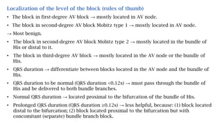
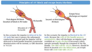
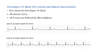
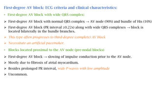
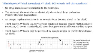
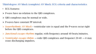
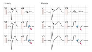
Conduction Defects
(Disturbances)
HOANG CUONG MS-V
HaNoi Medical University
HaNoi, November 03, 2018
Source: ECGwaves.com
Overview of Atrioventricular (AV) blocks
• Impulse conduction from the atria to the ventricles → abnormally delayed or even
blocked.
• Atrioventricular (AV) blocks → sub-divided according to the degree of block: First-,
second-,third-degree AV block.
• First-degree AV block (synonyms: AV block 1, AV block I, 1st degree AV block):
 Abnormally slow (PR interval >0.22s)
 All impulse are conducted to the ventricles
 Rareky serious and may be left untreated in the vast majority of cases.
• Second-degree AV block (synonyms: AV block 2, AV block II, 2nd degree AV block)
 Completely blocked
 Not all P-waves are followed by QRS complexes.
 Two variants: Mobitz type 1 (Wenckebach block) and Mobitz type 2.
 Second-degree AV block (particularly Mobitz type 2) mandates treatment.
• Third-degree AV block (synonyms: complete heart block, AV dissociation, AV
block III, AV block 3)
 No atrial impulses are conducted to the ventricles.
 The atrial and the ventricles → electrically dissociated from each other.
 Atrioventricular (AV) dissociation
 Ventricular rhythm – an escape rhythm must arise in an ectopic escape (located
distal to the block)
 Very serious condition, cardiac arrest occur if no escape rhthym arises.
Causes of AV blocks
• AV blocks occur due to functional or anatomical blcoks in the AV system.
• The block may be located in AV node, His bundle, bundle branches and/or fascicles.
• A wide range of conditions may cause AV blocks:
 Idiopathic fibrosis of the conduction system (50%)
 Ischemic heart disease (35%): Inferior MI → transient AV block (resolve within 7d),
anterior MI → permanent AV blocks.
 Vagal stimulation: carotid sinus massage, Valsalva maneuver, acute pain and
hypersensitive carotid sinus reflex.
 Structural heart disease: aortic stenosis, aortic regurgitation, mitral valve stenosis,
mitral valve regurgitation, myocarditis, perimyocarditis, myocardial infarction, heart
surgery and cardiomyopathy…
 Hyperkalemia, hypokalemia
 Digoxin
 Verapamil, amiodarone, beta-blockers.
Localization of the level of the block (rules of thumb)
• The block in first-degree AV block → mostly located in AV node.
• The block in second-degree AV block Mobitz type 1 → mostly located in AV node.
→ Most benign.
• The block in second-degree AV block Mobitz type 2 → mostly located in the bundle of
His or distal to it.
• The block in third-degree AV block → mostly located in the AV node or the bundle of
His.
• QRS duration → differentiate between blocks located in the AV node and the bundle of
His.
• QRS duration to be normal (QRS duration <0.12s) → must pass through the bundle of
His and be delivered to both bundle branches.
• Normal QRS duration → located proximal to the bifurcation of the bundle of His.
• Prolonged QRS duration (QRS duration ≥0.12s) → less helpful, because: (1) block located
distal to the bifurcation; (2) block located proximal to the bifurcation but with
concomitant (separate) bundle branch block.
In this scenario the impulse is delayed in the
AV node but once it passes the subsequent
conduction is normal and the impulse will be
delivered to both bundle branches (ventricular
depolarization will be normal, i.e QRS duration
is <0.12s)
In this scenario the impulse is blocked in the AV
node. Because the cells of His bundle possess
automaticity, they will discharge an impulse (and
establish an escape rhythm if necessary) which
spreads normally though the remained of the His
bundle. The QRS will be <0.12s. However, should be
block be located after the bifurcation, an ectopic
focus will yield wide QRS ≥0.12s
Principles of AV block and escape beats/rhythms
First-degree AV block: ECG criteria and clinical characteristics:
• ECG criteria for first-degree AV block:
 PR interval ≥0.22s.
 All P-waves are followed by QRS complexes.
First-degree AV block: ECG criteria and clinical characteristics:
• First-degree AV block with wide QRS complex:
 First-degree AV block with normal QRS complex → AV node (90%) and bundle of His (10%)
 First-degree AV block (PR interval ≥0.22s) along with wide QRS complexes → block is
located bilaterally in the bundle branches.
 This type often progresses to third-degree (complete) AV block
 Necessitate an artificial pacemaker.
• Blocks located proximal to the AV node (pre-nodal blocks)
 First-degree AV block → slowing of impulse conduction prior to the AV node.
 Mostly due to fibrosis of atrial myocardium.
 Besides prolonged PR interval, wide P-waves with low amplitude
 Uncommon.
Second-degree (2nd degree) AV block: ECG criteria and characteristics
• Some atrial impulses → completely blocked → not all P-waves are followed by QRS
complexes.
• Subdivided into type 1 and type 2 (Mobitz type 1 and Mobitz type 2)
• Second-degree AV block Mobitz type I exhibits the Wenckebach phenomenon: ECG signs
of gradual exhaustion of impulse conduction → Gradual increase of PR interval before a
block occurs.
• Second-degree AV block Mobitz type II → Sporadically occurring blocks, without any
Wenckebach phenomenon.
• Electrophysiology of second-degree AV block Mobitz type 1:
 A tendency to exhaust the conduction capacity.
 AV node is dysfunctional → not be able to repolarize adequately by the time the next
impulse arrives → more and more exhausted each time until it is completely refractory and
blocks the atrial impulse.
Second-degree (2nd degree) AV block: ECG criteria and characteristics
• Second-degree AV block Mobitz type I (Wenckebach block)
 Mobitz type 1 block is characterized by a gradual prolongation of the PR interval over a
few heart cycles until an atrial impulse is completely blocked → P-wave not followed by a
QRS complex.
 The degree of block → counting the number of P-waves before each block. If every third P-
wave is blocked → 3-to-2 block (most common).
 In 2-to-1 block → may be impossible to observe and PR prolongation.
 Very simple rule of thumb → varying PR intervals → Mobitz 1 (Wenckebach block)
Second-degree (2nd degree) AV block: ECG criteria and characteristics
• Second-degree AV block Mobitz type II
 Atrial impulse are blocked sporadically.
 PR interval → constant.
 Block located in the bundle of His (~20%), and bundle branches (~80%).
 More serious, chronic and tends to progress to third-degree AV block.
 Necessitates an artificial pacemaker.
Differentiate Mobitz type 1 block from Mobitz type 2 block:
 The hallmark of Mobitz type 1 block → gradual prolongation of PR intervals before a block
occurs.
 Mobitz type 2 → constant PR intervals before blocks occur.
 The following situations may pose a diagnostic challenge:
• If the PR prolongation is minimal before a P-wave is blocked.
• If many impulses are blocked (2-to-1 block), because the PR prolongation becomes difficult
to discern.
 Additional tools to differentiate the two:
• Atropin or physical activity → increases heart rate.
• Vagal stimulation: increased block.
• If the PR interval is prolonged → Mobitz type 1 block is more likely.
• If the QRS complexes are abnormal → Mobitz type 2 block is more likely.
Third-degree AV block (complete) AV block: ECG criteria and characteristics
• No atrial impulses are conducted to the ventricles.
• The atria and the ventricles → electrically dissociated from each other
(Atrioventricular dissociation)
• An escape rhythm must arise in an ectopic focus (located distal to the block)
• Third-degree AV block is a very serious condition because escape rhythms may (1)
not occur, (2) occur transiently, (3) occur but generate insufficient cardiac output.
• Third-degree AV block may be preceded by second-degree or (rarely) first-degree
AV block.
Third-degree AV block (complete) AV block: ECG criteria and characteristics
• ECG features:
 P-wave have no relation to the QRS complexes.
 QRS complexes may be normal or wide.
 P-waves have constant PP interval.
 Isoarrhythmic AV block – ventricular rate is equal and the P-waves occur right
before the QRS complexes.
 Junctional escape rhythm: regular, with frequency around 40 beats/minutes.
 Ventricular escape rhthm → wide QRS complexes and frequenct 20-40 → it may
cease discharging impulses.
Seperating AV block 3 from AV block 2 using ECG
• Irregular ventricular rhythm → Second-degree AV block because escape rhthms
in third-degree AV block are regular.
• Regular ventricular rhthm with association between P and QRS and constant PR
interval → second-degree AV block.
• Regular ventricular rhthm and varying PR interval → third-degree AV block.
Treatment of AV blocks:
• Treatment of AV block in the acute setting:
 Managing bradycardia and reduced cardiac output.
 Atropin 0.5 mg i.v (may be repeated). Atropin will have effect if the block is
located in the AV node, may aggravate the block if it is located distal to the AV
node.
 Isoprenaline (isoproterenol, 5μg/min)→ may be administered (caution in case of
acute coronary syndromes→ trigger ventricular tachycardia).
 If sinus bradycardia or asystole (despite attempts with atropine and isoprenaline)
→ Transcutaneous or transvenous pacemaker → implemented.
• Long-term treatment of AV block: permanent artificial pacemaker:
 First-degree AV block and second-degree AV block Mobitz type I: Only
necessitates pacemaker if symptomatic.
 Second-degree AV block Mobitz type II and third-degree AV block: almost
invariably receive a pacemaker.
Causes of AV blocks
• AV blocks occur due to functional or anatomical blcoks in the AV system.
• The block may be located in AV node, His bundle, bundle branches and/or fascicles.
• A wide range of conditions may cause AV blocks:
 Idiopathic fibrosis of the conduction system (50%)
 Ischemic heart disease (35%): Inferior MI → transient AV block (resolve within 7d),
anterior MI → permanent AV blocks.
 Vagal stimulation: carotid sinus massage, Valsalva maneuver, acute pain and
hypersensitive carotid sinus reflex.
 Structural heart disease: aortic stenosis, aortic regurgitation, mitral valve stenosis,
mitral valve regurgitation, myocarditis, perimyocarditis, myocardial infarction, heart
surgery and cardiomyopathy…
 Hyperkalemia, hypokalemia
 Digoxin
 Verapamil, amiodarone, beta-blockers.
Overview of intraventricular conduction delay/defect:
• Normal and abnormal intraventricular impulse conduction
 Composed of the bundle of His, the left and right bundle branch and the fascicles of
the left bundle branch (His-Purkinje system).
 The interventricular septum obtains Purkinje fibers from the left bundle branch, right
bundle branch does not give off any Purkinje fibers during its passage through the
septum.
 Impulse conduction through the Purkinje network is very fast (4m/s) which enables
the vast majority of ventricular myocardium to be depolarized approximately
simultaneously.
 Conduction defects in the bundle branches and/or fascicles → ECG changes:
 Widening of the QRS complex.
 Altered QRS morphology.
 Altered electrical axis.
Right bundle branch block (RBBB) Left bundle branch block (LBBB)
• ECG changes in right bundle branch block (RBBB) and left bundle branch block
(LBBB)
 Right bundle branch block → a second R wave (denoted R’) in V1 → lead V1: an rSR’
complex; V6 a broad and deep S wave.
 Left bundle branch block → V1: a deep S-wave and in V6 a broad and clumsy R-wave.
 Both bundle branch blocks → QRS duration ≥0.12s.
 ST-T segment is discordant to the QRS complex (QRS and ST-T have opposite
directions)
• Complete vs. incomplete bundle branch blocks:
 Complete bundle branch blocks → QRS duration ≥0.12s
 Incomplete bundle branch blocks → QRS duration <0.12s, they tend to progress to
complete blocks.
• Fascicular block (hemiblock)
 Left anterior fascicular block (LAFB) and Left posterior fascicular block (LPFB)
 Fascicular blocks only cause slight prolongation of the QRS duration (not reach ≥0,12
seconds) unless there is concomitant bundle branch blocks.
• Bifascicular block
 Fascicular block accompanied by right bundle branch block.
 Most common bifascicular block: RBBB + LAFB.
 RBBB + LPFB is uncommon.
 RBBB + LAFB: rather common.
o RBBB pattern in V1 and V6, along with LAFB pattern in lead II, III, and aVF.
o QRS duration ≥0.12s.
o Left axis deviation
 RBBB + LPFB: uncommon.
o May only be diagnosed in absence of right ventricular hypertrophy.
o Lead V1 shows RBBB pattern, lead aVL and I display LPFB pattern.
o QRS duration ≥0.12s.
o Right axis deviation.
Left bundle branch block (LBBB): ECG criteria and clinical implications
• Depolarization of the left ventricle will be carried out by impulses spreading from the
right ventricle: The impulse will spread (through the left ventricle) partly or entirely
outside of the conduction system → impulse conduction is slow → QRS duration
≥0.12s.
• ECG criteria for LBBB:
 QRS duration ≥0.12s.
 Leads V1-V2: deep and broad S-wave. The small r-wave is missing or smaller than
normal. If it’s missing→ a QS complex appears in V1 and occasionally V2, but rarely
V3. The S-wave in V1 may be notched and resemble the letter “W”.
 Leads V5-V6: Broad, clumpsy, completely positive and often notched R-wave.
 ST-T changes: Left sided leads (V5, V6, I and aVL) → T-wave inversions and ST
segment depressions. V1-V3 → ST-segment elevation and positive T-waves. ST-segment
elevation rarely exceeds 5mm.
• Electrophysiology of left bundle branch block (LBBB)
 Ventricular depolarization normally starts in the interventricular septum, obtains
Purkinje fibers from the left bundle branch → small r-waves in V1-V2, small q-waves in
V5-V6 (septal q-waves)
 In left bundle branch block → depolarization of septum spreading from the right
ventricle → small r-wave (V1-V2) and small q-wave (V5-V6): diminished or disappears.
 Depolarization continues (slowly) towards the left ventricular free wall → directed
leftward → a wide S-wave in V1-V2 (QS complex if the r-wave is absent) and broad and
clumpsy R-wave in V5-V6 (may be notched at the apex)
 Left ventricular depolarization is abnormal → repolarization will also be abnormal
and secondary ST-T changes are always present.
 Left axis deviation suggests a pronounced LBBB.
Diagnosis of acute ischemia and infarction in the setting of left bundle branch
block (LBBB)
Three reasons why LBBB complicates ECG diagnosis of acute myocardial infarction:
• LBBB may imitate acute STEMI:
 ST elevations, ST depressions and T-wave inversions → typical of acute STEMI → often
confuse LBBB and acute STEMI.
• LBBB may mask (conceal) ongoing ischemia:
 LBBB causes severe disturbance of ventricular repolarization → prevents other ST-T
changes (arising from ischemia) → Ischemic ST-T changes are typically concealed in
the setting of LBBB
 A patient with acute STEMI may therefore display a normal LBBB pattern.
• LBBB may be caused by ischemia/infarction:
 An acute myocardial infarction may actually result in LBBB → masks the ischemic ST-T
changes on ECG.
 Current guidelines recommend that patients with a clinical suspiction of going
myocardial ischemia and LBBB should be managed in a way similar to acute STEMI.
• Sgarbossa’s ECG criteria for detecting ischemia in the presence of LBBB:
 The sgarbossa criteria consist of three simple criteria and may be applied to all left
bundle branch blocks, regardless of time of onset.
 Studies show that a cut-off of ≥3 points yields a sensitivity of 20-36% and specificity of
90-98% for acute STEMI in the setting of LBBB.
Right bundle branch block (RBBB):
• ECG criteria for RBBB:
 QRS duration ≥0.12s.
 Leads V1-V2: The QRS complex appears as letter “M” → rsr’, rsR’, rSR’ (most common)
pattern
 Leads V5-V6: Broad S-wave. S-wave duration is greater than R-wave duration, or S-wave
duration is greater than 40 ms in V6 and I.
 ST-T changes: V1-V2 shows downsloping ST-segments and inverted T-waves. Leads
V5, V6, I and aVL shows positive T-waves.
• Incomplete RBBB: QRS duration € [0.110s; 0.12).
• A second r-wave (r’) may occur as a normal variant in lead V1
• Normal septal q-waves (V5, V6) are not affected by RBBB.
• Occasionally RBBB only displays a broad and notched R-wave in V1 (instead of two R-
waves)
Electrophysiology of right bundle branch block (RBBB)
 In right bundle branch block, the right ventricle is depolarized after the left ventricle,
the right ventricles electrical vector appears on the later part of the QRS complex, in
form of R’-wave (the vector yielding R’ is directed anteriorly and rightway).
 Depolarization of the right ventricle is abnormal → repolarization abnormal →
secondary ST-T changes (Discordant ST-T segments)
 Right bundle branch block does not alter the electrical axis of the heart. Axis deviation
→ concomitant fascicular block. LAD (left axis deviation) → concomitant left anterior
fascicular block. RAD (right axis deviation) → concomitant left posterior fascicular
block.
Causes of right bundle branch block (RBBB)
 Idiopathic fibrosis or degeneration in the right bundle branch.
 Congenital heart disease.
 Ischemic heart disease (coronary artery disease)
 Acute cor pulmonale (pulmonary embolism).
 Chronic obstructive pulmonary disease.
 Cardiac surgery may cause permanent or transient RBBB,…
Diagnosis of ischemia and infarction in the setting of RBBB
 RBBB does not interfere with diagnosis of ischemia/infarction.
 It is possible to diagnose pathological Q-waves
 Acute cor pulmonale with RBBB → may cause large Q-waves in V1-V3, II, III, and/or
aVF.
Fascicular block (hemiblock): Left anterior fascicular block & left posterior
fascicular block.
• LBBB is subdivided into the following two fascicles: (1) the anterior (anterosuporior)
fascicle; (2) The posterior (posteriorinferior) fascicle.
• The hallmark of fascicular blocks is deviation of the electrical axis. QRS duration is
only slightly prolonged but it does not reach 0.12s.
Left anterior fascicular block (LAFB) Left posterior fascicular block (LPFB)
Left anterior fascicular block (LAFB)
• Depolarization of the left ventricle will depend entirely on the posterior fascicle.
• The initial vector: directed inferiorly → a small r-wave in inferior leads (II, III, and aVF)
and small q-wave in lateral leads (aVL, I and –aVR).
• The second vector, considerably stronger → directed to the left, back and upwards → a
deep S-wave in inferior leads and large R-wave in left lateral leads.
• The electrical axis → shifted to the left (LAD)
• QRS duration will be slightly prolonged (prolongation ranges between 0.01 to 0,04s)
 ECG criteria for LAFB:
 Electrical axis between -45o to -90o. If the electrical axis is -30o to -45o→ probable LAFB.
 QRS duration <0.12s but slightly prolonged.
 aVL shows qR complex, V5, V6 usually also shows qR complexes.
 Leads II, III and aVF display rS complexes.
Left anterior fascicular block (LAFB)
Causes of LAFB:
 In persons who are otherwise healthy.
 Majority of LAFB → Significant heart diseas: Myocardial infarction, coronary artery
disease, left ventricular hypertrophy, dilated or hypertrophic cardiomyopathy,
degenerative disease, hypertension, hyperkalemia, myocarditis, amyloidosis.
Prognosis of LAFB:
 Isolated LAFB → a benign conduction defect.
 ~7% of cases progress to bifascicular block, 3% progress to third-degree AV
block(complete heart block).
Notewhorthy about LAFB:
 LAFB may imitate anteroseptal infarction
 rS complexes in leads II, III and aVF may mask Q-wave from a prior inferior infarction.
Left posterior fascicular block (LPFB)
• Much less common than LAFB.
• Depolarization of the left ventricle → depend entirely on impulses from the anterior
fascicle.
• Initial vector : directed upwards and to the left → q-wave in lead aVF and R-wave in lead I.
• Second vector: directed downwards and to the right → a prominent R-wave in lead aVF
and equally prominent S-wave in lead I..
• Electrical axis → more positive than 90o (right axis deviation)
• QRS duration (prolongation within 0.01-0.04) <0.12s
 ECG criteria for LPFB:
 Electrical axis +90o to +180o
 rS complexes in leads I and aVL
 qR complexes in inferior leads (II, III and aVF)
 Q-wave is mandatory in leads III and aVF.
 QRS duration <0.12s
Left posterior fascicular block (LPFB)
Causes of left posterior fascicular block (LPFB)
• Degenerate processes, ischemic heart disease, hyperkalemia, myocarditis, amyloidosis and
acute cor pulmonale.
• LPFB is highly unusual in otherwise healthy individuals.
Noteworthy about LPFB:
• LPFB may imitate inferior infarction.
• LPFB may mask lateral infarction.
• T-wave inversion may occur in inferior leads and simulate post-ischemic T-waves.

Conduction defects - Hoang Cuong HMU - Source: ECGwaves.com

  • 1.
    Conduction Defects (Disturbances) HOANG CUONGMS-V HaNoi Medical University HaNoi, November 03, 2018 Source: ECGwaves.com
  • 2.
    Overview of Atrioventricular(AV) blocks • Impulse conduction from the atria to the ventricles → abnormally delayed or even blocked. • Atrioventricular (AV) blocks → sub-divided according to the degree of block: First-, second-,third-degree AV block. • First-degree AV block (synonyms: AV block 1, AV block I, 1st degree AV block):  Abnormally slow (PR interval >0.22s)  All impulse are conducted to the ventricles  Rareky serious and may be left untreated in the vast majority of cases. • Second-degree AV block (synonyms: AV block 2, AV block II, 2nd degree AV block)  Completely blocked  Not all P-waves are followed by QRS complexes.  Two variants: Mobitz type 1 (Wenckebach block) and Mobitz type 2.  Second-degree AV block (particularly Mobitz type 2) mandates treatment.
  • 3.
    • Third-degree AVblock (synonyms: complete heart block, AV dissociation, AV block III, AV block 3)  No atrial impulses are conducted to the ventricles.  The atrial and the ventricles → electrically dissociated from each other.  Atrioventricular (AV) dissociation  Ventricular rhythm – an escape rhythm must arise in an ectopic escape (located distal to the block)  Very serious condition, cardiac arrest occur if no escape rhthym arises.
  • 5.
    Causes of AVblocks • AV blocks occur due to functional or anatomical blcoks in the AV system. • The block may be located in AV node, His bundle, bundle branches and/or fascicles. • A wide range of conditions may cause AV blocks:  Idiopathic fibrosis of the conduction system (50%)  Ischemic heart disease (35%): Inferior MI → transient AV block (resolve within 7d), anterior MI → permanent AV blocks.  Vagal stimulation: carotid sinus massage, Valsalva maneuver, acute pain and hypersensitive carotid sinus reflex.  Structural heart disease: aortic stenosis, aortic regurgitation, mitral valve stenosis, mitral valve regurgitation, myocarditis, perimyocarditis, myocardial infarction, heart surgery and cardiomyopathy…  Hyperkalemia, hypokalemia  Digoxin  Verapamil, amiodarone, beta-blockers.
  • 6.
    Localization of thelevel of the block (rules of thumb) • The block in first-degree AV block → mostly located in AV node. • The block in second-degree AV block Mobitz type 1 → mostly located in AV node. → Most benign. • The block in second-degree AV block Mobitz type 2 → mostly located in the bundle of His or distal to it. • The block in third-degree AV block → mostly located in the AV node or the bundle of His. • QRS duration → differentiate between blocks located in the AV node and the bundle of His. • QRS duration to be normal (QRS duration <0.12s) → must pass through the bundle of His and be delivered to both bundle branches. • Normal QRS duration → located proximal to the bifurcation of the bundle of His. • Prolonged QRS duration (QRS duration ≥0.12s) → less helpful, because: (1) block located distal to the bifurcation; (2) block located proximal to the bifurcation but with concomitant (separate) bundle branch block.
  • 7.
    In this scenariothe impulse is delayed in the AV node but once it passes the subsequent conduction is normal and the impulse will be delivered to both bundle branches (ventricular depolarization will be normal, i.e QRS duration is <0.12s) In this scenario the impulse is blocked in the AV node. Because the cells of His bundle possess automaticity, they will discharge an impulse (and establish an escape rhythm if necessary) which spreads normally though the remained of the His bundle. The QRS will be <0.12s. However, should be block be located after the bifurcation, an ectopic focus will yield wide QRS ≥0.12s Principles of AV block and escape beats/rhythms
  • 8.
    First-degree AV block:ECG criteria and clinical characteristics: • ECG criteria for first-degree AV block:  PR interval ≥0.22s.  All P-waves are followed by QRS complexes.
  • 9.
    First-degree AV block:ECG criteria and clinical characteristics: • First-degree AV block with wide QRS complex:  First-degree AV block with normal QRS complex → AV node (90%) and bundle of His (10%)  First-degree AV block (PR interval ≥0.22s) along with wide QRS complexes → block is located bilaterally in the bundle branches.  This type often progresses to third-degree (complete) AV block  Necessitate an artificial pacemaker. • Blocks located proximal to the AV node (pre-nodal blocks)  First-degree AV block → slowing of impulse conduction prior to the AV node.  Mostly due to fibrosis of atrial myocardium.  Besides prolonged PR interval, wide P-waves with low amplitude  Uncommon.
  • 10.
    Second-degree (2nd degree)AV block: ECG criteria and characteristics • Some atrial impulses → completely blocked → not all P-waves are followed by QRS complexes. • Subdivided into type 1 and type 2 (Mobitz type 1 and Mobitz type 2) • Second-degree AV block Mobitz type I exhibits the Wenckebach phenomenon: ECG signs of gradual exhaustion of impulse conduction → Gradual increase of PR interval before a block occurs. • Second-degree AV block Mobitz type II → Sporadically occurring blocks, without any Wenckebach phenomenon. • Electrophysiology of second-degree AV block Mobitz type 1:  A tendency to exhaust the conduction capacity.  AV node is dysfunctional → not be able to repolarize adequately by the time the next impulse arrives → more and more exhausted each time until it is completely refractory and blocks the atrial impulse.
  • 11.
    Second-degree (2nd degree)AV block: ECG criteria and characteristics • Second-degree AV block Mobitz type I (Wenckebach block)  Mobitz type 1 block is characterized by a gradual prolongation of the PR interval over a few heart cycles until an atrial impulse is completely blocked → P-wave not followed by a QRS complex.  The degree of block → counting the number of P-waves before each block. If every third P- wave is blocked → 3-to-2 block (most common).  In 2-to-1 block → may be impossible to observe and PR prolongation.  Very simple rule of thumb → varying PR intervals → Mobitz 1 (Wenckebach block)
  • 12.
    Second-degree (2nd degree)AV block: ECG criteria and characteristics • Second-degree AV block Mobitz type II  Atrial impulse are blocked sporadically.  PR interval → constant.  Block located in the bundle of His (~20%), and bundle branches (~80%).  More serious, chronic and tends to progress to third-degree AV block.  Necessitates an artificial pacemaker.
  • 13.
    Differentiate Mobitz type1 block from Mobitz type 2 block:  The hallmark of Mobitz type 1 block → gradual prolongation of PR intervals before a block occurs.  Mobitz type 2 → constant PR intervals before blocks occur.  The following situations may pose a diagnostic challenge: • If the PR prolongation is minimal before a P-wave is blocked. • If many impulses are blocked (2-to-1 block), because the PR prolongation becomes difficult to discern.  Additional tools to differentiate the two: • Atropin or physical activity → increases heart rate. • Vagal stimulation: increased block. • If the PR interval is prolonged → Mobitz type 1 block is more likely. • If the QRS complexes are abnormal → Mobitz type 2 block is more likely.
  • 14.
    Third-degree AV block(complete) AV block: ECG criteria and characteristics • No atrial impulses are conducted to the ventricles. • The atria and the ventricles → electrically dissociated from each other (Atrioventricular dissociation) • An escape rhythm must arise in an ectopic focus (located distal to the block) • Third-degree AV block is a very serious condition because escape rhythms may (1) not occur, (2) occur transiently, (3) occur but generate insufficient cardiac output. • Third-degree AV block may be preceded by second-degree or (rarely) first-degree AV block.
  • 15.
    Third-degree AV block(complete) AV block: ECG criteria and characteristics • ECG features:  P-wave have no relation to the QRS complexes.  QRS complexes may be normal or wide.  P-waves have constant PP interval.  Isoarrhythmic AV block – ventricular rate is equal and the P-waves occur right before the QRS complexes.  Junctional escape rhythm: regular, with frequency around 40 beats/minutes.  Ventricular escape rhthm → wide QRS complexes and frequenct 20-40 → it may cease discharging impulses.
  • 16.
    Seperating AV block3 from AV block 2 using ECG • Irregular ventricular rhythm → Second-degree AV block because escape rhthms in third-degree AV block are regular. • Regular ventricular rhthm with association between P and QRS and constant PR interval → second-degree AV block. • Regular ventricular rhthm and varying PR interval → third-degree AV block.
  • 17.
    Treatment of AVblocks: • Treatment of AV block in the acute setting:  Managing bradycardia and reduced cardiac output.  Atropin 0.5 mg i.v (may be repeated). Atropin will have effect if the block is located in the AV node, may aggravate the block if it is located distal to the AV node.  Isoprenaline (isoproterenol, 5μg/min)→ may be administered (caution in case of acute coronary syndromes→ trigger ventricular tachycardia).  If sinus bradycardia or asystole (despite attempts with atropine and isoprenaline) → Transcutaneous or transvenous pacemaker → implemented. • Long-term treatment of AV block: permanent artificial pacemaker:  First-degree AV block and second-degree AV block Mobitz type I: Only necessitates pacemaker if symptomatic.  Second-degree AV block Mobitz type II and third-degree AV block: almost invariably receive a pacemaker.
  • 18.
    Causes of AVblocks • AV blocks occur due to functional or anatomical blcoks in the AV system. • The block may be located in AV node, His bundle, bundle branches and/or fascicles. • A wide range of conditions may cause AV blocks:  Idiopathic fibrosis of the conduction system (50%)  Ischemic heart disease (35%): Inferior MI → transient AV block (resolve within 7d), anterior MI → permanent AV blocks.  Vagal stimulation: carotid sinus massage, Valsalva maneuver, acute pain and hypersensitive carotid sinus reflex.  Structural heart disease: aortic stenosis, aortic regurgitation, mitral valve stenosis, mitral valve regurgitation, myocarditis, perimyocarditis, myocardial infarction, heart surgery and cardiomyopathy…  Hyperkalemia, hypokalemia  Digoxin  Verapamil, amiodarone, beta-blockers.
  • 19.
    Overview of intraventricularconduction delay/defect: • Normal and abnormal intraventricular impulse conduction  Composed of the bundle of His, the left and right bundle branch and the fascicles of the left bundle branch (His-Purkinje system).  The interventricular septum obtains Purkinje fibers from the left bundle branch, right bundle branch does not give off any Purkinje fibers during its passage through the septum.  Impulse conduction through the Purkinje network is very fast (4m/s) which enables the vast majority of ventricular myocardium to be depolarized approximately simultaneously.  Conduction defects in the bundle branches and/or fascicles → ECG changes:  Widening of the QRS complex.  Altered QRS morphology.  Altered electrical axis.
  • 21.
    Right bundle branchblock (RBBB) Left bundle branch block (LBBB)
  • 22.
    • ECG changesin right bundle branch block (RBBB) and left bundle branch block (LBBB)  Right bundle branch block → a second R wave (denoted R’) in V1 → lead V1: an rSR’ complex; V6 a broad and deep S wave.  Left bundle branch block → V1: a deep S-wave and in V6 a broad and clumsy R-wave.  Both bundle branch blocks → QRS duration ≥0.12s.  ST-T segment is discordant to the QRS complex (QRS and ST-T have opposite directions) • Complete vs. incomplete bundle branch blocks:  Complete bundle branch blocks → QRS duration ≥0.12s  Incomplete bundle branch blocks → QRS duration <0.12s, they tend to progress to complete blocks. • Fascicular block (hemiblock)  Left anterior fascicular block (LAFB) and Left posterior fascicular block (LPFB)  Fascicular blocks only cause slight prolongation of the QRS duration (not reach ≥0,12 seconds) unless there is concomitant bundle branch blocks.
  • 23.
    • Bifascicular block Fascicular block accompanied by right bundle branch block.  Most common bifascicular block: RBBB + LAFB.  RBBB + LPFB is uncommon.  RBBB + LAFB: rather common. o RBBB pattern in V1 and V6, along with LAFB pattern in lead II, III, and aVF. o QRS duration ≥0.12s. o Left axis deviation  RBBB + LPFB: uncommon. o May only be diagnosed in absence of right ventricular hypertrophy. o Lead V1 shows RBBB pattern, lead aVL and I display LPFB pattern. o QRS duration ≥0.12s. o Right axis deviation.
  • 25.
    Left bundle branchblock (LBBB): ECG criteria and clinical implications • Depolarization of the left ventricle will be carried out by impulses spreading from the right ventricle: The impulse will spread (through the left ventricle) partly or entirely outside of the conduction system → impulse conduction is slow → QRS duration ≥0.12s. • ECG criteria for LBBB:  QRS duration ≥0.12s.  Leads V1-V2: deep and broad S-wave. The small r-wave is missing or smaller than normal. If it’s missing→ a QS complex appears in V1 and occasionally V2, but rarely V3. The S-wave in V1 may be notched and resemble the letter “W”.  Leads V5-V6: Broad, clumpsy, completely positive and often notched R-wave.  ST-T changes: Left sided leads (V5, V6, I and aVL) → T-wave inversions and ST segment depressions. V1-V3 → ST-segment elevation and positive T-waves. ST-segment elevation rarely exceeds 5mm.
  • 27.
    • Electrophysiology ofleft bundle branch block (LBBB)  Ventricular depolarization normally starts in the interventricular septum, obtains Purkinje fibers from the left bundle branch → small r-waves in V1-V2, small q-waves in V5-V6 (septal q-waves)  In left bundle branch block → depolarization of septum spreading from the right ventricle → small r-wave (V1-V2) and small q-wave (V5-V6): diminished or disappears.  Depolarization continues (slowly) towards the left ventricular free wall → directed leftward → a wide S-wave in V1-V2 (QS complex if the r-wave is absent) and broad and clumpsy R-wave in V5-V6 (may be notched at the apex)  Left ventricular depolarization is abnormal → repolarization will also be abnormal and secondary ST-T changes are always present.  Left axis deviation suggests a pronounced LBBB.
  • 28.
    Diagnosis of acuteischemia and infarction in the setting of left bundle branch block (LBBB) Three reasons why LBBB complicates ECG diagnosis of acute myocardial infarction: • LBBB may imitate acute STEMI:  ST elevations, ST depressions and T-wave inversions → typical of acute STEMI → often confuse LBBB and acute STEMI. • LBBB may mask (conceal) ongoing ischemia:  LBBB causes severe disturbance of ventricular repolarization → prevents other ST-T changes (arising from ischemia) → Ischemic ST-T changes are typically concealed in the setting of LBBB  A patient with acute STEMI may therefore display a normal LBBB pattern. • LBBB may be caused by ischemia/infarction:  An acute myocardial infarction may actually result in LBBB → masks the ischemic ST-T changes on ECG.  Current guidelines recommend that patients with a clinical suspiction of going myocardial ischemia and LBBB should be managed in a way similar to acute STEMI.
  • 29.
    • Sgarbossa’s ECGcriteria for detecting ischemia in the presence of LBBB:  The sgarbossa criteria consist of three simple criteria and may be applied to all left bundle branch blocks, regardless of time of onset.  Studies show that a cut-off of ≥3 points yields a sensitivity of 20-36% and specificity of 90-98% for acute STEMI in the setting of LBBB.
  • 31.
    Right bundle branchblock (RBBB): • ECG criteria for RBBB:  QRS duration ≥0.12s.  Leads V1-V2: The QRS complex appears as letter “M” → rsr’, rsR’, rSR’ (most common) pattern  Leads V5-V6: Broad S-wave. S-wave duration is greater than R-wave duration, or S-wave duration is greater than 40 ms in V6 and I.  ST-T changes: V1-V2 shows downsloping ST-segments and inverted T-waves. Leads V5, V6, I and aVL shows positive T-waves. • Incomplete RBBB: QRS duration € [0.110s; 0.12). • A second r-wave (r’) may occur as a normal variant in lead V1 • Normal septal q-waves (V5, V6) are not affected by RBBB. • Occasionally RBBB only displays a broad and notched R-wave in V1 (instead of two R- waves)
  • 33.
    Electrophysiology of rightbundle branch block (RBBB)  In right bundle branch block, the right ventricle is depolarized after the left ventricle, the right ventricles electrical vector appears on the later part of the QRS complex, in form of R’-wave (the vector yielding R’ is directed anteriorly and rightway).  Depolarization of the right ventricle is abnormal → repolarization abnormal → secondary ST-T changes (Discordant ST-T segments)  Right bundle branch block does not alter the electrical axis of the heart. Axis deviation → concomitant fascicular block. LAD (left axis deviation) → concomitant left anterior fascicular block. RAD (right axis deviation) → concomitant left posterior fascicular block.
  • 34.
    Causes of rightbundle branch block (RBBB)  Idiopathic fibrosis or degeneration in the right bundle branch.  Congenital heart disease.  Ischemic heart disease (coronary artery disease)  Acute cor pulmonale (pulmonary embolism).  Chronic obstructive pulmonary disease.  Cardiac surgery may cause permanent or transient RBBB,… Diagnosis of ischemia and infarction in the setting of RBBB  RBBB does not interfere with diagnosis of ischemia/infarction.  It is possible to diagnose pathological Q-waves  Acute cor pulmonale with RBBB → may cause large Q-waves in V1-V3, II, III, and/or aVF.
  • 35.
    Fascicular block (hemiblock):Left anterior fascicular block & left posterior fascicular block. • LBBB is subdivided into the following two fascicles: (1) the anterior (anterosuporior) fascicle; (2) The posterior (posteriorinferior) fascicle. • The hallmark of fascicular blocks is deviation of the electrical axis. QRS duration is only slightly prolonged but it does not reach 0.12s.
  • 36.
    Left anterior fascicularblock (LAFB) Left posterior fascicular block (LPFB)
  • 37.
    Left anterior fascicularblock (LAFB) • Depolarization of the left ventricle will depend entirely on the posterior fascicle. • The initial vector: directed inferiorly → a small r-wave in inferior leads (II, III, and aVF) and small q-wave in lateral leads (aVL, I and –aVR). • The second vector, considerably stronger → directed to the left, back and upwards → a deep S-wave in inferior leads and large R-wave in left lateral leads. • The electrical axis → shifted to the left (LAD) • QRS duration will be slightly prolonged (prolongation ranges between 0.01 to 0,04s)  ECG criteria for LAFB:  Electrical axis between -45o to -90o. If the electrical axis is -30o to -45o→ probable LAFB.  QRS duration <0.12s but slightly prolonged.  aVL shows qR complex, V5, V6 usually also shows qR complexes.  Leads II, III and aVF display rS complexes.
  • 38.
    Left anterior fascicularblock (LAFB) Causes of LAFB:  In persons who are otherwise healthy.  Majority of LAFB → Significant heart diseas: Myocardial infarction, coronary artery disease, left ventricular hypertrophy, dilated or hypertrophic cardiomyopathy, degenerative disease, hypertension, hyperkalemia, myocarditis, amyloidosis. Prognosis of LAFB:  Isolated LAFB → a benign conduction defect.  ~7% of cases progress to bifascicular block, 3% progress to third-degree AV block(complete heart block). Notewhorthy about LAFB:  LAFB may imitate anteroseptal infarction  rS complexes in leads II, III and aVF may mask Q-wave from a prior inferior infarction.
  • 39.
    Left posterior fascicularblock (LPFB) • Much less common than LAFB. • Depolarization of the left ventricle → depend entirely on impulses from the anterior fascicle. • Initial vector : directed upwards and to the left → q-wave in lead aVF and R-wave in lead I. • Second vector: directed downwards and to the right → a prominent R-wave in lead aVF and equally prominent S-wave in lead I.. • Electrical axis → more positive than 90o (right axis deviation) • QRS duration (prolongation within 0.01-0.04) <0.12s  ECG criteria for LPFB:  Electrical axis +90o to +180o  rS complexes in leads I and aVL  qR complexes in inferior leads (II, III and aVF)  Q-wave is mandatory in leads III and aVF.  QRS duration <0.12s
  • 40.
    Left posterior fascicularblock (LPFB) Causes of left posterior fascicular block (LPFB) • Degenerate processes, ischemic heart disease, hyperkalemia, myocarditis, amyloidosis and acute cor pulmonale. • LPFB is highly unusual in otherwise healthy individuals. Noteworthy about LPFB: • LPFB may imitate inferior infarction. • LPFB may mask lateral infarction. • T-wave inversion may occur in inferior leads and simulate post-ischemic T-waves.