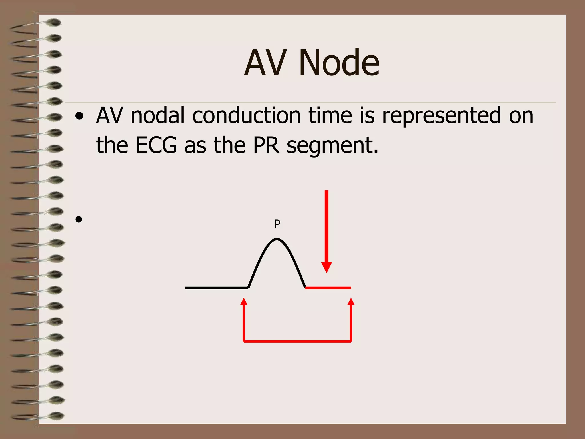
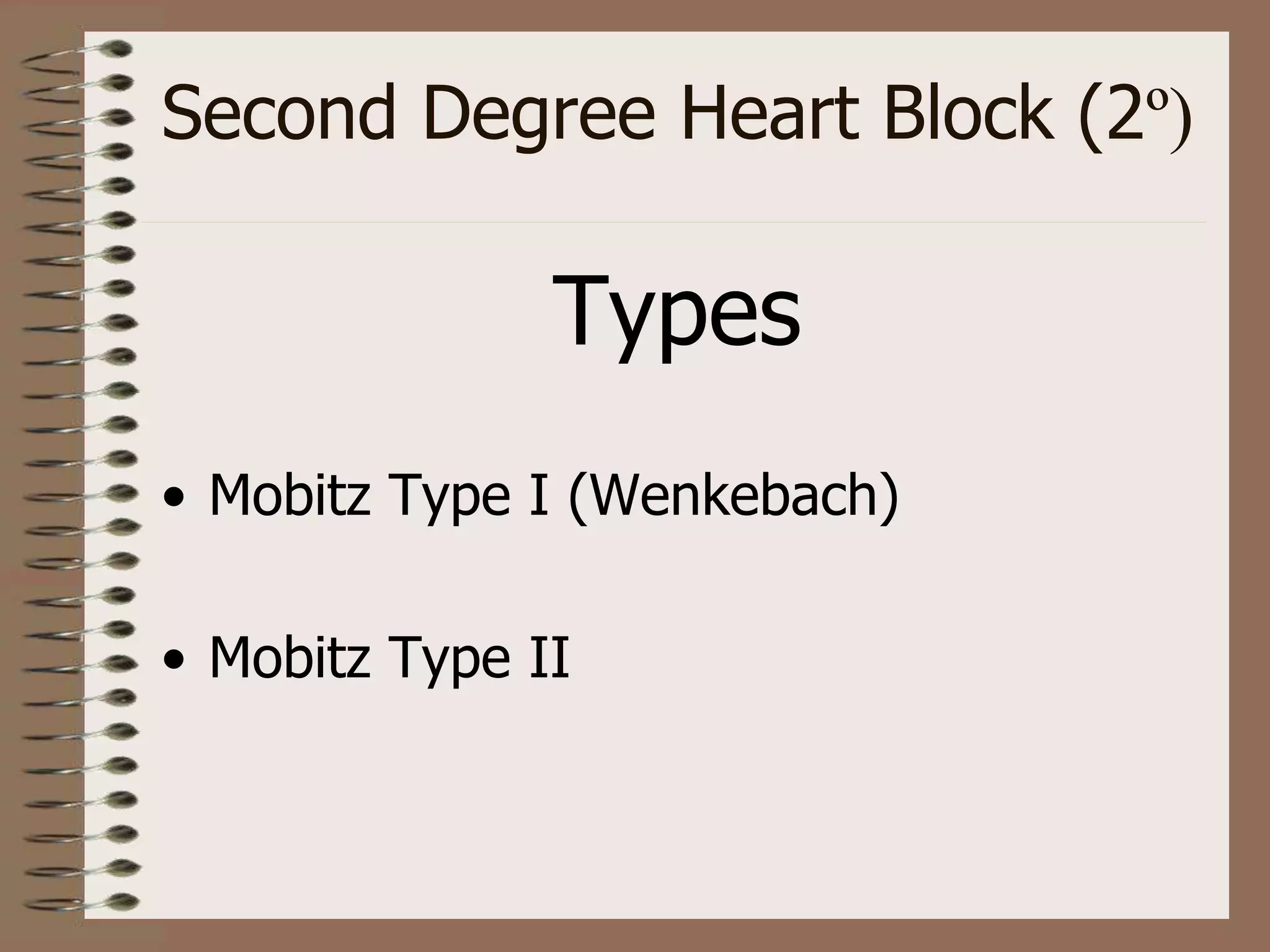
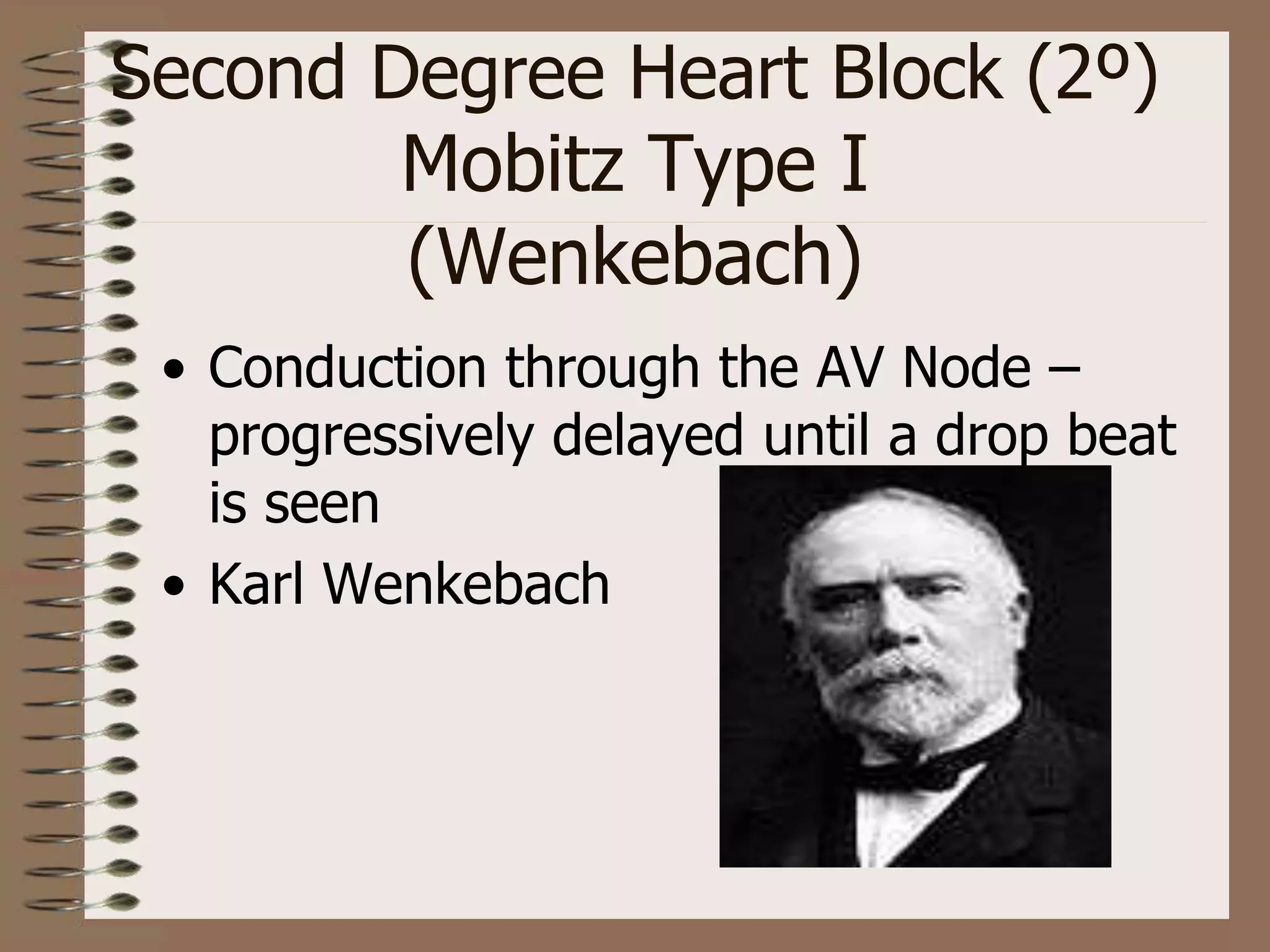
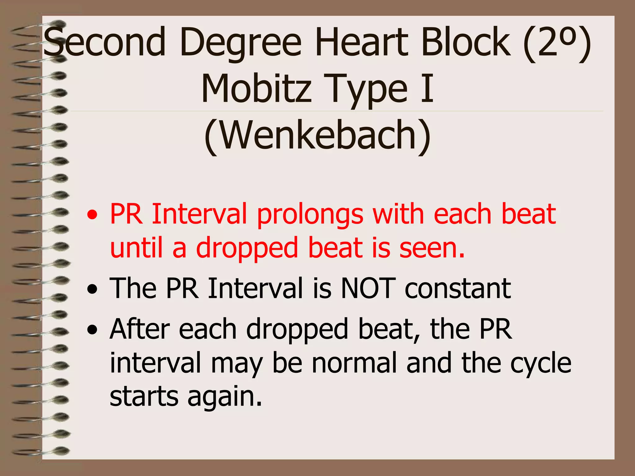
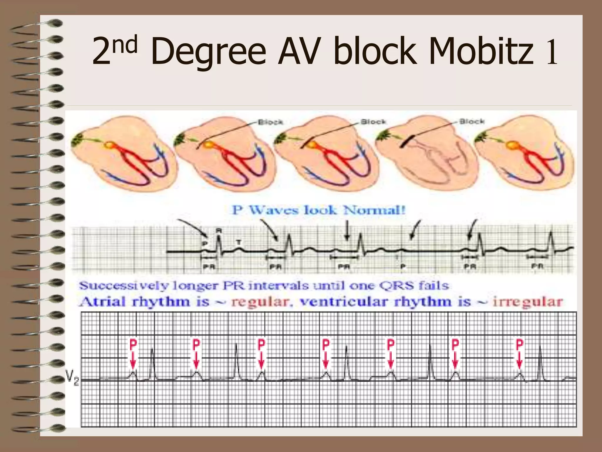
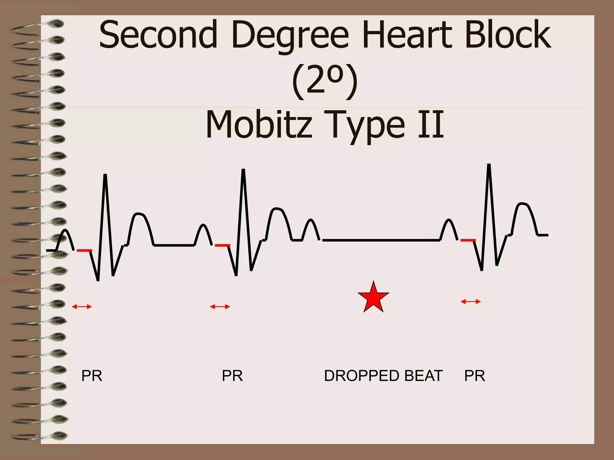
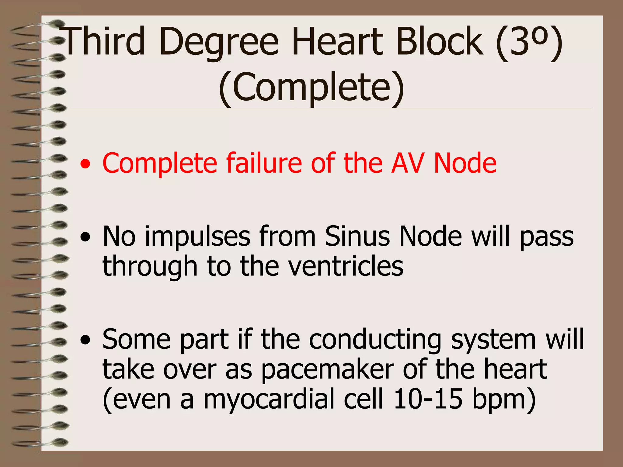
AV Heart Blocks
Rami AL-Khatib
AV Heart Blocks
Causes
• Drug therapy
• IHD (MI)
• Degeneration (age)
• Sclerosis (Aortic)
• Cardiac surgery
AV Node
• AV nodal conduction time is represented on
the ECG as the PR segment.
• P
First Degree Heart Block (1º)
• SA Node – normal
•Normal P wave
• AV Node conducts more slowly than
normal
•Prolonged constant PR Interval
• Rest of conduction is normal
•Normal QRS
First Degree Heart Block (1º)
• PR Interval > 0.2 seconds (5 small sq)
• Note – the PR Interval is constant
Significance
• Clinical significance
• None
• Treatment
• None
• Note – this can progress to 2º or 3º
heart block
Second Degree Heart Block (2º)
Types
• Mobitz Type I (Wenkebach)
• Mobitz Type II
Second Degree Heart Block (2º)
Mobitz Type I
(Wenkebach)
• Conduction through the AV Node –
progressively delayed until a drop beat
is seen
• Karl Wenkebach
Second Degree Heart Block (2º)
Mobitz Type I
(Wenkebach)
• PR Interval prolongs with each beat
until a dropped beat is seen.
• The PR Interval is NOT constant
• After each dropped beat, the PR
interval may be normal and the cycle
starts again.
Second Degree Heart Block (2º)
Mobitz Type I
(Wenkebach)
PR PR PR DROPPED BEAT
2nd Degree AV block Mobitz 1
Significance
• Clinical Significance
•Slight symptoms
e.g. Lethargy, Confusion
• Treatment
•Pacemaker
• Note – this can progress to 3º Heart Block
Second Degree Heart Block
(2º)
Mobitz Type II
• Conduction through the AV node is
constant.
• PR interval is constant
• Occasionally more than one a dropped
beat is seen
Second Degree Heart Block
(2º)
Mobitz Type II
PR PR DROPPED BEAT PR
Significance
• Clinical significance – this is more
significant disease
• Treatment – pacemaker
• Note – this can progress to 3º Heart
Block
Third Degree Heart Block (3º)
(Complete)
• Complete failure of the AV Node
• No impulses from Sinus Node will pass
through to the ventricles
• Some part if the conducting system will
take over as pacemaker of the heart
(even a myocardial cell 10-15 bpm)
Third Degree Heart Block (3º)
(Complete)
• P wave rate – normal
• Ventricular rate – slow
• Ventricular complex may be broad
•Idioventricular rhythm
• Complete dissociation between P
waves & QRS
Third Degree Heart Block (3º)
(Complete)
P P P P P
QRS QRS
3rd degree AV block
Significance
• clinical significance
•Symptoms LOC, Confusion,
Dizziness, Low BP
•Can lead to standstill, VT or VF
• Treatment - pacemaker
Summary
• 1º - prolongation of PR Interval & costant
• 2º - Mobitz I – Increasing PR Interval until dropped
beat is seen
• Mobitz II – Constant PR Interval with more P
waves to QRS
• 3º - Complete dissociation between P waves & QRS
References
• Hampton, J. R. (2013). The ECG made
easy. Elsevier Health Sciences.
• Hampton, J. R. (2013). 150 ECG problems.
Elsevier Health Sciences.
• O'Keefe Jr, J. H., Hammill, S. C., Freed,
M. S., & Pogwizd, S. M. (2010). The
complete guide to ECGs. Jones & Bartlett
Publishers.
Thank You

Heart blocks

  • 1.
  • 2.
  • 3.
    Causes • Drug therapy •IHD (MI) • Degeneration (age) • Sclerosis (Aortic) • Cardiac surgery
  • 4.
    AV Node • AVnodal conduction time is represented on the ECG as the PR segment. • P
  • 5.
    First Degree HeartBlock (1º) • SA Node – normal •Normal P wave • AV Node conducts more slowly than normal •Prolonged constant PR Interval • Rest of conduction is normal •Normal QRS
  • 6.
    First Degree HeartBlock (1º) • PR Interval > 0.2 seconds (5 small sq) • Note – the PR Interval is constant
  • 8.
    Significance • Clinical significance •None • Treatment • None • Note – this can progress to 2º or 3º heart block
  • 9.
    Second Degree HeartBlock (2º) Types • Mobitz Type I (Wenkebach) • Mobitz Type II
  • 10.
    Second Degree HeartBlock (2º) Mobitz Type I (Wenkebach) • Conduction through the AV Node – progressively delayed until a drop beat is seen • Karl Wenkebach
  • 11.
    Second Degree HeartBlock (2º) Mobitz Type I (Wenkebach) • PR Interval prolongs with each beat until a dropped beat is seen. • The PR Interval is NOT constant • After each dropped beat, the PR interval may be normal and the cycle starts again.
  • 12.
    Second Degree HeartBlock (2º) Mobitz Type I (Wenkebach) PR PR PR DROPPED BEAT
  • 13.
    2nd Degree AVblock Mobitz 1
  • 14.
    Significance • Clinical Significance •Slightsymptoms e.g. Lethargy, Confusion • Treatment •Pacemaker • Note – this can progress to 3º Heart Block
  • 15.
    Second Degree HeartBlock (2º) Mobitz Type II • Conduction through the AV node is constant. • PR interval is constant • Occasionally more than one a dropped beat is seen
  • 16.
    Second Degree HeartBlock (2º) Mobitz Type II PR PR DROPPED BEAT PR
  • 18.
    Significance • Clinical significance– this is more significant disease • Treatment – pacemaker • Note – this can progress to 3º Heart Block
  • 19.
    Third Degree HeartBlock (3º) (Complete) • Complete failure of the AV Node • No impulses from Sinus Node will pass through to the ventricles • Some part if the conducting system will take over as pacemaker of the heart (even a myocardial cell 10-15 bpm)
  • 20.
    Third Degree HeartBlock (3º) (Complete) • P wave rate – normal • Ventricular rate – slow • Ventricular complex may be broad •Idioventricular rhythm • Complete dissociation between P waves & QRS
  • 21.
    Third Degree HeartBlock (3º) (Complete) P P P P P QRS QRS
  • 22.
  • 23.
    Significance • clinical significance •SymptomsLOC, Confusion, Dizziness, Low BP •Can lead to standstill, VT or VF • Treatment - pacemaker
  • 24.
    Summary • 1º -prolongation of PR Interval & costant • 2º - Mobitz I – Increasing PR Interval until dropped beat is seen • Mobitz II – Constant PR Interval with more P waves to QRS • 3º - Complete dissociation between P waves & QRS
  • 25.
    References • Hampton, J.R. (2013). The ECG made easy. Elsevier Health Sciences. • Hampton, J. R. (2013). 150 ECG problems. Elsevier Health Sciences. • O'Keefe Jr, J. H., Hammill, S. C., Freed, M. S., & Pogwizd, S. M. (2010). The complete guide to ECGs. Jones & Bartlett Publishers.
  • 26.