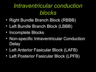
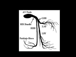
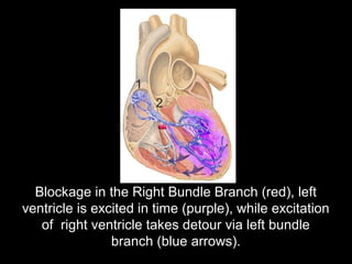
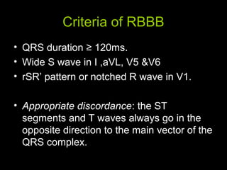
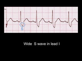
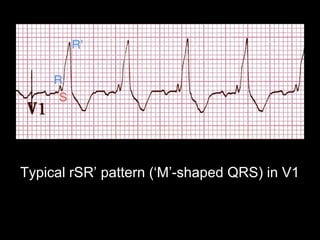
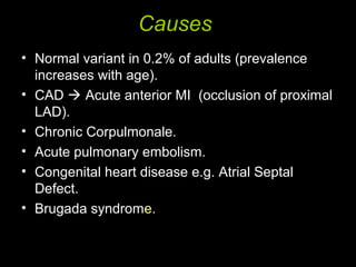
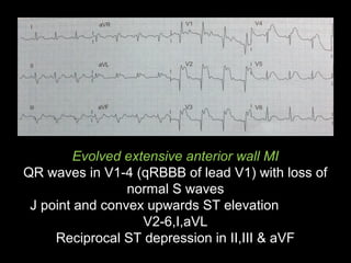
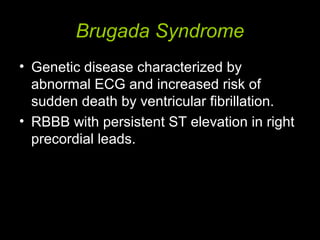
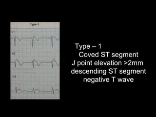
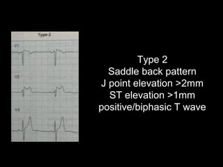
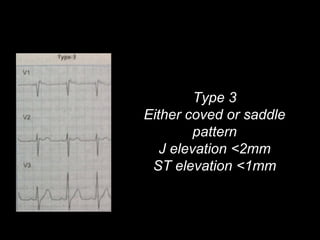
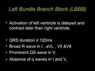
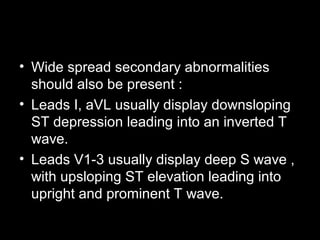
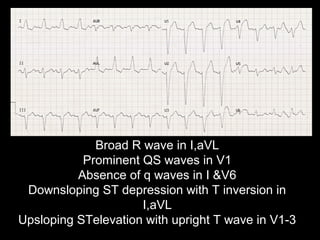
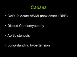
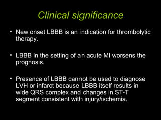
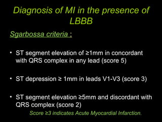
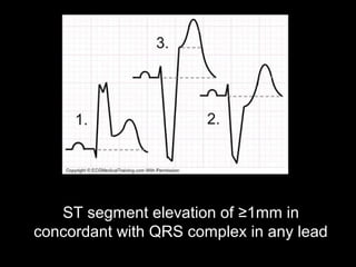
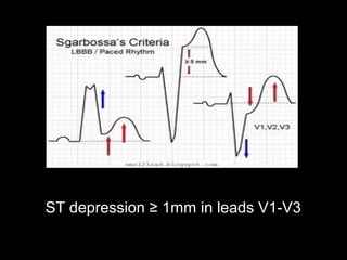
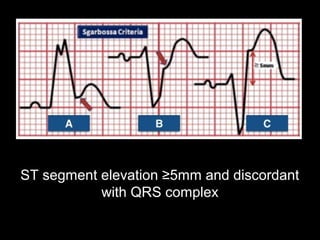
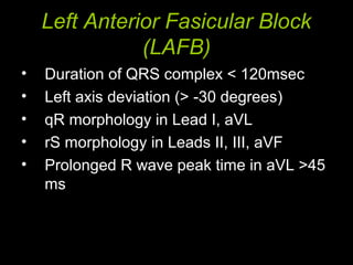
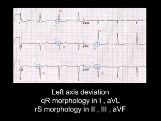
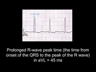
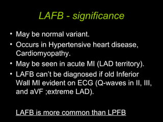
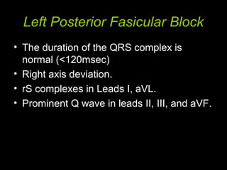
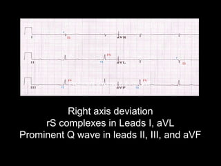
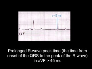
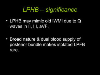
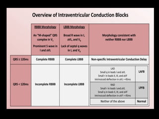
Intraventricular Conduction
Blocks
Dr. Sujith Chadala
(MD)(Gen. Medicine)
Upgraded Dept. Of Medicine
Osmania General Hospital
Intraventricular conduction
blocks
• Right Bundle Branch Block (RBBB)
• Left Bundle Branch Block (LBBB)
• Incomplete Blocks
• Non-specific Intraventricular Conduction
Delay
• Left Anterior Fasicular Block (LAFB)
• Left Posterior Fasicular Block (LPFB)
Right Bundle Branch Block (RBBB)
• Impulses travel through myocardium of left
ventricle to right ventricle and depolarise
right ventricle.
• Conduction through myocardium is slower
than that of Bundle of His , QRS complex
is seen to be widened.
Blockage in the Right Bundle Branch (red), left
ventricle is excited in time (purple), while excitation
of right ventricle takes detour via left bundle
branch (blue arrows).
Criteria of RBBB
• QRS duration ≥ 120ms.
• Wide S wave in I ,aVL, V5 &V6
• rSR’ pattern or notched R wave in V1.
• Appropriate discordance: the ST
segments and T waves always go in the
opposite direction to the main vector of the
QRS complex.
Wide S wave in lead I
Typical rSR’ pattern (‘M’-shaped QRS) in V1
Causes
• Normal variant in 0.2% of adults (prevalence
increases with age).
• CAD  Acute anterior MI (occlusion of proximal
LAD).
• Chronic Corpulmonale.
• Acute pulmonary embolism.
• Congenital heart disease e.g. Atrial Septal
Defect.
• Brugada syndrome.
Clinical significance
• Concordant T wave with Bundle Branch
Block may suggest ischemia or infarction.
• RBBB in the setting of an acute MI
worsens the prognosis , indicates proximal
Left Anterior Descending artery occlusion
( infarction of Interventricular septum ).
• Both LBBB and qRBBB signify large area
of infarction ,hence associated with high
risk.
Evolved extensive anterior wall MI
QR waves in V1-4 (qRBBB of lead V1) with loss of
normal S waves
J point and convex upwards ST elevation
V2-6,I,aVL
Reciprocal ST depression in II,III & aVF
Brugada Syndrome
• Genetic disease characterized by
abnormal ECG and increased risk of
sudden death by ventricular fibrillation.
• RBBB with persistent ST elevation in right
precordial leads.
Type – 1
Coved ST segment
J point elevation >2mm
descending ST segment
negative T wave
Type 2
Saddle back pattern
J point elevation >2mm
ST elevation >1mm
positive/biphasic T wave
Type 3
Either coved or saddle
pattern
J elevation <2mm
ST elevation <1mm
Left Bundle Branch Block (LBBB)
• Activation of left ventricle is delayed and
contract later than right ventricle.
• QRS duration ≥ 120ms
• Broad R wave in I , aVL , V5 &V6
• Prominent QS wave in V1
• Absence of q waves in I and V6
• Wide spread secondary abnormalities
should also be present :
• Leads I, aVL usually display downsloping
ST depression leading into an inverted T
wave.
• Leads V1-3 usually display deep S wave ,
with upsloping ST elevation leading into
upright and prominent T wave.
Broad R wave in I,aVL
Prominent QS waves in V1
Absence of q waves in I &V6
Downsloping ST depression with T inversion in
I,aVL
Upsloping STelevation with upright T wave in V1-3
Causes
• CAD  Acute AWMI (new onset LBBB)
• Dilated Cardiomyopathy
• Aortic stenosis
• Long-standing hypertension
Clinical significance
• New onset LBBB is an indication for thrombolytic
therapy.
• LBBB in the setting of an acute MI worsens the
prognosis.
• Presence of LBBB cannot be used to diagnose
LVH or infarct because LBBB itself results in
wide QRS complex and changes in ST-T
segment consistent with injury/ischemia.
Diagnosis of MI in the presence of
LBBB
Sgarbossa criteria :
• ST segment elevation of ≥1mm in concordant
with QRS complex in any lead (score 5)
• ST depression ≥ 1mm in leads V1-V3 (score 3)
• ST segment elevation ≥5mm and discordant with
QRS complex (score 2)
Score ≥3 indicates Acute Myocardial Infarction.
ST segment elevation of ≥1mm in
concordant with QRS complex in any lead
ST depression ≥ 1mm in leads V1-V3
ST segment elevation ≥5mm and discordant
with QRS complex
Incomplete Blocks
• Intraventricular conduction abnormalities
in which conduction is slowed in bundle,
but not completely blocked.
• Generally manifests as similar morphology
to complete right or left bundle branch
block but with QRS duration100 – 120ms.
• Compared to complete blocks , etiologies
are the same , but clinical significance is
reduced.
Nonspecific Intraventricular
Conduction delay
• Occurs when QRS duration is prolonged
>120ms,but criteria for neither RBBB nor
LBBB is met.
• It is a marker of non-specific cardiac
pathology.
Left Anterior Fasicular Block
(LAFB)
• Duration of QRS complex < 120msec
• Left axis deviation (> -30 degrees)
• qR morphology in Lead I, aVL
• rS morphology in Leads II, III, aVF
• Prolonged R wave peak time in aVL >45
ms
Left axis deviation
qR morphology in I , aVL
rS morphology in II , III , aVF
Prolonged R-wave peak time (the time from
onset of the QRS to the peak of the R wave)
in aVL > 45 ms
LAFB - significance
• May be normal variant.
• Occurs in Hypertensive heart disease,
Cardiomyopathy.
• May be seen in acute MI (LAD territory).
• LAFB can’t be diagnosed if old Inferior
Wall MI evident on ECG (Q-waves in II, III,
and aVF ;extreme LAD).
LAFB is more common than LPFB
Left Posterior Fasicular Block
• The duration of the QRS complex is
normal (<120msec)
• Right axis deviation.
• rS complexes in Leads I, aVL.
• Prominent Q wave in leads II, III, and aVF.
Right axis deviation
rS complexes in Leads I, aVL
Prominent Q wave in leads II, III, and aVF
Prominent Q waves in II , III , aVF
Prolonged R-wave peak time (the time from
onset of the QRS to the peak of the R wave)
in aVF > 45 ms
LPHB – significance
• LPHB may mimic old IWMI due to Q
waves in II, III, aVF.
• Broad nature & dual blood supply of
posterior bundle makes isolated LPFB
rare.
Bifascicular block
• Two of three fasicles of His / Purkinje
system are blocked.
• Most commonly RBBB + either LAFB OR
LPFB.
Etiologies:
• CAD (most common)
• Hypertension
• Aortic stenosis
• Congenital heart diseases
Trifasicular Block
• Presence of conducting disease in all
three fasicles.
• Has three features
• Prolongation of PR interval ( First degree
AV block ).
• RBBB.
• Either LAFB/LPFB.
Thank you

Bundle branch blocks by Dr Sujith Chadala

  • 1.
    Intraventricular Conduction Blocks Dr. SujithChadala (MD)(Gen. Medicine) Upgraded Dept. Of Medicine Osmania General Hospital
  • 2.
    Intraventricular conduction blocks • RightBundle Branch Block (RBBB) • Left Bundle Branch Block (LBBB) • Incomplete Blocks • Non-specific Intraventricular Conduction Delay • Left Anterior Fasicular Block (LAFB) • Left Posterior Fasicular Block (LPFB)
  • 4.
    Right Bundle BranchBlock (RBBB) • Impulses travel through myocardium of left ventricle to right ventricle and depolarise right ventricle. • Conduction through myocardium is slower than that of Bundle of His , QRS complex is seen to be widened.
  • 5.
    Blockage in theRight Bundle Branch (red), left ventricle is excited in time (purple), while excitation of right ventricle takes detour via left bundle branch (blue arrows).
  • 6.
    Criteria of RBBB •QRS duration ≥ 120ms. • Wide S wave in I ,aVL, V5 &V6 • rSR’ pattern or notched R wave in V1. • Appropriate discordance: the ST segments and T waves always go in the opposite direction to the main vector of the QRS complex.
  • 7.
    Wide S wavein lead I
  • 8.
    Typical rSR’ pattern(‘M’-shaped QRS) in V1
  • 9.
    Causes • Normal variantin 0.2% of adults (prevalence increases with age). • CAD  Acute anterior MI (occlusion of proximal LAD). • Chronic Corpulmonale. • Acute pulmonary embolism. • Congenital heart disease e.g. Atrial Septal Defect. • Brugada syndrome.
  • 10.
    Clinical significance • ConcordantT wave with Bundle Branch Block may suggest ischemia or infarction. • RBBB in the setting of an acute MI worsens the prognosis , indicates proximal Left Anterior Descending artery occlusion ( infarction of Interventricular septum ). • Both LBBB and qRBBB signify large area of infarction ,hence associated with high risk.
  • 11.
    Evolved extensive anteriorwall MI QR waves in V1-4 (qRBBB of lead V1) with loss of normal S waves J point and convex upwards ST elevation V2-6,I,aVL Reciprocal ST depression in II,III & aVF
  • 12.
    Brugada Syndrome • Geneticdisease characterized by abnormal ECG and increased risk of sudden death by ventricular fibrillation. • RBBB with persistent ST elevation in right precordial leads.
  • 13.
    Type – 1 CovedST segment J point elevation >2mm descending ST segment negative T wave
  • 14.
    Type 2 Saddle backpattern J point elevation >2mm ST elevation >1mm positive/biphasic T wave
  • 15.
    Type 3 Either covedor saddle pattern J elevation <2mm ST elevation <1mm
  • 16.
    Left Bundle BranchBlock (LBBB) • Activation of left ventricle is delayed and contract later than right ventricle. • QRS duration ≥ 120ms • Broad R wave in I , aVL , V5 &V6 • Prominent QS wave in V1 • Absence of q waves in I and V6
  • 17.
    • Wide spreadsecondary abnormalities should also be present : • Leads I, aVL usually display downsloping ST depression leading into an inverted T wave. • Leads V1-3 usually display deep S wave , with upsloping ST elevation leading into upright and prominent T wave.
  • 18.
    Broad R wavein I,aVL Prominent QS waves in V1 Absence of q waves in I &V6 Downsloping ST depression with T inversion in I,aVL Upsloping STelevation with upright T wave in V1-3
  • 19.
    Causes • CAD Acute AWMI (new onset LBBB) • Dilated Cardiomyopathy • Aortic stenosis • Long-standing hypertension
  • 20.
    Clinical significance • Newonset LBBB is an indication for thrombolytic therapy. • LBBB in the setting of an acute MI worsens the prognosis. • Presence of LBBB cannot be used to diagnose LVH or infarct because LBBB itself results in wide QRS complex and changes in ST-T segment consistent with injury/ischemia.
  • 21.
    Diagnosis of MIin the presence of LBBB Sgarbossa criteria : • ST segment elevation of ≥1mm in concordant with QRS complex in any lead (score 5) • ST depression ≥ 1mm in leads V1-V3 (score 3) • ST segment elevation ≥5mm and discordant with QRS complex (score 2) Score ≥3 indicates Acute Myocardial Infarction.
  • 22.
    ST segment elevationof ≥1mm in concordant with QRS complex in any lead
  • 23.
    ST depression ≥1mm in leads V1-V3
  • 24.
    ST segment elevation≥5mm and discordant with QRS complex
  • 25.
    Incomplete Blocks • Intraventricularconduction abnormalities in which conduction is slowed in bundle, but not completely blocked. • Generally manifests as similar morphology to complete right or left bundle branch block but with QRS duration100 – 120ms. • Compared to complete blocks , etiologies are the same , but clinical significance is reduced.
  • 26.
    Nonspecific Intraventricular Conduction delay •Occurs when QRS duration is prolonged >120ms,but criteria for neither RBBB nor LBBB is met. • It is a marker of non-specific cardiac pathology.
  • 27.
    Left Anterior FasicularBlock (LAFB) • Duration of QRS complex < 120msec • Left axis deviation (> -30 degrees) • qR morphology in Lead I, aVL • rS morphology in Leads II, III, aVF • Prolonged R wave peak time in aVL >45 ms
  • 28.
    Left axis deviation qRmorphology in I , aVL rS morphology in II , III , aVF
  • 29.
    Prolonged R-wave peaktime (the time from onset of the QRS to the peak of the R wave) in aVL > 45 ms
  • 30.
    LAFB - significance •May be normal variant. • Occurs in Hypertensive heart disease, Cardiomyopathy. • May be seen in acute MI (LAD territory). • LAFB can’t be diagnosed if old Inferior Wall MI evident on ECG (Q-waves in II, III, and aVF ;extreme LAD). LAFB is more common than LPFB
  • 31.
    Left Posterior FasicularBlock • The duration of the QRS complex is normal (<120msec) • Right axis deviation. • rS complexes in Leads I, aVL. • Prominent Q wave in leads II, III, and aVF.
  • 32.
    Right axis deviation rScomplexes in Leads I, aVL Prominent Q wave in leads II, III, and aVF Prominent Q waves in II , III , aVF
  • 33.
    Prolonged R-wave peaktime (the time from onset of the QRS to the peak of the R wave) in aVF > 45 ms
  • 34.
    LPHB – significance •LPHB may mimic old IWMI due to Q waves in II, III, aVF. • Broad nature & dual blood supply of posterior bundle makes isolated LPFB rare.
  • 36.
    Bifascicular block • Twoof three fasicles of His / Purkinje system are blocked. • Most commonly RBBB + either LAFB OR LPFB. Etiologies: • CAD (most common) • Hypertension • Aortic stenosis • Congenital heart diseases
  • 37.
    Trifasicular Block • Presenceof conducting disease in all three fasicles. • Has three features • Prolongation of PR interval ( First degree AV block ). • RBBB. • Either LAFB/LPFB.
  • 38.