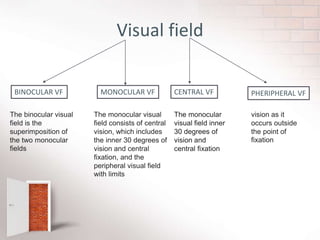
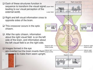
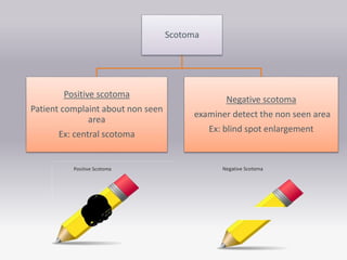
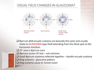
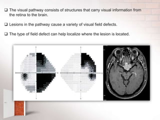
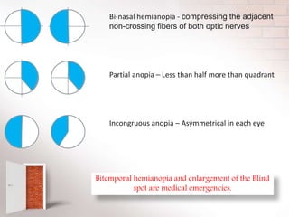
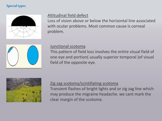
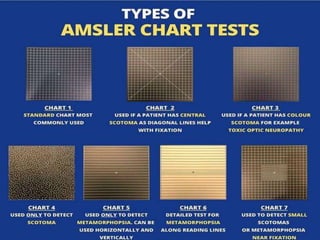
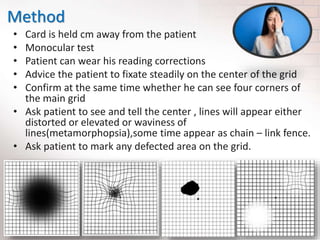
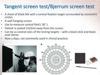
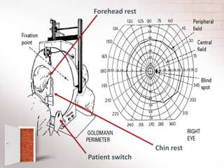
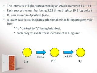
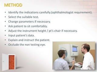
3. What is Visual field ?
• Definition
Visual field can be defined as the entire area that can be seen when an
eye is fixed straight at a point
or
It is the portion of the subject’s surrounding that can be seen at one time.
4. Optic disc nasal to the fovea – seen in temporal VF as a blind spot
Nasal Retina – Temporal Side vision
Temporal Retina – Nasal Side Vision
Inferior Retina– Superior Side Vision
Superior Retina–Inferior Side Retina
5. Boundaries of the visual field
Normal vision is limited to the following
physical limitations ,
Monocular:
Temporal(towards the temple) – 100
Nasal(towards the nose) - 60 degrees
Superior(towards the head ) - 60
Inferior(towards the feet ) – 70 degrees
In monocular field – Blind area is called BLIND SPOT
Binocular:
vertical field - 110˚
Horizontal field -
200˚
6. Location Of Blind Spot
• Corresponding to the optic nerve head
10-20 degree temporal to point of fixation
1.5 degree below the horizontal meridian
It is 5.5 degree wide by 7.5 degree high in diameter
10˚ 20˚
1.5˚
7.5˚
5.5˚
Size
7. Visual field
BINOCULAR VF MONOCULAR VF CENTRAL VF PHERIPHERAL VF
The binocular visual
field is the
superimposition of
the two monocular
fields
The monocular visual
field consists of central
vision, which includes
the inner 30 degrees of
vision and central
fixation, and the
peripheral visual field
with limits
The monocular
visual field inner
30 degrees of
vision and
central fixation
vision as it
occurs outside
the point of
fixation
8. Hill of vision
• Visual sensitivity is variable
central to peripheral in the
retina
• Macula is the highest
sensitive area
• Peripheral retinal
sensitivity is lower than the
central
• We can graph the visual
sensitivity against the
distance
Under photopic condition , the sharp of hill of vision is closely
related to the packing density of the cones and receptive field size.
9. TRAQUAIR- “HILL OF VISION IN THE SEA OF DARKNESS”
3D Graphical representation
Number of hill of vision get together
360˚ hill of vision
Sea represent the area of no light perception
10. Visual path
Visual cortex
Cones
Bipolar neurons
Ganglion cell’s axon forms the
optic nerve
Optic nerve to
the optic chiasm
Optic tract
Lateral geniculate nuclei of
the thalamus
Optic radiations
Primary visual areas of the
occipital lobes
11. Each of these structures function in
sequence to transform the visual signal,
leading to our visual perception of the
external world.
Right and left visual information cross to
opposite sides of the brain.
This crossover occurs in the optic
chiasm.
After the optic chiasm, information
about the right visual field is on the left
side of the brain, and information about
the left visual field is on the right side.
Images formed in the eye
are inverted but the brain inverts them
once more to make them seem upright.
12. Disorders of the visual pathway
A scotoma is an area of partial alteration in the field of vision
consisting of a partially diminished or entirely degenerated
visual acuity that is surrounded by a field of normal or
relatively well perceived vision
scotoma
13. Scotoma
Absolute scotoma
When increasing light
intensity, the scotoma size is
not change , its called absolute
scotoma
Relative scotoma
when increasing light intensity,
the scotoma size is changing. Its
called relative scotoma
15. Every normal
mammalian eye has a
scotoma in its field
vision. Usually termed as
Blind spot. Not a defect.
Blind spot size is in
abnormal size. Its a
defect
No visual sensation
with fixation point
Include the blind
spot and fixation
point
Arcuate scotoma
Usually include the blind
spot and arches in to the
nasal field. Arcuate fibers
damage cause to field
defect
Lies adjuscent to the
fixation point with
the greater part
being on one side
16. Bjerrum and arcuate scotoma are basically the same and usually
relate to GLAUCOMA type field extending from the blind spot to the
horizontal meridian.
25˚ area is Bjerrum area
Bjerrum screen VF test – not common
Small Bjerrum scotoma collected together – double arcuate scotoma
Ring scotoma – glaucoma pattern
Ring scotoma cause to Tunnel vision
18. The visual pathway consists of structures that carry visual information from
the retina to the brain.
Lesions in the pathway cause a variety of visual field defects.
The type of field defect can help localize where the lesion is located.
19. 1. Complete loss of vision in the
right eye (right eye blind)- Right
eye optic nerve full damage
2. Bitemporal hemianopia
(common) – central nerve fibers
easy to compress and damage due
to pituitary gland enlargement
3. Homonymous hemianopia
4. Quadrantanopia (quarter)
5.& 6. Quadrantanopia
with macular sparing
20. Bi-nasal hemianopia - compressing the adjacent
non-crossing fibers of both optic nerves
Partial anopia – Less than half more than quadrant
Incongruous anopia – Asymmetrical in each eye
Bitemporal hemianopia and enlargement of the Blind
spot are medical emergencies.
21. Altitudinal field defect
Loss of vision above or below the horizontal line associated
with ocular problems. Most common cause is corneal
problem.
Junctional scotoma
This pattern of field loss involves the entire visual field of
one eye and portion( usually superior temporal )of visual
field of the opposite eye.
Specialtypes
Zig zag scotoma/scintillating scotoma
Transient flashes of bright lights and or zig zag line which
may produce the migraine headache. we cant mark the
clear margin of the scotoma.
22. VF evaluation techniques
A visual field examination can detect dysfunction in central and
peripheral vision which may be caused by various medical
conditions such as glaucoma , stroke , brain tumors or other
neurological defects.
VISUAL FIELD TESTING METHODS/TOOLS
CENTRAL
AMSLER GRID: (macular field test)
TANGENT (Bjerrum screen)
GOLDMANN PERIMETRY TESTS
AUTOMATED PERIMETRY TEST (OCTOPUS / HUMPHERY)
PERIPHERAL
CONFRONTATION TEST ( gross method)
GOLDMANN PERIMETRY TEST
AUTOMATED 900 PROGRAMME PERIMETRY TEST
23. Stimulus for the VF tests…..
• Apparent size ( diameter ) of the spot.
• Intensity of the spot.
• Background illumination.
• Other factors,
Color
Movement of the stimulus
Duration of the stimulus
Attentiveness of the patient
Refractive state of the eye
24. STATIC
• Light spot not moving
• Static on off stimuli
• Stationary test objects
• Constant size
• Variable intensity
• Project increasing
intense stimuli at one
location until it is first
seen
• Target is just flash point
to point though out the
field
KINETIC
• Light spot moving
• Constant size
• Constant intensity
• Moves from non seeing
area to seeing area
• Move a single intense
stimulus until it no longer
seen
• Automated testing with
kinetic target is more
difficult to run with
computer
VF testing can be divided in to two types
25. Mainly focus on………
1) Confrontation test
2) Amsler grid test
3) Tangent screen test
4) Hemisphere perimetry tests
Goldman perimetry tests
Allegan Humphrey VF tests
26. CONFRONTATION TEST
It’s a Very simple method
It use to have gross idea
This test Highly recommended for bedridden people, children
as in emergency room test.
The easiest and most rudimentary method.
Use fingers , Hat pins as test objects.
It’s a monocular test, each eye is tested in turn
Examiner compare the patient VF with his own VF.
There can be inter – examiner variability.
Red cap
target
27. How to perform confrontation test
1.Sit or stand about 50 cm away from the patient ask the patient
to cover or close one eye and you close the one opposite ask
the subject to stare at your eye or nose and you do the same .
2.Starting at the top outer quadrant, move your fingers (or the
pin( in from the side, wiggling them as they go, and ask the
patient to tell you when he first see your fingers and, as you
move towards the center, whether they disappear.
3.Repeat the process in each quadrant and for each eye
separately.
4.If you detect a defect, reexamine that area and define it
further.
5.This technique compares the patient's visual field with yours,
so it does assume normal examiner visual fields
28. Examine non seen area to seen area/ peripheral area to central area
29. How to record confrontation test results
According to patient responses..……
R) L)
R) L)
R) L)
OR
OR
30. Amsler grid testing
• This assesses the central 10˚ the visual field. It detects
central and paracentral scotomas.
• There are seven charts of which chart is suitable for
most patient.
• It consist of a 20×20 grid of 5mmsquirs each
representing 1 degrees of central field.
• Testing distance 33 cm away from the eye.
• Qualitative measurement take within 1 minute.
31. When we do Amsler test,
• Evidence of macular disturbance seen on ophthalmoscopy in either
eye.
• Unrespectable loss of central visual acuity
• Reduction of acuity through a pinhole
• Symptoms of central visual disturbance, such as distortion.
• History of systemic disease or, more commonly, drugs which may
predispose to a maculopathy( such as tamoxifen or cloroquine)
• For the mapping of a central scotoma already detected
• This is useful for monitoring any progression of scotoma.
• History of poor photo stress recovery.
32.
33. Method
• Card is held cm away from the patient
• Monocular test
• Patient can wear his reading corrections
• Advice the patient to fixate steadily on the center of the grid
• Confirm at the same time whether he can see four corners of
the main grid
• Ask patient to see and tell the center , lines will appear either
distorted or elevated or waviness of
lines(metamorphopsia),some time appear as chain – link fence.
• Ask patient to mark any defected area on the grid.
34. How record Amsler chart test….
• Ask patient to mark his abnormal views on the amsler chart
We can give this chart to the patient to practice and check any
decreases or increases of disease.
35. Tangent screen test/Bjerrum screen test
• A sheet of black felt with a central fixation target surrounded by concentric
circles.
• A wall hanging screen.
• Use to measure central field ( 30˚ ).
• Patient is seated 1m(2m) away from the screen.
• Can be us several sizes of the testing targets – with a black stick and black
coat sleeve.
• Now a days, not commonly used in clinical practice.
36. Method….
1. Patient is seated 1m away from the screen.
2. Patient eye level is at the center fixation target.
3. Wears the corrective for distance if necessary.
4. Monocular test-non testing eye is occluded in turn.
5. The patient fixates at the central target spot.
6. Instruction are given to the patient.
7. The examiner moves a white(or red)disk target either 1mm or
2mm diameter, peripheral to the central until the patient first
see it.
8. Repeat the process along the whole meridians.
9. The results are plotted on a chart as the patient sees them
10.The target colour and size is the nominator(1W or2r) and the
denominator is the distance (eg:2000mm)at the patient from the
chart.
E.g.:- 1W/2000 OR 2r/2000
37. How To Record Tangent Screen Test
Results ?
• Mark each boundaries (meridians and isopters ) on the report chart
according to the patients responses.
• Joint each points together.
• Notice –
*date
*Patient data
*Target size.Colour / pt.S distance from the chart.
*Patients Hbrx
*Comments
38. • New one……
Auto plot tangent screen,
1. Patient switch
2. Chin rest
3. Head rest
4. Eye piece
5. Targets are moving
39. Arc perimeter
• Used for measuring the peripheral visual field.
• Arc can be rotated around the horizontal axis, target also can
be moved.
• Telescope is mounted.
• Examiner can see the patient’s eye through the telescope.
• Move the target along the arc.
• There is a lever in the backside ,when we move the lever the
target is also moving.
41. Manual perimetry
• Goldman perimetry
The most commonest type of kinetic perimetry in
clinical practice.
Usually kinetic test.(however static perimetry can
use central field)
Skills persons are required.
Perimetrist moves the target randomly.( non seen
area to seen area)
Repeatable.
Better to useful for patient who need significant
supervision to produce a VF.
42. How to perform the test….
• Calibrate the perimeter.(standard values)
• Insert a standard test paper – making sure aligned.
• Adjust the viewing telescope to see patient eye and observe whether patient fix
his eye during the test.
• Perimetrist directs the stimulus to map out their field of vision to successive
stimuli.
• Move stimulus slowly and steadily from unseen to seen.
- Inward for periphery
- Outward for mapping the blind spot/ central scotoma
• To move the stimulus arm from one side to the other , it must be swung around
the bottom line of the chart.
• Once the peripheral isopters are plotted, the central area is examine for scotoma.
usually peripheral stimulus – III-4e
central stimulus - I-4e
• Perimetrist should monitor patient fixation via viewing telescope.
• Perimetrist can recheck any suspected area.
43. The machine should be calibrated at the start of each
sessions.
Distance and near addition with wide aperture lenses
should be used (prevent ring scotoma ).
Aphakic eyes should wear possible be corrected with
contact lens.
Patient should be seated comfortably and adjust the
machine height according to the patient height.
44. Before the examination………..
Explain “ what is the field examination?” .
What is going to do be done with the test object.
What is expected from the patient.
It is easy to do , if the patient thoroughly understand the
process.
Seat patient with chin on the chin rest and forehead against
the forehead rest.
Occlude the non testing eye.
Ask patient to fix on central target and to press buzzer(pt
switch)whenever they see the light stimulus.
48. • The target sizes are indicated by roman numbers
( o – v) representing the size of the target in
squire millimeters.
• Each successive number being equivalent to a
four – fold increase in area.
i ii iii (standard size)
4 4 4
4
49. • The intensity of light represented by an Arabic numerals ( 1 – 4 )
• Each successive number being 3.15 times brighter (0.5 log units )
• It is measured in Apostilbs (asb).
• A lower case letter indicates additional minor filters progressively
from;
* “ a” darkest to “e” being brightest.
* each progressive letter is increase of 0.1 log unit.
1,a 2,b 3,c
× 3.15 × 3.15
50. Caution For Errors
Potential sources of error / artifact include ,
• Miosis
• Media opacities un corrective refractive errors
• Rim of the trial frame.
• Ptosis
• Dermatochalasis
• Incomprehension of the test
• Tremor
• Inadequate Retinal adaptation
52. • This machines are usually configured to test static
perimetry.
• The stationary stimulus is used, but change its intensity
until the sensitivity of the eye at that point is found.
• Used to measured at pre selected location of the VF.
• Programme selection is possible,
• central 30,24,10 or full field.
53. Humphrey cont…….
• Sensitive and reproducible.
• Need the patients attention and concentration during
the test.
• Monitoring is essential.
• Save the skilled Perimetrist time.
• Expensive
• Environmental sensitive.( A/C , dust free , dark free )
• Printed test result ( report ) is presented.
• Usually static test.
• Screening test are available.
• Data storage / transfer ability is available.
54. METHOD
• Identify the indications carefully.(ophthalmologist requirement).
• Select the suitable test.
• Change parameters if necessary.
• Ask patient to sit comfortably.
• Adjust the instrument height / pt’s chair if necessary.
• Input patient’s data.
• Explain and instruct the patient.
• Occlude the non testing eye.
55. • Position and align the patient.
• Place the correcting power if
necessary.
• Start the test with demonstration.
• Monitor the process.
( fixation losses , falls positive , falls
negative ,gaze tracking, blind spot
,vertex monitor and head tracking ,
video eye monitor )
• Encourage the patient to get
better responses.
• Turn the eye and repeat the test.
• Print the report.
56. PERIMETRIST DUTY
• Check necessary requirements.
• Select the suitable test.
• Instruct patient well- before starting the
test.
• Enter accurate details.
• Start from the good eye.
• Position and align.
• Correction if any.
• Monitor whole process during the test.
• Encourage the patient to get better
responses.
• Present a reliable, accurate report.