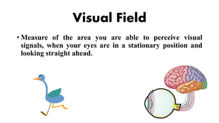
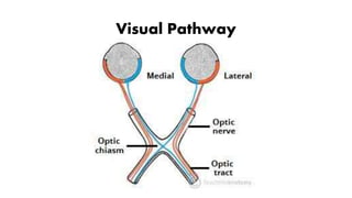
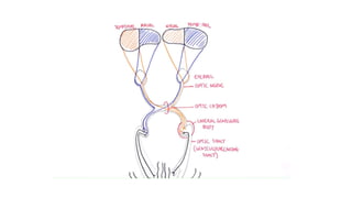
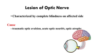
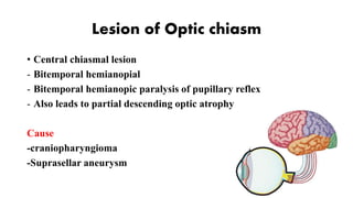
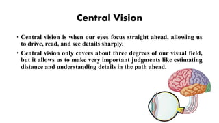
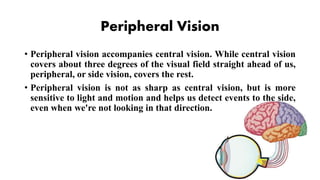
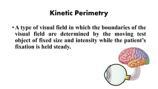
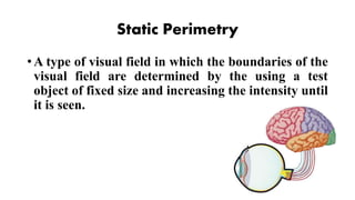
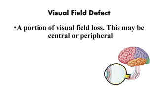
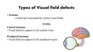
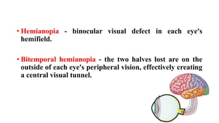
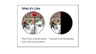
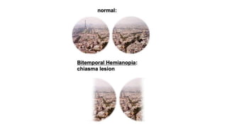
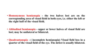
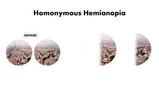
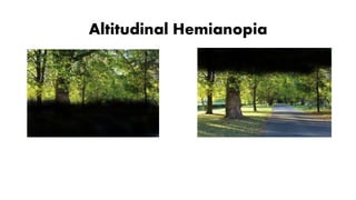
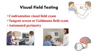
Visual Field/
Field of Vision
By: Aubrey Vale Sagun
Jeanette Ronquillo
Visual Field
• Measure of the area you are able to perceive visual
signals, when your eyes are in a stationary position and
looking straight ahead.
Visual Pathway
Lesions of Visual
Pathway
Lesion of Optic Nerve
• Characterized by complete blindness on affected side
Cause
- traumatic optic avulsion, acute optic neuritis, optic atrophy
Lesion through proximal part of Optic
nerve
• Ipsilateral blindess in affected side
• Absence of light reflex in ipsilateral side and consensual on
contralateral side.
Lesion of Optic chiasm
• Central chiasmal lesion
- Bitemporal hemianopial
- Bitemporal hemianopic paralysis of pupillary reflex
- Also leads to partial descending optic atrophy
Cause
-craniopharyngioma
-Suprasellar aneurysm
Lateral chiasma lesion
• Binasal hemianopia
• Binasal hemianopic paralysis of pupillary reflex
• Also leads to partial descending optic atrophy
Cause
Internal carotid aneurysm
Lesions causing distension of third ventricle
Lesion of Optic tract
• Incongruous homonymous hemianopia
• Contralateral hemianopia pupillary reaction (Wernicke’s
reaction)
Causes
Syphilitic meningitis or gumma, tuberculosis and tumors of optic
thalamus
Lesion of Visual Cortex
• Anterior Occipital cortex
• Homonymous hemianopia
Cause
Occlusion of posterior cerebral artery
Central Vision
• Central vision is when our eyes focus straight ahead, allowing us
to drive, read, and see details sharply.
• Central vision only covers about three degrees of our visual field,
but it allows us to make very important judgments like estimating
distance and understanding details in the path ahead.
Peripheral Vision
• Peripheral vision accompanies central vision. While central vision
covers about three degrees of the visual field straight ahead of us,
peripheral, or side vision, covers the rest.
• Peripheral vision is not as sharp as central vision, but is more
sensitive to light and motion and helps us detect events to the side,
even when we're not looking in that direction.
Perimetry
Kinetic Perimetry
• A type of visual field in which the boundaries of the
visual field are determined by the moving test
object of fixed size and intensity while the patient’s
fixation is held steady.
Static Perimetry
• A type of visual field in which the boundaries of the
visual field are determined by the using a test
object of fixed size and increasing the intensity until
it is seen.
Visual Field Defect
•A portion of visual field loss. This may be
central or peripheral
Types of Visual field defects
• Scotoma
- a blind spot surrounded by normal visual fields.
TYPES
Central Scotoma
- Visual field loss appears in the central vision
Peripheral Scotoma
- Visual field loss appears in the peripheral region
Central Scotoma
Peripheral Scotoma
• Hemianopia - binocular visual defect in each eye's
hemifield.
• Bitemporal hemianopia - the two halves lost are on the
outside of each eye's peripheral vision, effectively creating
a central visual tunnel.
• Homonymous hemianopia - the two halves lost are on the
corresponding area of visual field in both eyes, i.e. either the left or
the right half of the visual field.
• Altitudinal hemianopia - upper or lower halves of visual field are
lost; may be unilateral or bilateral.
• Quadrantanopia - ( incomplete hemianopia) Visual field loss in a
quarter of the visual field of the eye. The defect is usually bilateral.
Homonymous Hemianopia
Altitudinal Hemianopia
Quadrantanopia
Visual Field Testing
• Confrontation visual field exam
• Tangent screen or Goldmann field exam
• Automated perimetry

Visual field report

  • 1.
    Visual Field/ Field ofVision By: Aubrey Vale Sagun Jeanette Ronquillo
  • 2.
    Visual Field • Measureof the area you are able to perceive visual signals, when your eyes are in a stationary position and looking straight ahead.
  • 3.
  • 5.
  • 6.
    Lesion of OpticNerve • Characterized by complete blindness on affected side Cause - traumatic optic avulsion, acute optic neuritis, optic atrophy
  • 7.
    Lesion through proximalpart of Optic nerve • Ipsilateral blindess in affected side • Absence of light reflex in ipsilateral side and consensual on contralateral side.
  • 8.
    Lesion of Opticchiasm • Central chiasmal lesion - Bitemporal hemianopial - Bitemporal hemianopic paralysis of pupillary reflex - Also leads to partial descending optic atrophy Cause -craniopharyngioma -Suprasellar aneurysm
  • 9.
    Lateral chiasma lesion •Binasal hemianopia • Binasal hemianopic paralysis of pupillary reflex • Also leads to partial descending optic atrophy Cause Internal carotid aneurysm Lesions causing distension of third ventricle
  • 10.
    Lesion of Optictract • Incongruous homonymous hemianopia • Contralateral hemianopia pupillary reaction (Wernicke’s reaction) Causes Syphilitic meningitis or gumma, tuberculosis and tumors of optic thalamus
  • 11.
    Lesion of VisualCortex • Anterior Occipital cortex • Homonymous hemianopia Cause Occlusion of posterior cerebral artery
  • 12.
    Central Vision • Centralvision is when our eyes focus straight ahead, allowing us to drive, read, and see details sharply. • Central vision only covers about three degrees of our visual field, but it allows us to make very important judgments like estimating distance and understanding details in the path ahead.
  • 13.
    Peripheral Vision • Peripheralvision accompanies central vision. While central vision covers about three degrees of the visual field straight ahead of us, peripheral, or side vision, covers the rest. • Peripheral vision is not as sharp as central vision, but is more sensitive to light and motion and helps us detect events to the side, even when we're not looking in that direction.
  • 14.
  • 15.
    Kinetic Perimetry • Atype of visual field in which the boundaries of the visual field are determined by the moving test object of fixed size and intensity while the patient’s fixation is held steady.
  • 16.
    Static Perimetry • Atype of visual field in which the boundaries of the visual field are determined by the using a test object of fixed size and increasing the intensity until it is seen.
  • 17.
    Visual Field Defect •Aportion of visual field loss. This may be central or peripheral
  • 18.
    Types of Visualfield defects • Scotoma - a blind spot surrounded by normal visual fields. TYPES Central Scotoma - Visual field loss appears in the central vision Peripheral Scotoma - Visual field loss appears in the peripheral region
  • 19.
  • 20.
  • 21.
    • Hemianopia -binocular visual defect in each eye's hemifield. • Bitemporal hemianopia - the two halves lost are on the outside of each eye's peripheral vision, effectively creating a central visual tunnel.
  • 24.
    • Homonymous hemianopia- the two halves lost are on the corresponding area of visual field in both eyes, i.e. either the left or the right half of the visual field. • Altitudinal hemianopia - upper or lower halves of visual field are lost; may be unilateral or bilateral. • Quadrantanopia - ( incomplete hemianopia) Visual field loss in a quarter of the visual field of the eye. The defect is usually bilateral.
  • 25.
  • 26.
  • 27.
  • 28.
    Visual Field Testing •Confrontation visual field exam • Tangent screen or Goldmann field exam • Automated perimetry