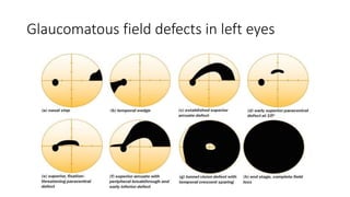
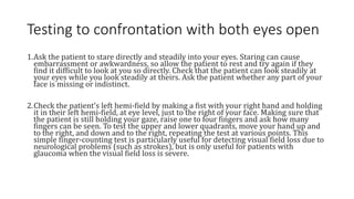
IMAGING TESTS IN GLAUC0MA :
VISUAL FIELD TESTS ,OCT SCAN
The purpose of a visual field test, often called a perimetry exam, is to detect changes in peripheral vision.
Peripheral vision test, Humphrey visual field exam, and the Goldmann visual field exam are among the other
names that you may hear regarding visual field testing.
What is the Purpose of the Visual Field Test?
Glaucoma is a disease that initially affects your peripheral (side) vision. Often, it is difficult to recognize that
peripheral vision is changing during normal daily activities because the deficits can be subtle and one eye can
compensate for the other.
One of the key tests that your eye doctor will perform as part of the diagnosis for glaucoma is the visual field test,
which is repeated periodically to determine if the disease is stable or getting worse. There are several different
types of visual field machines.
As the disease progresses, more and more of the peripheral vision is lost until eventually, in very late and
advanced disease, the central vision is also affected. Sometimes there are patients with glaucoma who
have their central vision affected early in the course of the disease, which is another reason that formal
visual field testing is so important.
What happens during a visual field test?
• You don’t have to prepare for a visual field test. It’s not
invasive, so you aren’t likely to have any side effects.
• There are several types of visual field tests, but they all
have one thing in common: you look straight ahead at
one point and signal when you see an object or a light
somewhere off to the side.
• Your provider will explain to you exactly where to look so
that the test is accurate.
• The two most basic types of visual field tests are very
simple:
The two most basic types of visual field tests are very simple
•Amsler grid: The Amsler grid is a pattern of straight lines that make perfect squares.
• You look at a large dot in the middle of the grid and describe any areas where the lines look
blurry, wavy or broken.
•The Amsler grid is a quick test that only measures the middle of the visual field (your central
vision) and provides your doctor with a small amount of information.
•Confrontation visual field: The term “confrontation” in this test just means that the person
giving the test sits facing the person having the test, about 3 or 4 feet (around 1 meter) away.
•The provider holds their arms straight out to the sides. You look straight ahead, and the
tester moves one hand and then the other inward toward you.
•You give a signal as soon as you see their hand.
The confrontation visual field test measures only the outer edge of the visual field. It’s not
very exact.
Key facts about visual field testing
1.Manual and/or automated visual field testing is subjective: it is totally dependent on
the co-operation and responses of the patient. Poor results that are difficult to
interpret are often due to the fact that the patient either did not understand what
was required, or did understand but was unable to respond.
2.Abnormalities in the visual field are a sign of damage anywhere in the visual system
from the retina through to the brain's visual cortex. Visual field defects are,
therefore, not limited to glaucoma. It is very important to examine the retina and
optic disc carefully to assess whether or not a visual field defect matches the
appearance of the disc and retina, or fits with other clinical signs. One should be very
wary of the person with extensive field loss, which seems genuine, where
examination of the retina and optic disc are normal. This person may have a
neurological condition (e.g. a brain tumour) or they may have had a stroke and not
have glaucoma at all.
3.The combination of good visual acuities with full visual fields leads to excellent functional vision and
both are very important. Loss of visual acuity can be very disabling, but so too can extensive loss of
peripheral visual field. Loss of visual field, particularly the lower field, makes walking around difficult
and slow, and people can easily lose confidence. Even in advanced glaucoma, when only a small area of
peripheral visual field remains, health care workers should do all they can to preserve this remnant of
vision, which can be very important for the patient's independence and dignity.
4.For diagnostic purposes, it is important to test each eye separately. This is because non-congruous
defects in each eye (e.g. a superior defect in the visual field of the right eye and an inferior defect in the
left eye) could be missed when testing both eyes together as the normal areas of field in one eye
overlap the defects in the other eye. This leads to a normal binocular field of vision when both eyes are
open . This is clearly a good situation as far as the patient is concerned, but means that further
extensive loss of field can occur without the person being aware of it. So, test the field of vision in each
eye separately – at every visit!
5.Early glaucomatous visual field defects are subtle and easily missed. Even with modern automated
and sensitive visual field analysers, glaucomatous visual field loss is not evident until at least 30% of
the retinal ganglion cell axons that make up the optic nerve have been lost. Progression of visual field
loss in untreated glaucoma can be quite slow, and signs of deteriorating disease can therefore be
missed quite easily.
6.There are two major types of perimetry. Kinetic perimetry involves the detection of moving
targets and static perimetry involves the detection of a stationary target. Static testing in general is
superior to kinetic perimetry in detecting slopes and scotomata (field defects), and tends to be
more reliable and consistent, particularly for detecting glaucomatous visual field loss.
7.In order to detect glaucomatous field loss, it is important to test for differences in the superior
and inferior hemi-fields (above or below the horizontal raphé) and hunt for defects such as a nasal
step, which is typical of glaucoma.
8.In order to detect neurological field loss, test for differences either side of the vertical meridian.
Careful detection of visual field defects can be diagnostic of many ophthalmic and/or neurological
conditions, including glaucoma. In glaucoma – as well as other conditions – it is vital to repeat visual
field testing to track any changes over time.
Ideally, the same method of testing should be used for baseline and subsequent follow-up. In glaucoma,
if visual field loss is progressive, it may mean that control of intraocular pressure (IOP) is inadequate.
Be aware, though, that patients may remember to use their eye drops just before they come to the
clinic, so that their IOP may appear to be controlled. It is far better to monitor control by assessment of
the optic disc and visual fields than to rely on IOP alone.
Visual field loss can be diffuse (as with cataract or corneal opacification), but more commonly there are
isolated defects. The visual field defects associated with glaucoma appear to be fairly non-specific, although
typical loss fits with the arrangement of the retinal ganglion cell axons within the retinal nerve fibre layer of
the retina.
Relatively specific glaucomatous visual field defects include:
1.a nasal step defect obeying the horizontal meridian
2.a temporal wedge defect
3.the classic arcuate defect, which is a comma-shaped extension of the blind spot
4.a paracentral defect 10–20° from the blind spot
5.an arcuate defect with peripheral breakthrough
6.generalised constriction (tunnel vision)
7.temporal-sparing severe visual field loss
8.total loss of field.
Glaucomatous field defects in left eyes
Non-glaucomatous bilateral visual field
defects
Testing to confrontation with both eyes open
1.Ask the patient to stare directly and steadily into your eyes. Staring can cause
embarrassment or awkwardness, so allow the patient to rest and try again if they
find it difficult to look at you so directly. Check that the patient can look steadily at
your eyes while you look steadily at theirs. Ask the patient whether any part of your
face is missing or indistinct.
2.Check the patient's left hemi-field by making a fist with your right hand and holding
it in their left hemi-field, at eye level, just to the right of your face. Making sure that
the patient is still holding your gaze, raise one to four fingers and ask how many
fingers can be seen. To test the upper and lower quadrants, move your hand up and
to the right, and down and to the right, repeating the test at various points. This
simple finger-counting test is particularly useful for detecting visual field loss due to
neurological problems (such as strokes), but is only useful for patients with
glaucoma when the visual field loss is severe.
Testing to confrontation with both eyes open
3.To test the patient's right hemi-field and upper and lower quadrants,
repeat the finger-counting test using your left hand, starting just to the
left of your face and moving up and left and then down and left.
4.A useful, additional test to perform in patients with a suspected
homonymous hemianopsia (i.e. loss of either the right or left field of
vision in both eyes, often from a stroke) is to test for sensory inattention.
Hold both hands up and wiggle the fingers of the right hand, followed by
those of the left hand in each hemi-field. If the patient sees the moving
fingers, then wiggle one finger of each hand at the same time – if the
patient can only see movement on one side then they may have a subtle
hemianopia.
Testing each eye to confrontation
1.Ask the patient to cover their own eye with the palm of their hand (not their fingers, as it is easy to peep between
fingers). Remember that you should close your eyes in turn too, so that you are comparing the field in your right eye
with the field of the patient's left eye, for example
2.Do the finger counting test first (static testing). Be sure to test on both the left and the right for each eye tested.
3.Next, bring your target finger from the far periphery in towards the central region (kinetic testing). Ask the patient to
say when they first see the target. Repeat from several different directions, ensuring that the full 360° for each eye is
tested. The examiner should remember to perform kinetic testing at a speed appropriate for the patient's responses.
4.Next, test the peripheral field with a white-headed neurological pin (beyond a central 30° radius) and the central field
with a red-headed neurological pin (within a 30° radius). Testing with neurological pin targets gives much more
accurate results than testing with fingers, and can detect earlier visual field loss. Red-headed neurological pins are also
useful for assessing the size of the blind spot (e.g., with papilloedema), again by comparing the size of your blind spot
with that of the patient's. In addition, red-headed neurological pins can be used to test for red-desaturation in early
optic nerve disease.
Testing visual fields to confrontation. The examiner's left eye is closed, so he can
compare the field of his right eye with the field of the patient's left eye. T
Other visual field tests
• Amsler chart testing
• A printed grid (Amsler chart) can be used to detect subtle central
defects (uncommon in patients with glaucoma) as well as paracentral
defects (fairly common in patients with glaucoma -especially those
with normal tension glaucoma). Test one eye at a time, correcting for
any near refractive errors. Patients should hold the chart at a
comfortable reading distance from their uncovered eye, and stare at
the central spot of the grid. Ask them to identify and then point out
any areas where the grid is missing or distorted. Missing areas may
suggest paracentral glaucomatous visual field loss, whereas distortion
is more common with macular disorders
Amsler grid, when viewed by someone with normal central vision (a) and by people with a
problem with their central visual field (b and c)
OCT
• Optical coherence tomography (OCT) is an imaging technology
that employs low-coherence interferometry to obtain cross-
sectional images of the ocular tissues.
• The principle of OCT is analogous to that of ultrasonography, but
uses light instead of sound to acquire high resolution images of the
ocular structures. A beam of light is shone on the eye and
reflections returning from the structures are analyzed to produce
real time images.
PRINCIPLES OF OCT
• A light source is directed to a partially reflecting mirror that splits the
light into two beams: One is directed toward a mirror placed at a known
distance (reference mirror) and the other is directed toward the eye,
from where it will reflect back.
• The back-reflected light from the eye is combined with the back-
reflected light from the reference mirror and coherent light is
compared. Interference is produced when two light pulses coincide.
• The reference mirror is then moved so that the time delay of the
reference light pulse can change accordingly and therefore other
intraocular structures can be measured.
• The laser beam is passed throughout the tissue and a series of scans
are obtained to produce a two-dimensional map.
APPLICATION OF OCT IN GLAUCOMA
Anterior Segment-OCT
• Anterior segment OCT (AS-OCT) uses light of longer wavelength (1310 nm) to obtain images of
the anterior segment. Light-induced damage of the retina at this high wavelength is prevented by
the absorption of light by the water in the ocular tissues.
Applications of AS-OCT
1.Evaluation of angle structures. It causes minimal distortion of the angle anatomy because no
visible light is shone on the eye and there is no pressure on the globe.
2.AS-OCT provides detailed calculations of parameters, such as angle opening distance, angle
recess area, and the trabecular―iris space area, thus introducing new levels of precision for
approaching PAC disease
To evaluate the effect of laser peripheral iridotomy and other
interventions on the angle anatomy, which helps the clinician
plan further interventions if necessary.
Assessment of bleb morphology and patency of ostium post-
filtering surgery helps the clinician to plan further interventions
as necessary.
AS-OCT showing 180° narrow angles

IMAGING TESTS IN GLAUC0MA___VISUAL FIELD TESTS.pptx

  • 1.
    IMAGING TESTS INGLAUC0MA : VISUAL FIELD TESTS ,OCT SCAN
  • 2.
    The purpose ofa visual field test, often called a perimetry exam, is to detect changes in peripheral vision. Peripheral vision test, Humphrey visual field exam, and the Goldmann visual field exam are among the other names that you may hear regarding visual field testing. What is the Purpose of the Visual Field Test? Glaucoma is a disease that initially affects your peripheral (side) vision. Often, it is difficult to recognize that peripheral vision is changing during normal daily activities because the deficits can be subtle and one eye can compensate for the other. One of the key tests that your eye doctor will perform as part of the diagnosis for glaucoma is the visual field test, which is repeated periodically to determine if the disease is stable or getting worse. There are several different types of visual field machines. As the disease progresses, more and more of the peripheral vision is lost until eventually, in very late and advanced disease, the central vision is also affected. Sometimes there are patients with glaucoma who have their central vision affected early in the course of the disease, which is another reason that formal visual field testing is so important.
  • 3.
    What happens duringa visual field test? • You don’t have to prepare for a visual field test. It’s not invasive, so you aren’t likely to have any side effects. • There are several types of visual field tests, but they all have one thing in common: you look straight ahead at one point and signal when you see an object or a light somewhere off to the side. • Your provider will explain to you exactly where to look so that the test is accurate. • The two most basic types of visual field tests are very simple:
  • 4.
    The two mostbasic types of visual field tests are very simple •Amsler grid: The Amsler grid is a pattern of straight lines that make perfect squares. • You look at a large dot in the middle of the grid and describe any areas where the lines look blurry, wavy or broken. •The Amsler grid is a quick test that only measures the middle of the visual field (your central vision) and provides your doctor with a small amount of information. •Confrontation visual field: The term “confrontation” in this test just means that the person giving the test sits facing the person having the test, about 3 or 4 feet (around 1 meter) away. •The provider holds their arms straight out to the sides. You look straight ahead, and the tester moves one hand and then the other inward toward you. •You give a signal as soon as you see their hand. The confrontation visual field test measures only the outer edge of the visual field. It’s not very exact.
  • 5.
    Key facts aboutvisual field testing 1.Manual and/or automated visual field testing is subjective: it is totally dependent on the co-operation and responses of the patient. Poor results that are difficult to interpret are often due to the fact that the patient either did not understand what was required, or did understand but was unable to respond. 2.Abnormalities in the visual field are a sign of damage anywhere in the visual system from the retina through to the brain's visual cortex. Visual field defects are, therefore, not limited to glaucoma. It is very important to examine the retina and optic disc carefully to assess whether or not a visual field defect matches the appearance of the disc and retina, or fits with other clinical signs. One should be very wary of the person with extensive field loss, which seems genuine, where examination of the retina and optic disc are normal. This person may have a neurological condition (e.g. a brain tumour) or they may have had a stroke and not have glaucoma at all.
  • 6.
    3.The combination ofgood visual acuities with full visual fields leads to excellent functional vision and both are very important. Loss of visual acuity can be very disabling, but so too can extensive loss of peripheral visual field. Loss of visual field, particularly the lower field, makes walking around difficult and slow, and people can easily lose confidence. Even in advanced glaucoma, when only a small area of peripheral visual field remains, health care workers should do all they can to preserve this remnant of vision, which can be very important for the patient's independence and dignity. 4.For diagnostic purposes, it is important to test each eye separately. This is because non-congruous defects in each eye (e.g. a superior defect in the visual field of the right eye and an inferior defect in the left eye) could be missed when testing both eyes together as the normal areas of field in one eye overlap the defects in the other eye. This leads to a normal binocular field of vision when both eyes are open . This is clearly a good situation as far as the patient is concerned, but means that further extensive loss of field can occur without the person being aware of it. So, test the field of vision in each eye separately – at every visit! 5.Early glaucomatous visual field defects are subtle and easily missed. Even with modern automated and sensitive visual field analysers, glaucomatous visual field loss is not evident until at least 30% of the retinal ganglion cell axons that make up the optic nerve have been lost. Progression of visual field loss in untreated glaucoma can be quite slow, and signs of deteriorating disease can therefore be missed quite easily.
  • 7.
    6.There are twomajor types of perimetry. Kinetic perimetry involves the detection of moving targets and static perimetry involves the detection of a stationary target. Static testing in general is superior to kinetic perimetry in detecting slopes and scotomata (field defects), and tends to be more reliable and consistent, particularly for detecting glaucomatous visual field loss. 7.In order to detect glaucomatous field loss, it is important to test for differences in the superior and inferior hemi-fields (above or below the horizontal raphé) and hunt for defects such as a nasal step, which is typical of glaucoma. 8.In order to detect neurological field loss, test for differences either side of the vertical meridian.
  • 8.
    Careful detection ofvisual field defects can be diagnostic of many ophthalmic and/or neurological conditions, including glaucoma. In glaucoma – as well as other conditions – it is vital to repeat visual field testing to track any changes over time. Ideally, the same method of testing should be used for baseline and subsequent follow-up. In glaucoma, if visual field loss is progressive, it may mean that control of intraocular pressure (IOP) is inadequate. Be aware, though, that patients may remember to use their eye drops just before they come to the clinic, so that their IOP may appear to be controlled. It is far better to monitor control by assessment of the optic disc and visual fields than to rely on IOP alone. Visual field loss can be diffuse (as with cataract or corneal opacification), but more commonly there are isolated defects. The visual field defects associated with glaucoma appear to be fairly non-specific, although typical loss fits with the arrangement of the retinal ganglion cell axons within the retinal nerve fibre layer of the retina.
  • 9.
    Relatively specific glaucomatousvisual field defects include: 1.a nasal step defect obeying the horizontal meridian 2.a temporal wedge defect 3.the classic arcuate defect, which is a comma-shaped extension of the blind spot 4.a paracentral defect 10–20° from the blind spot 5.an arcuate defect with peripheral breakthrough 6.generalised constriction (tunnel vision) 7.temporal-sparing severe visual field loss 8.total loss of field.
  • 10.
  • 11.
  • 12.
    Testing to confrontationwith both eyes open 1.Ask the patient to stare directly and steadily into your eyes. Staring can cause embarrassment or awkwardness, so allow the patient to rest and try again if they find it difficult to look at you so directly. Check that the patient can look steadily at your eyes while you look steadily at theirs. Ask the patient whether any part of your face is missing or indistinct. 2.Check the patient's left hemi-field by making a fist with your right hand and holding it in their left hemi-field, at eye level, just to the right of your face. Making sure that the patient is still holding your gaze, raise one to four fingers and ask how many fingers can be seen. To test the upper and lower quadrants, move your hand up and to the right, and down and to the right, repeating the test at various points. This simple finger-counting test is particularly useful for detecting visual field loss due to neurological problems (such as strokes), but is only useful for patients with glaucoma when the visual field loss is severe.
  • 13.
    Testing to confrontationwith both eyes open 3.To test the patient's right hemi-field and upper and lower quadrants, repeat the finger-counting test using your left hand, starting just to the left of your face and moving up and left and then down and left. 4.A useful, additional test to perform in patients with a suspected homonymous hemianopsia (i.e. loss of either the right or left field of vision in both eyes, often from a stroke) is to test for sensory inattention. Hold both hands up and wiggle the fingers of the right hand, followed by those of the left hand in each hemi-field. If the patient sees the moving fingers, then wiggle one finger of each hand at the same time – if the patient can only see movement on one side then they may have a subtle hemianopia.
  • 14.
    Testing each eyeto confrontation 1.Ask the patient to cover their own eye with the palm of their hand (not their fingers, as it is easy to peep between fingers). Remember that you should close your eyes in turn too, so that you are comparing the field in your right eye with the field of the patient's left eye, for example 2.Do the finger counting test first (static testing). Be sure to test on both the left and the right for each eye tested. 3.Next, bring your target finger from the far periphery in towards the central region (kinetic testing). Ask the patient to say when they first see the target. Repeat from several different directions, ensuring that the full 360° for each eye is tested. The examiner should remember to perform kinetic testing at a speed appropriate for the patient's responses. 4.Next, test the peripheral field with a white-headed neurological pin (beyond a central 30° radius) and the central field with a red-headed neurological pin (within a 30° radius). Testing with neurological pin targets gives much more accurate results than testing with fingers, and can detect earlier visual field loss. Red-headed neurological pins are also useful for assessing the size of the blind spot (e.g., with papilloedema), again by comparing the size of your blind spot with that of the patient's. In addition, red-headed neurological pins can be used to test for red-desaturation in early optic nerve disease.
  • 15.
    Testing visual fieldsto confrontation. The examiner's left eye is closed, so he can compare the field of his right eye with the field of the patient's left eye. T
  • 16.
    Other visual fieldtests • Amsler chart testing • A printed grid (Amsler chart) can be used to detect subtle central defects (uncommon in patients with glaucoma) as well as paracentral defects (fairly common in patients with glaucoma -especially those with normal tension glaucoma). Test one eye at a time, correcting for any near refractive errors. Patients should hold the chart at a comfortable reading distance from their uncovered eye, and stare at the central spot of the grid. Ask them to identify and then point out any areas where the grid is missing or distorted. Missing areas may suggest paracentral glaucomatous visual field loss, whereas distortion is more common with macular disorders
  • 17.
    Amsler grid, whenviewed by someone with normal central vision (a) and by people with a problem with their central visual field (b and c)
  • 18.
    OCT • Optical coherencetomography (OCT) is an imaging technology that employs low-coherence interferometry to obtain cross- sectional images of the ocular tissues. • The principle of OCT is analogous to that of ultrasonography, but uses light instead of sound to acquire high resolution images of the ocular structures. A beam of light is shone on the eye and reflections returning from the structures are analyzed to produce real time images.
  • 19.
    PRINCIPLES OF OCT •A light source is directed to a partially reflecting mirror that splits the light into two beams: One is directed toward a mirror placed at a known distance (reference mirror) and the other is directed toward the eye, from where it will reflect back. • The back-reflected light from the eye is combined with the back- reflected light from the reference mirror and coherent light is compared. Interference is produced when two light pulses coincide. • The reference mirror is then moved so that the time delay of the reference light pulse can change accordingly and therefore other intraocular structures can be measured. • The laser beam is passed throughout the tissue and a series of scans are obtained to produce a two-dimensional map.
  • 20.
    APPLICATION OF OCTIN GLAUCOMA Anterior Segment-OCT • Anterior segment OCT (AS-OCT) uses light of longer wavelength (1310 nm) to obtain images of the anterior segment. Light-induced damage of the retina at this high wavelength is prevented by the absorption of light by the water in the ocular tissues. Applications of AS-OCT 1.Evaluation of angle structures. It causes minimal distortion of the angle anatomy because no visible light is shone on the eye and there is no pressure on the globe. 2.AS-OCT provides detailed calculations of parameters, such as angle opening distance, angle recess area, and the trabecular―iris space area, thus introducing new levels of precision for approaching PAC disease
  • 21.
    To evaluate theeffect of laser peripheral iridotomy and other interventions on the angle anatomy, which helps the clinician plan further interventions if necessary. Assessment of bleb morphology and patency of ostium post- filtering surgery helps the clinician to plan further interventions as necessary.
  • 22.
    AS-OCT showing 180°narrow angles