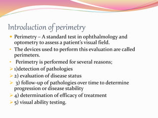
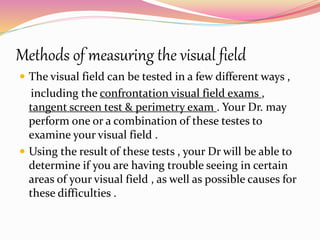
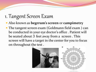
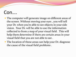
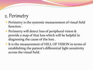
Magnification is a method of increasing the size of the image
so that enough of the retina is stimulated to send an impulse
through the optic nerve allowing an object to be perceived .
Accommodation/ Accommodation of Eye, Measurement of Accommodation of Eye (hea...Bikash Sapkota
CLICK HERE TO DOWNLOAD FULL PPT ❤❤ https://healthkura.com/measurement-of-accommodation-of-eye/ ❤❤
Dear viewers Check Out my other piece of works at ❤❤❤ https://healthkura.com ❤❤❤
Measurement of Accommodation of eye:
Amplitude, Facility,
Relative Accommodation, Fatigue, Lag,
Dynamic Retinoscopy
Presentation Layout:
-Introduction to accommodation of eye
-Mechanism
-Components
-Measurement of accommodation of eye
- Amplitude
- Facility
- Relative accommodation
- Lag
-Dynamic Retinoscopy
Accommodation
-dioptric adjustment of the crystalline lens of the eye
- to obtain clear vision for a given target of regard
-process by which the refractive power of eye is altered
- to ensure a clear retinal image
For further reading
-Clinical Procedures in Optometry by J.D. Bartlett, J.B. Eskridge, J.F. Amos
-Primary Care Optometry by Theodere Grosvenor
-Borish’s Clinical Refraction by W.J. Benjamin
-Clinical Procedures for Ocular examination by Carlson et al
-American Academy of Ophthalmology
-Optometric Clinical Practice Guideline by American Optometric Association
-Internet
Follow me to get in touch with optometric and ophthalmic updates
It contains Examination Protocol for Contact Lenses along with information about pre-requisites for fitting a Contact Lens. A helpful guide for all Students, Eye Care Practitioners (Optometrist, Ophthalmologist).
How to interpret the visual field printout
Learn basic terms of visual field analysis
How to diagnose glaucomatous field defect
How to diagnose neurological field defect
To know Humphrey visual field analyser
To know about various types of perimetry
To identify field defect
To recognize that field defect is due to glaucoma or neurological lesion
To know that field defect is progressive or not
Interpretation of HVFA
The dimensions of healthcare quality refer to various attributes or aspects that define the standard of healthcare services. These dimensions are used to evaluate, measure, and improve the quality of care provided to patients. A comprehensive understanding of these dimensions ensures that healthcare systems can address various aspects of patient care effectively and holistically. Dimensions of Healthcare Quality and Performance of care include the following; Appropriateness, Availability, Competence, Continuity, Effectiveness, Efficiency, Efficacy, Prevention, Respect and Care, Safety as well as Timeliness.
CRISPR-Cas9, a revolutionary gene-editing tool, holds immense potential to reshape medicine, agriculture, and our understanding of life. But like any powerful tool, it comes with ethical considerations.
Unveiling CRISPR: This naturally occurring bacterial defense system (crRNA & Cas9 protein) fights viruses. Scientists repurposed it for precise gene editing (correction, deletion, insertion) by targeting specific DNA sequences.
The Promise: CRISPR offers exciting possibilities:
Gene Therapy: Correcting genetic diseases like cystic fibrosis.
Agriculture: Engineering crops resistant to pests and harsh environments.
Research: Studying gene function to unlock new knowledge.
The Peril: Ethical concerns demand attention:
Off-target Effects: Unintended DNA edits can have unforeseen consequences.
Eugenics: Misusing CRISPR for designer babies raises social and ethical questions.
Equity: High costs could limit access to this potentially life-saving technology.
The Path Forward: Responsible development is crucial:
International Collaboration: Clear guidelines are needed for research and human trials.
Public Education: Open discussions ensure informed decisions about CRISPR.
Prioritize Safety and Ethics: Safety and ethical principles must be paramount.
CRISPR offers a powerful tool for a better future, but responsible development and addressing ethical concerns are essential. By prioritizing safety, fostering open dialogue, and ensuring equitable access, we can harness CRISPR's power for the benefit of all. (2998 characters)
One of the most developed cities of India, the city of Chennai is the capital of Tamilnadu and many people from different parts of India come here to earn their bread and butter. Being a metropolitan, the city is filled with towering building and beaches but the sad part as with almost every Indian city
Leading the Way in Nephrology: Dr. David Greene's Work with Stem Cells for Ki...Dr. David Greene Arizona
As we watch Dr. Greene's continued efforts and research in Arizona, it's clear that stem cell therapy holds a promising key to unlocking new doors in the treatment of kidney disease. With each study and trial, we step closer to a world where kidney disease is no longer a life sentence but a treatable condition, thanks to pioneers like Dr. David Greene.
Defecation
Normal defecation begins with movement in the left colon, moving stool toward the anus. When stool reaches the rectum, the distention causes relaxation of the internal sphincter and an awareness of the need to defecate. At the time of defecation, the external sphincter relaxes, and abdominal muscles contract, increasing intrarectal pressure and forcing the stool out
The Valsalva maneuver exerts pressure to expel faeces through a voluntary contraction of the abdominal muscles while maintaining forced expiration against a closed airway. Patients with cardiovascular disease, glaucoma, increased intracranial pressure, or a new surgical wound are at greater risk for cardiac dysrhythmias and elevated blood pressure with the Valsalva maneuver and need to avoid straining to pass the stool.
Normal defecation is painless, resulting in passage of soft, formed stool
CONSTIPATION
Constipation is a symptom, not a disease. Improper diet, reduced fluid intake, lack of exercise, and certain medications can cause constipation. For example, patients receiving opiates for pain after surgery often require a stool softener or laxative to prevent constipation. The signs of constipation include infrequent bowel movements (less than every 3 days), difficulty passing stools, excessive straining, inability to defecate at will, and hard feaces
IMPACTION
Fecal impaction results from unrelieved constipation. It is a collection of hardened feces wedged in the rectum that a person cannot expel. In cases of severe impaction the mass extends up into the sigmoid colon.
DIARRHEA
Diarrhea is an increase in the number of stools and the passage of liquid, unformed feces. It is associated with disorders affecting digestion, absorption, and secretion in the GI tract. Intestinal contents pass through the small and large intestine too quickly to allow for the usual absorption of fluid and nutrients. Irritation within the colon results in increased mucus secretion. As a result, feces become watery, and the patient is unable to control the urge to defecate. Normally an anal bag is safe and effective in long-term treatment of patients with fecal incontinence at home, in hospice, or in the hospital. Fecal incontinence is expensive and a potentially dangerous condition in terms of contamination and risk of skin ulceration
HEMORRHOIDS
Hemorrhoids are dilated, engorged veins in the lining of the rectum. They are either external or internal.
FLATULENCE
As gas accumulates in the lumen of the intestines, the bowel wall stretches and distends (flatulence). It is a common cause of abdominal fullness, pain, and cramping. Normally intestinal gas escapes through the mouth (belching) or the anus (passing of flatus)
FECAL INCONTINENCE
Fecal incontinence is the inability to control passage of feces and gas from the anus. Incontinence harms a patient’s body image
PREPARATION AND GIVING OF LAXATIVESACCORDING TO POTTER AND PERRY,
An enema is the instillation of a solution into the rectum and sig
CHAPTER 1 SEMESTER V - ROLE OF PEADIATRIC NURSE.pdfSachin Sharma
Pediatric nurses play a vital role in the health and well-being of children. Their responsibilities are wide-ranging, and their objectives can be categorized into several key areas:
1. Direct Patient Care:
Objective: Provide comprehensive and compassionate care to infants, children, and adolescents in various healthcare settings (hospitals, clinics, etc.).
This includes tasks like:
Monitoring vital signs and physical condition.
Administering medications and treatments.
Performing procedures as directed by doctors.
Assisting with daily living activities (bathing, feeding).
Providing emotional support and pain management.
2. Health Promotion and Education:
Objective: Promote healthy behaviors and educate children, families, and communities about preventive healthcare.
This includes tasks like:
Administering vaccinations.
Providing education on nutrition, hygiene, and development.
Offering breastfeeding and childbirth support.
Counseling families on safety and injury prevention.
3. Collaboration and Advocacy:
Objective: Collaborate effectively with doctors, social workers, therapists, and other healthcare professionals to ensure coordinated care for children.
Objective: Advocate for the rights and best interests of their patients, especially when children cannot speak for themselves.
This includes tasks like:
Communicating effectively with healthcare teams.
Identifying and addressing potential risks to child welfare.
Educating families about their child's condition and treatment options.
4. Professional Development and Research:
Objective: Stay up-to-date on the latest advancements in pediatric healthcare through continuing education and research.
Objective: Contribute to improving the quality of care for children by participating in research initiatives.
This includes tasks like:
Attending workshops and conferences on pediatric nursing.
Participating in clinical trials related to child health.
Implementing evidence-based practices into their daily routines.
By fulfilling these objectives, pediatric nurses play a crucial role in ensuring the optimal health and well-being of children throughout all stages of their development.
R3 Stem Cells and Kidney Repair A New Horizon in Nephrology.pptxR3 Stem Cell
R3 Stem Cells and Kidney Repair: A New Horizon in Nephrology" explores groundbreaking advancements in the use of R3 stem cells for kidney disease treatment. This insightful piece delves into the potential of these cells to regenerate damaged kidney tissue, offering new hope for patients and reshaping the future of nephrology.
Global launch of the Healthy Ageing and Prevention Index 2nd wave – alongside...ILC- UK
The Healthy Ageing and Prevention Index is an online tool created by ILC that ranks countries on six metrics including, life span, health span, work span, income, environmental performance, and happiness. The Index helps us understand how well countries have adapted to longevity and inform decision makers on what must be done to maximise the economic benefits that comes with living well for longer.
Alongside the 77th World Health Assembly in Geneva on 28 May 2024, we launched the second version of our Index, allowing us to track progress and give new insights into what needs to be done to keep populations healthier for longer.
The speakers included:
Professor Orazio Schillaci, Minister of Health, Italy
Dr Hans Groth, Chairman of the Board, World Demographic & Ageing Forum
Professor Ilona Kickbusch, Founder and Chair, Global Health Centre, Geneva Graduate Institute and co-chair, World Health Summit Council
Dr Natasha Azzopardi Muscat, Director, Country Health Policies and Systems Division, World Health Organisation EURO
Dr Marta Lomazzi, Executive Manager, World Federation of Public Health Associations
Dr Shyam Bishen, Head, Centre for Health and Healthcare and Member of the Executive Committee, World Economic Forum
Dr Karin Tegmark Wisell, Director General, Public Health Agency of Sweden
2. 1. Introduction of Visual Field
2. Hill of vision
3. Method of studying visual fields
4. History of perimetry
5. Indications of perimetry
6. Terminologies
7. How to perform perimetry test
8. Perimetry interpretation
9. Advance techniques for visual field measurement
3. Introduction of perimetry
Perimetry – A standard test in ophthalmology and
optometry to assess a patient’s visual field.
• The devices used to perform this evaluation are called
perimeters.
• Perimetry is performed for several reasons;
1)detection of pathologies
2) evaluation of disease status
3) follow-up of pathologies over time to determine
progression or disease stability
4) determination of efficacy of treatment
5) visual ability testing.
4. •Perimetry is most commonly used to diagnose glaucoma, but it is also often
used to assess visual loss resulting from retinal diseases, as well as optic nerve,
chiasmal or post-chiasmal damage due to trauma, stroke, compression and
tumors.
•Perimetry is used regularly for visual ability testing. Its most common use is
to test a person’s visual ability to drive.
5. Introduction of visual field
The visual field of a person is defined as the area in which a
person can see at a given moment relative to the direction
of fixation, without head or eye movement.
The entire expanse of space visible at a given instant
without moving the eyes.
The monocular visual field consist of ….
superiorly : 50 deg , nasally : 60 deg , inferiorly : 70 deg &
temporally : 90 deg
6.
7. THE HILL OF VISION – A VISULZATION OF VISUAL FUNCTON
Its a 3D representation of the retinal light sensitivity.
Traquair reflects the visual field in the light-adapted
or photoptic visual field.
The highest concentration of cones is in the fovea ,
most of these cones project to their own ganglion cell.
The contour of the island
of vision changes greatly
in the mesopic(twlight)
8. Con…
and scotoptic (dark-adapted) states .
In the dark-adapted island of vision , the contour is
flatter than in the light-adapted state .
9. Methods of measuring the visual field
The visual field can be tested in a few different ways ,
including the confrontation visual field exams ,
tangent screen test & perimetry exam . Your Dr. may
perform one or a combination of these testes to
examine your visual field .
Using the result of these tests , your Dr will be able to
determine if you are having trouble seeing in certain
areas of your visual field , as well as possible causes for
these difficulties .
10. 1. Tangent Screen Exam
Also known as begrrum’s screen or campimetry
The tangent screen exam (Goldmann field exam ) can
be conducted in your eye doctor’s office . Patient will
be seated about 3 feet away from a screen . This
screen will have a target in the center for you to focus
on throughout the test .
11. Con….
The computer will generate image on different areas of
the screen .Without moving your eyes , you will tell
your Dr. when you’re able to see objects in your side
vision . Your Dr. will be able to use the information
collected to from a map of your visual field . This will
help them determine if there are certain areas in your
visual field that you are not able to see .
The location of these areas can help your Dr. diagnose
the cause of the visual field problems .
12.
13. 2. Perimetry
Perimetry is the systemic measurement of visual field
function .
Perimetry will detect loss of peripheral vision &
provide a map of that loss which will be helpful in
diagnosing the cause of the loss .
It is the measurement of HILL OF VISION in terms of
establishing the patient’s differential light sensitivity
across the visual field.
14. When is perimetry called for?
Perimetry is essential in glaucoma management .
It also is frequently useful in diagnosing & managing
neurological diseases and it has a role in the
diagnosing & managing of some retinal diseases .
15. HISTORY
1970 – The original octopus perimeter was first
introduced . Bucz , of its room size & high expensive
nature it become unpopular .
1980-, BJERRUM developed tangent screen
1982 – Humphrey field analyzer was first displayed at
American Academy of Ophthalmology .
1983 – Michal Patella showed its first clinical trail .
1984 – started production & become very popular bucz
, of its small size & affordable price .
16. Indications of Perimetry
Detection of glaucoma , progression
Chorioretinal lesions
Optic disc & optic nerve lesions
Neuro-ophthelmological diseases
Abnormal color vision
Reduction in visual acuity that can’t be improved with a
pinhole , stenopaic slit or refractive correction
17. Types of Perimetry
According to the principal :
1. Kinetic
2. Static
Clinically
1. Automated static perimetry
2. Manual kinetic & static perimetry using a (Goldmann type bowl
perimeter )
3. Potable perimetry
18. Kinetic perimetry
• Outer visual field is determined by moving objects from the non-seeing area
to the center .
• Uses a moving object of a fixed size & intensity .
• It is manual
Advantages :
• Allows large areas to be traversed in a fairly short period.
• Less expensive & durable .
• Perimetrist is constantly communing with the patient so it is more comfortable .
Disadvantage :
• Reproducibility & reliability is not constant in the manual kinetic perimetry .
• Early changes can overlooked
Eg : Goldmann perimeter
19. WHAT IT IS BEST AT DETECTING:-
Small changes in spatial extent of a defect
Peripheral changes
Remaining vision in advanced diseases
USES:-
Neuro-ophthalmological conditions{vision loss or disturbance
,double vision unequal pupils etc…}
Peripheral retina diseases
Low vision
Patient with cognitive impairment
20.
21. Static perimetry
•The outer boundary of island of vision determined by measuring the retinal
sensitivity at each point .
•The test location is fixed while the intensity of the test object of known size
is varied.
•It’s automated
Advantage:
•Reliability and reproducibility
• Clinical gold standard
•High precision sensitivity thresholds
•Fully automated
Disadvantage:
•Manual static perimetry is tedious
Eg: Octopus or the Humphrey Field Analyzer
22. WHAT IT IS BEST AT DETECTING:-
•Small changes in sensitivity thresholds
•Changes in the central area
USES:-
•Glaucoma
•Macular diseases
•Visual ability testing
25. Portable perimetry
palsamScan VF2000 is potable , battery operated , virtual based visual field analyzer
developed to be able to measure the pt’s visual field defect accurately and reliably .
The VF2000 is composed of 3 major sections that are connected to each other
wirelessly & there are no wires to deal with when using this system .
The 3 major components are:
1. The test goggles that are worn by the patients
2. The controller device that is used by the health care provider to set the parameters
and to monitor the progression of the test
3 . The clicker that the patient will use to notify the system that a stimulus has been
detected .
26. Why portable is best ?
No Need For Dedicated dark room
No need to patch the fellow eye
No need for trial lenses on majority of patients
Measure any patient anywhere
Proven accuracy
Increased your office revenue
Disposable and protective facemasks
Can be used for pediatric patients
Disadvantage :
Don’t provide enough clinical data to manage glaucoma patient
27.
28. Few Terminologies
Threshold :
-Differential light sensitivity at which a stimulus of a given size and duration
of presentation is seen 50%
-The threshold is the physiological capacity to detect a stimulus at a given location
under specific condition.
Suprathreshold :
-95% of the projected times
-stimulus- made suprathreshold – increasing size or duration
Infrathreshold :
-Low intensity stimulus – 5%
Isopter :
a line on a visual field representation – connecting points the same
threshold .
29. -Depression:
a decrease in retinal sensitivity
-Scotoma :
an area of decrease retinal sensitivity within the visual field
surrounded by an area of greater sensitivity .
Decibel (dB):
-Its value depends on the max. illumination of the perimeter
40 dB = 1 asb unit
0 dB = 10,000 asb unit
-The conversion from asb to dB is log & not a simple
multiplication factor .
-Good retinal sensitivity = 40 dB
-Poor retinal sensitivity = 0dB
-Dimmest light = 1 asb
-Brightest light = 10,000 asb
30. 30-2 central threshold test pattern
no. of test points: 76
Dist. b/t each two points : 6 deg
Indications : suspect cases of glaucoma
31. 24-2 central threshold test pattern
No. of test points : 54
Dist. b/t each two points : 6 deg
Indications : establish cases of glaucoma
32. 10-2 Central threshold test pattern
No. of test points : 68
Point density : 2 deg
Indication : advance case of glaucoma
33. Macular program
No. of test pints : 16
Point density : 2 deg
Indications : advance case of
glaucoma
34. How to perform perimetry test
Perimetry should be performed in a Distraction-free
environment, to enable the patient to concentrate on
the perimetric test .The room should be quiet, with no
activity distracting the patient, and should be at a
comfortable room temperature.
The room should be kept clean and free of dust and
particles.
The perimeter is automatically calibrated each time it
is turned on.
35. A perimeter should be set up in a distraction-free, dimly-lit
environment.
36. STEP-BY-STEP PATIENT INSTRUCTIONS
1 Perimetry tests your central and peripheral vision.
2 Be relatively still once positioned.
3 Always look straight ahead at the fixation target. Do not
look around the bowl for stimuli.
4 Press the response button whenever you see the stimulus.
a. The stimulus is a flash of light.
b. Only one stimulus is presented at a time.
c. The stimulus might appear anywhere.
d. Some stimuli are very bright, some are very dim,
and sometimes no stimulus is presented.
e. You are not expected to see all stimuli.
f. Do not worry about making mistakes.
37. Con…..
5. Blink regularly to avoid discomfort.
a. Don’t worry about missing a point, the device does not
measure while you blink.
6. If you feel uncomfortable or are getting tired
a. Close your eye for a moment, the test will
automatically stop.
b. The test will resume once you open your eye.
7. If you have a question
a. Keep the response button pressed; this will pause the
test.
38. CORRECT PATIENT POSITION
The patient should be seated in a comfortable position that
can be easily maintained throughout the test.
A height-adjustable chair with a backrest and, if available,
armrests should therefore be used.
39. CORRECT EYE PATCH POSITION:
Before fully positioning the patient, the eye not being tested should be
covered with an eye patch that allows the patient to blink freely.
40. PLACING AN ADEQUATE TRIAL LENS
The trial lens calculator is helpful in determining the
adequate spherical and cylindrical trial lenses, based on
the patient’s current refraction and age.
Trial lenses with a narrow metal rim should be used, to
prevent the rim of the trial lens from blocking the
patient’s field of view.
We use full aperture trial
lenses.
41. CORRECT EYE POSITION
Once the patient is correctly positioned in the device, it is important
to ensure that the eye is also correctly positioned.
The left-hand panel shows an eye in the video monitor that is
correctly positioned ,with the cross-hair target located within the
boundaries of the pupil.
The right-hand panel shows an eye that is incorrectly positioned, with
the cross-hair target located outside the boundaries of the pupil.
42. SELECTING A TEST PATTERN
INDICATION RECOMMENDATION COMMON
ALTERNATIVES
GLAUCOMA/CENTRAL FIELD G (Glaucoma) 32, 30-2, 24-2
MACULA M (Macula) 10-2
FULL FIELD (NEURO, RETINA) 07 Kinetic
FOVEA F (Fovea)
BLIND SPOT B (Blind spot) Kinetic
LOW VISION M, G, 07 depending on
pathology
Kinetic
SCREENING FOR ABNORMAL
VISION
Screening 28
DRIVING ET (Esterman) FG (Führerscheingutachten),
Kinetic
BLEPHAROPTOSIS BT (Blepharoptosis) Kinetic
BLINDNESS BLINDNESS
43. 1.)TEST PATTERNS FOR GLAUCOMA:-
•Glaucoma is a disease resulting in the degeneration of
retinal nerve fiber bundles in the eye.
•Typical defect patterns follow the distribution of the retinal nerve fiber bundles
and there is a clear separation along the superior and inferior hemifields at the
horizontal meridian.
•Since glaucomatous visual field defects typically occur within the central visual
field, the best trade-off between test duration and accuracy is achieved by using
a central 30° test pattern.
•In very advanced glaucoma, the visual field and therefore, it is common to
switch to a 10° macular test pattern in advanced glaucoma
.
44. . 32/30-2 AND 24-2 PATTERNS
•The 32, 30-2 and 24-2 patterns are similar to the G pattern in that they
cover the central visual field and respect the vertical and horizontal
meridians. In contrast however, they are not optimized for specific
pathologies.
•Instead, all test locations are equidistant from each other and separated by
6°.
45. 2.)TEST PATTERNS FOR NEUROLOGICAL VISUAL FIELD LOSS
•Neurological conditions lead to a large variety of typical visual field defect
patterns which are very specific, depending on the location at which the
visual pathways are affected
•Lesions of the optic disc and optic nerve lead to unilateral (i.e., only
affecting one eye) visual field defects. Common optic nerve and nerve head
diseases include disc edema, optic neuropathies, optic neuritis,
compressive lesions such as those caused by idiopathic intracranial
hypertension, and a number of congenital abnormalities, such as optic
nerve head drusen.
•Chiasmal lesions resulting from diseases such as pituitary adenomas and
related lesions typically result in bitemporal (i.e., left and right eye defects
are mirrored) hemianopias, which progress from the superior to the
inferior hemifield, but always respect the vertical midline.
46.
47. 3)TEST PATTERNS FOR THE MACULA:
M PATTERN
The M pattern is most commonly used for the testing of drug-induced
maculopathies, to follow up advanced-stage glaucoma patients, and for visual
function testing in patients with AMD or other macular dysfunction.
48. 4)TEST PATTERNS FOR VISUAL ABILITY TO DRIVE:-
•Safe driving requires a large horizontal field of view, to be able to notice other
cars coming from the side, and a fairly intact central field of view, to be able to
notice obstacles ahead.
•As driving is performed with both eyes open, the binocular field of view is
relevant for safe driving.
49.
50. BT PATTERN FOR BLEPHAROPTOSIS(BT test):-
•The BT pattern is designed speciically for blepharoptosis testing and
covers the area of the lid lines in the superior Some legislations also
accept driving ability tests performed with kinetic perimetry.
•As there is no vision underneath the lid
line, qualitative testing (seen, not seen) is sufficient.
The BT pattern for blepharoptosis testing covers the area of the potential
lid line.
52. Zones of field analysis
1. Independent of normative data:-
zone 1 : Patients data/ test data
zone 2: Reliability indices/ foveal threshold
zone 3: Raw data
zone 4: Gray scale
2. Dependant of normative data:-
Zone 5 : Total deviation numerical plot
zone 6 : Total deviation probability plot
zone 7 : Pattern deviation numerical plot
Zone 8 : Pattern deviation probability plot
Zone 9 : Global indices
Zone 10 : Glaucoma hemifield test
Gaze tracker(Eye tracking)
53. Zone1: Patients data/ test data
Fixation monitor : Blind spot
Color of stimulus : White
Background illumination : 31.5 asb
1.Age of the patient:
it is very important to enter the age of patient accuracy.
2. Size of the pupil :
Normally the size of the pupil should be b/t 3-4 mm
3. Refractive error :
The pt’s near vision refractive error must be properly corrected.
otherwise the visual field will show generalized depresstion.
4.Stimulus size
The standard size of the stimulus is size III for all routine tests & size V for
advance glaucoma
CONSTANT
54.
55. Zone 2 : Reliability indices/ foveal threshold
Reliability indices
fixation loss : >20% is unreliable
false positive : >33% is unreliable
false negative : > 33% is unreliable
56. Zone 3: Raw data
Exact retinal sensitivity in dB units of the selected points
Only numerical value of the retinal sensitivity is displayed
in the raw data & dB units are omitted.
in the raw data ,
‘0’ indicates Absolute Scotoma(no response to
the highest light sensitivity i.e. 10000 asb unit )
‘40’ indicates highest retinal sensitivity i.e. 1 asb
unit
57. Zone 4: Gray scale
Based on actual threshold value at each locating
Areas of high sensitivity are denoted by lighter shades
& areas of low sensitivity are denoted by darker shades.
Quickly identification overall depression
Not use for diagnose
Good for pt’s education
58. Zone 5 : Total deviation numerical plot
Words O indicates retinal sensitivity is equal to mean
value of same age group
Value without sign (positive) the measured retinal
sensitivity is better than mean normal values of the
same age group
Numerical value with negative sign indicates the
measured retinal sensitivity is worse than the
mean normal values of the same age group
60. zone 6 : Total deviation probability plot
Aim to be field chart is show that the field loss in scotomatous form
Probability value (P value ) : Indicates the significance of the defects
lower the p value the grater is its clinical significance
Clinical significance : if there are abnormal points in total deviation
plot that persist in pattern
TDNP TDPP
P value
61. Zone 7: pattern deviation numerical plot
It is a modified from of TDNP to bring out scotomas
7th best retinal sensitivity of TDNP is converted to zero
Addition of the threshold value that converts the 7th
best retinal sensitivity of TDNP to all points in TDNP
The pattern deviation numerical plot is also the basis
for glaucoma hemifield test analysis
The TDNP is converted to PDNP without changing the
contour of hill of vision
63. Zone 8 : pattern deviation probability plot
It is similar to formation of TDPP from TDNP
It bring out deep scotomas in a generalized depression
The main deference b/t TDPP & PDPP is that it
differentiate b/t actual field defects and loss of
sensitivity due to media opacities such as cataract,
corneal haze etc….
65. ZONE 9: Global indices
These are develop to express the height of hill of vison
and contour of hill of vision
It include : 1. mean deviation (MD)
2. Short term fluctuation (SF)
3. Corrected pattern standard deviation (CPSD)
4. Pattern standard deviation (PSD)
66. 1. MEAN DEVITION:
- Indicates overall depression or elevation
- Positive indicates better than normal field and vice-versa
- Negative value indicates that the pt’s overall sensitivity is
worse than the average normal
- Expressed in dB units with p value
67. 2. Short term fluctuation
-It measures the variation at each point on repeated
thresholding in the same test
-Value is almost always less than 3 dB
-Use to correct PSD to produce CPSD
-Indicates reliability and pathology
3. Corrected pattern standard deviation (CPSD)
- Intra testing variability (SF) is removed from PSD for produce
CPSD
-CPSD is not calculated if SF is not estimated during the test
68. 4 Pattern standard deviation :-
-Derived from total deviation plot
-Tells us how different each point is from one and other
- Actual irregularity in the field
69. Zone:10 Glaucoma hemifield test
5 sectors in the upper field are compared to 5 mirror images in the
lower
Five sectors are:
1. OUTSIDE NORMAL LIMITE
If, difference found in 1% population
2. BORDERLINE
If difference found in up to 3% population
3. ABNORMALLY LOW SENSITIVE
Best sensitive part is seen in less than 5% of
the population
4. ABNORMALLY HIGH SENSITIVE
Best sensitive part seen is more than that found in
99 – 5% population
70. Gaze tracker :
Heiji-Kraku method of fixation monitoring – it provides
index of quality of patient fixation during examination by
periodically exposing stimuli in blind spot
Infra red gaze monitors
Upward spikes indicates that the patient has lost fixation
Short reaching the top line indicate 5 degree off fixation
Long spikes indicate pt blink at the time of fixation check
79. VISUAL FIELDS EXAMINATION AND
INTERPRETATION – THOMAS J. WALSH
A visual field evaluation with automated devices by GR
Readdy
Ophthalmology - Yanoff and Duker