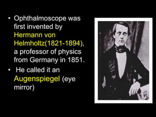
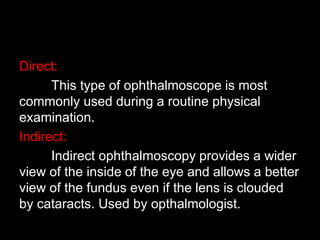
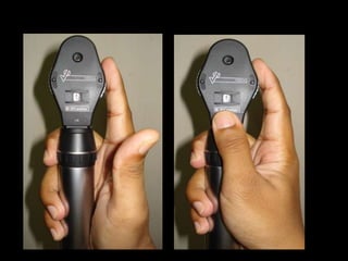
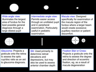
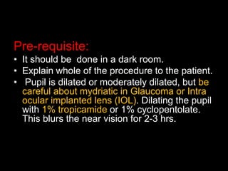
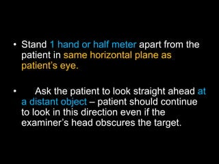
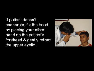
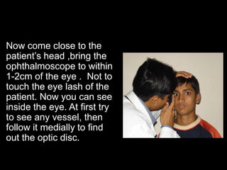
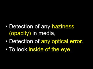
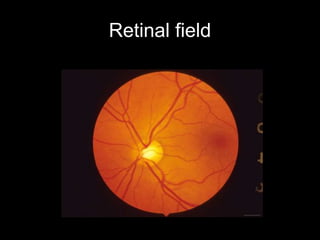
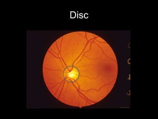
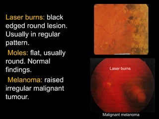
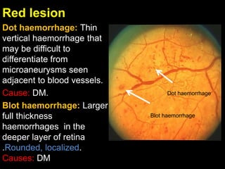
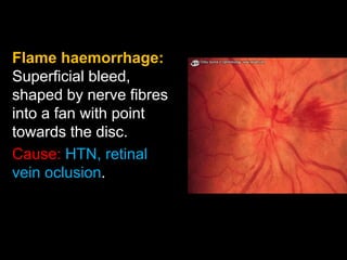
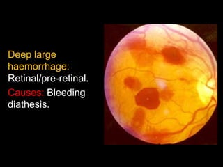
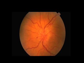
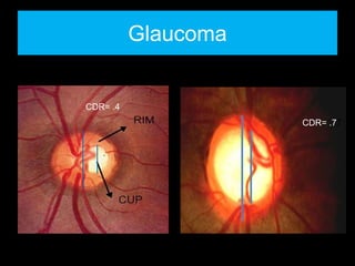
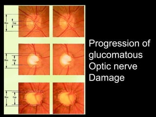
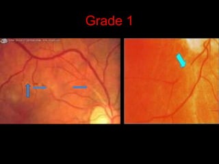
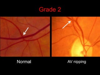
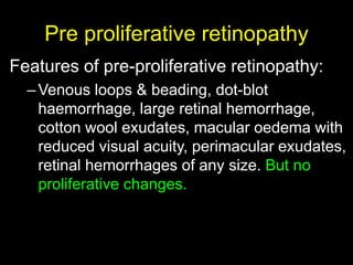
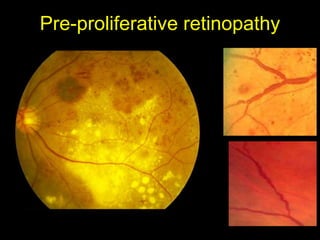
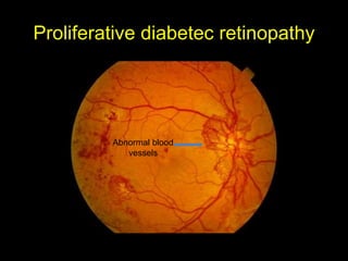
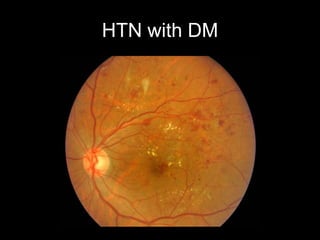
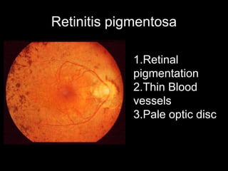
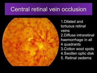
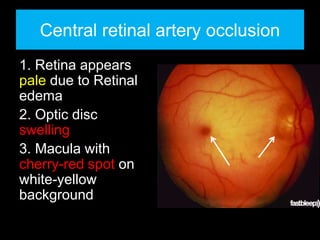
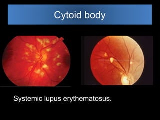
Ophthalmoscopy allows examination of the inside of the eye. It is done using an ophthalmoscope to view the retina and optic disc. It was invented in 1851 and has since improved. During the exam, the pupil is dilated and the ophthalmologist views the retina through different aperture settings and filters on the ophthalmoscope. They examine the optic disc, retina, blood vessels and look for any abnormalities. Common findings include signs of diabetes, hypertension, glaucoma, or other eye conditions. The ophthalmoscopy exam is important for evaluating eye health and detecting underlying diseases.


![CTEV [ clubfoot] DR ARUN LAL ,DR MOHAMED ASHRAF travancore medical college k...](https://cdn.slidesharecdn.com/ss_thumbnails/ctevclubfootdrarunlaldrmohamedashraftravancoremedicalcollegekollamkeralaindia-260208063247-18fc466c-thumbnail.jpg?width=640&height=640&fit=bounds)


