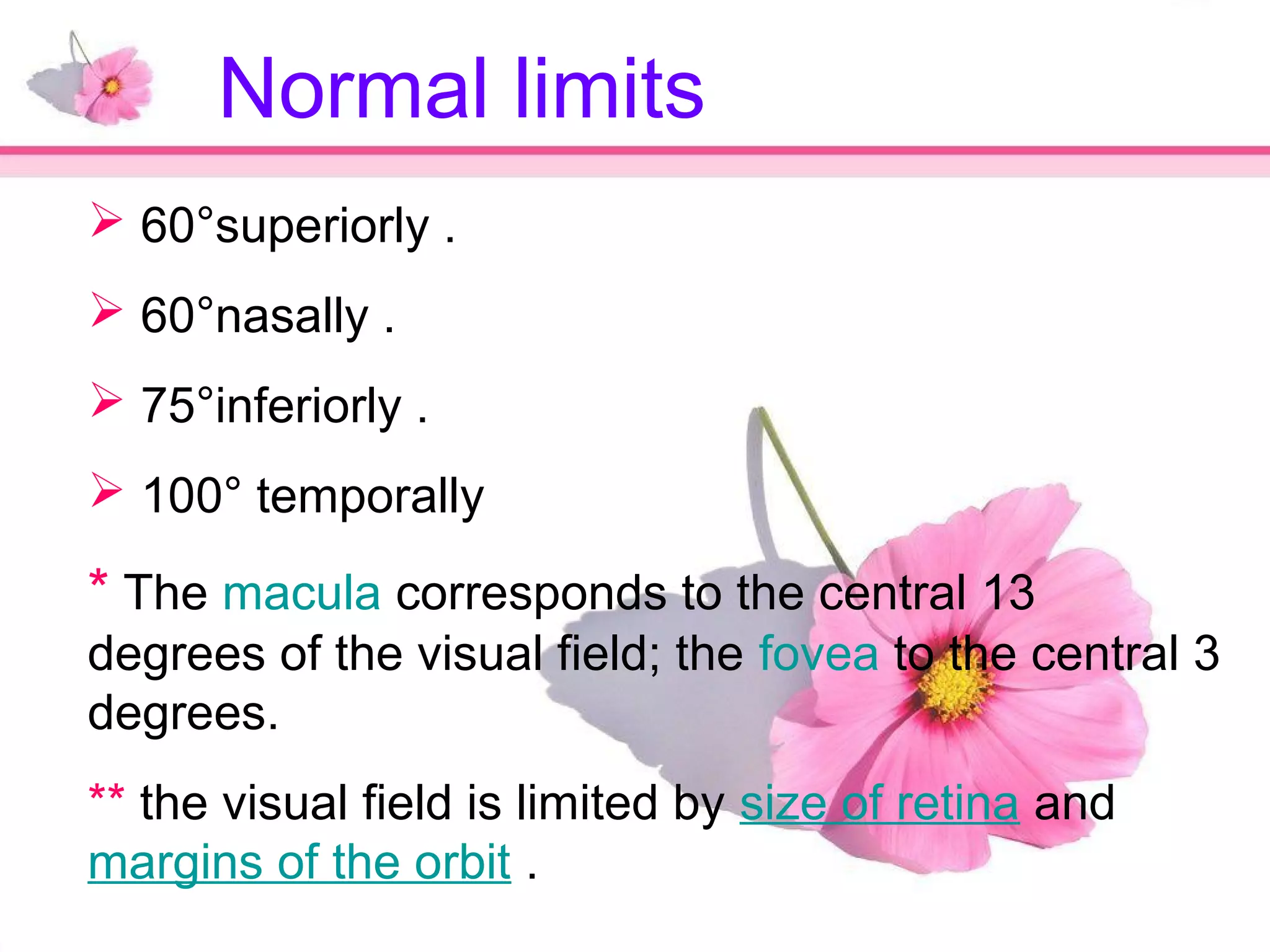
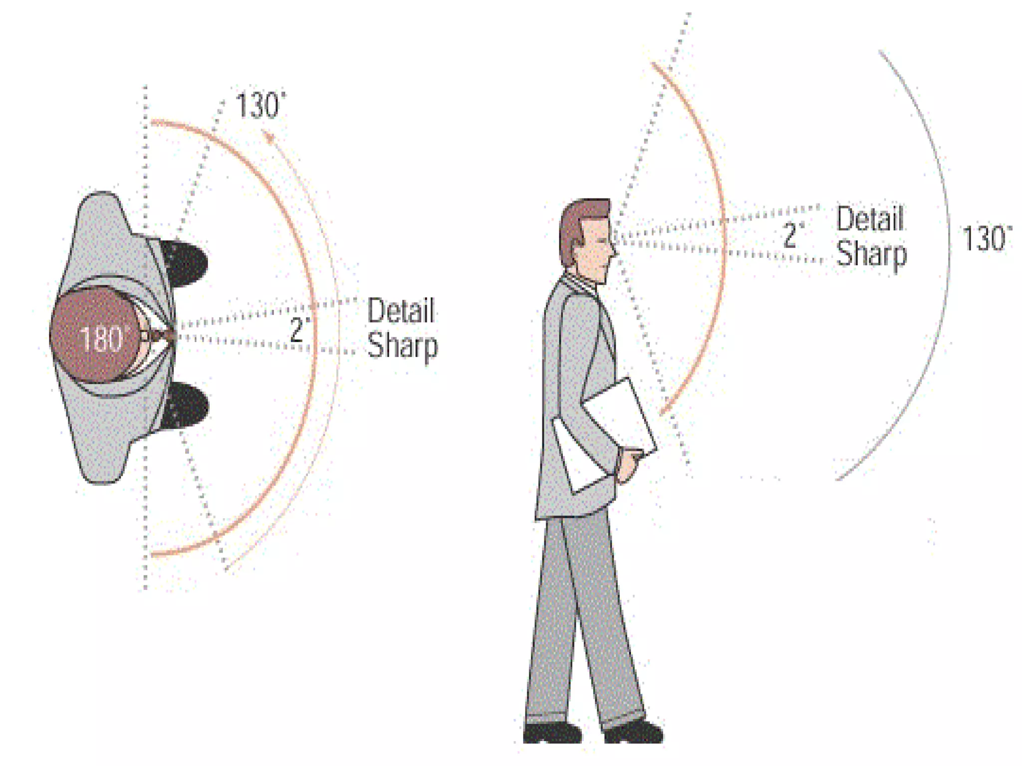
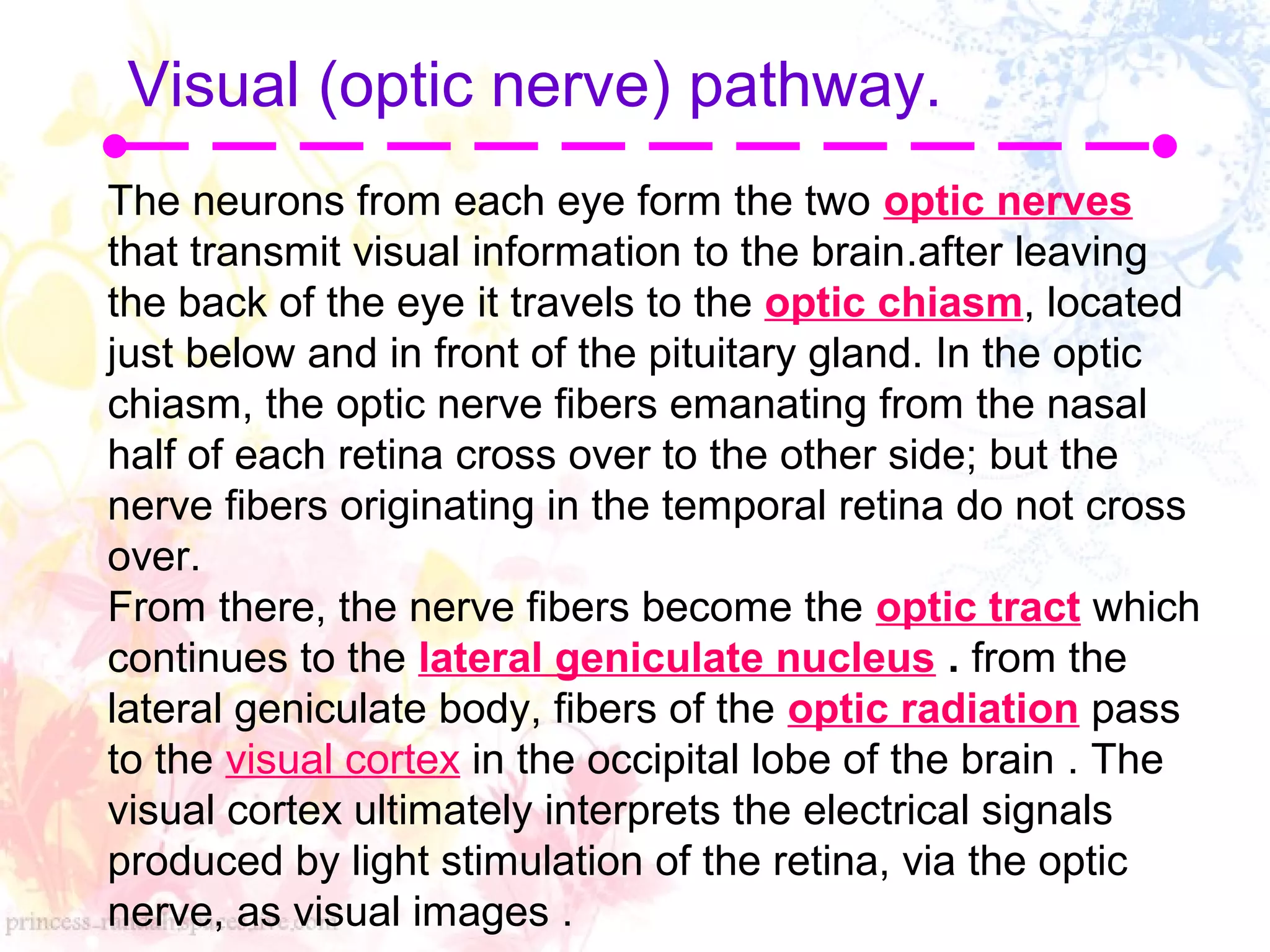
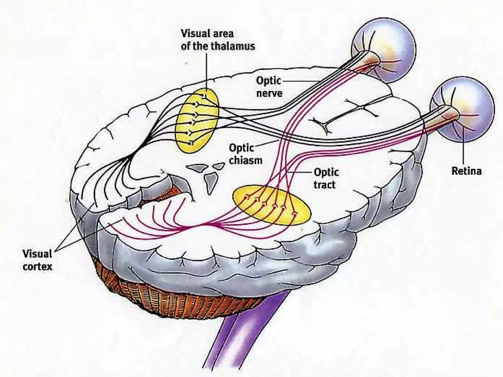
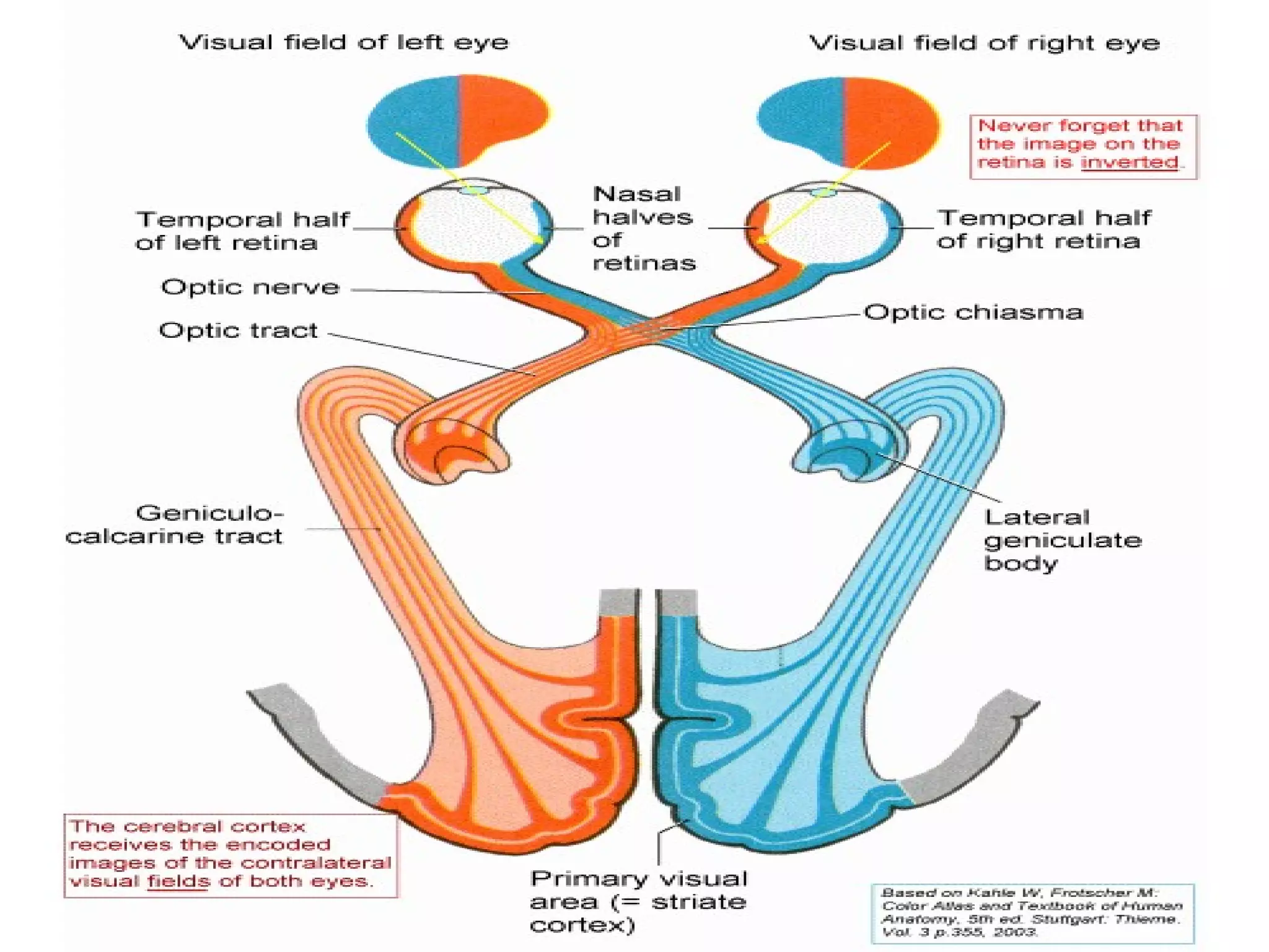
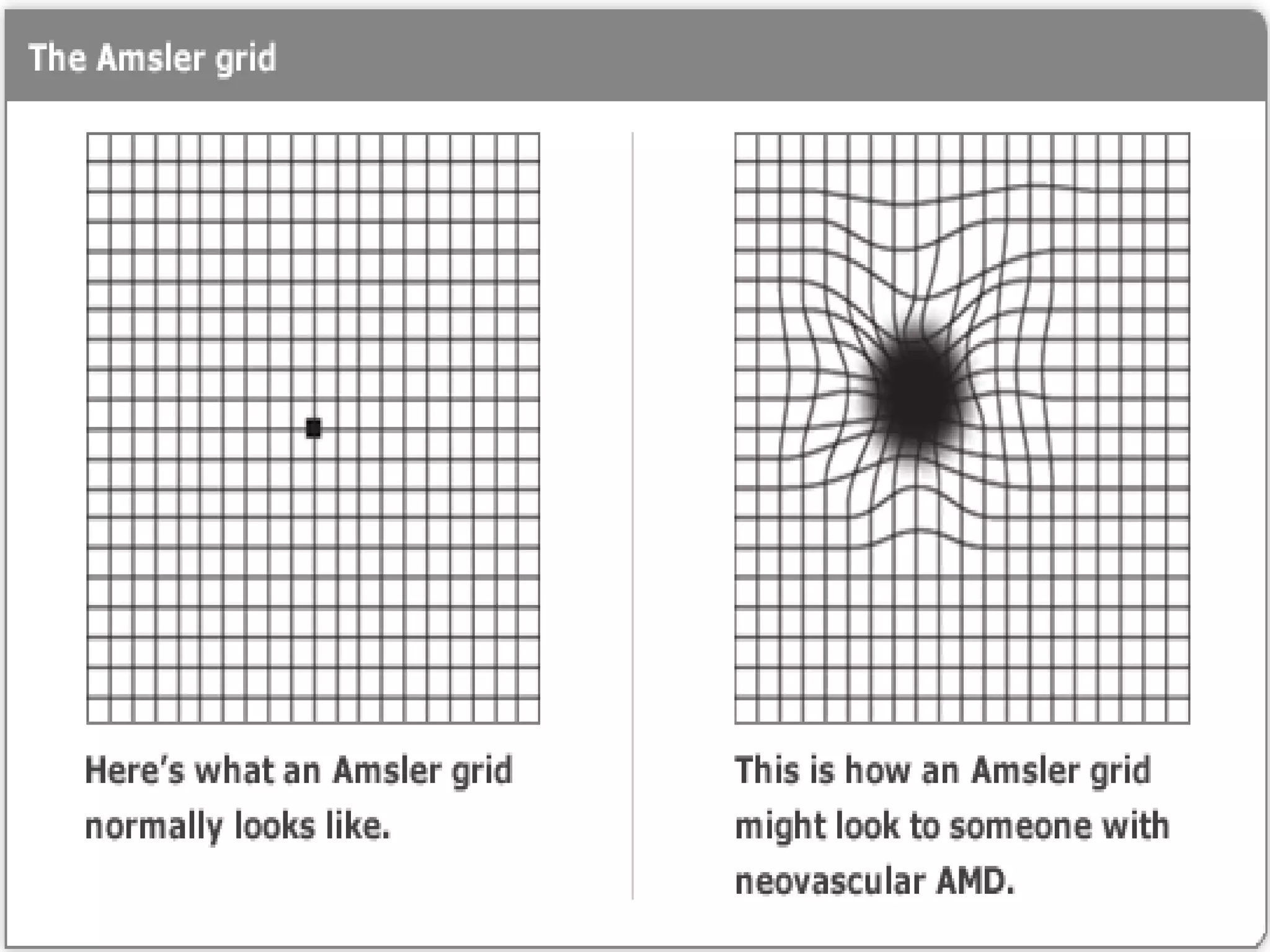
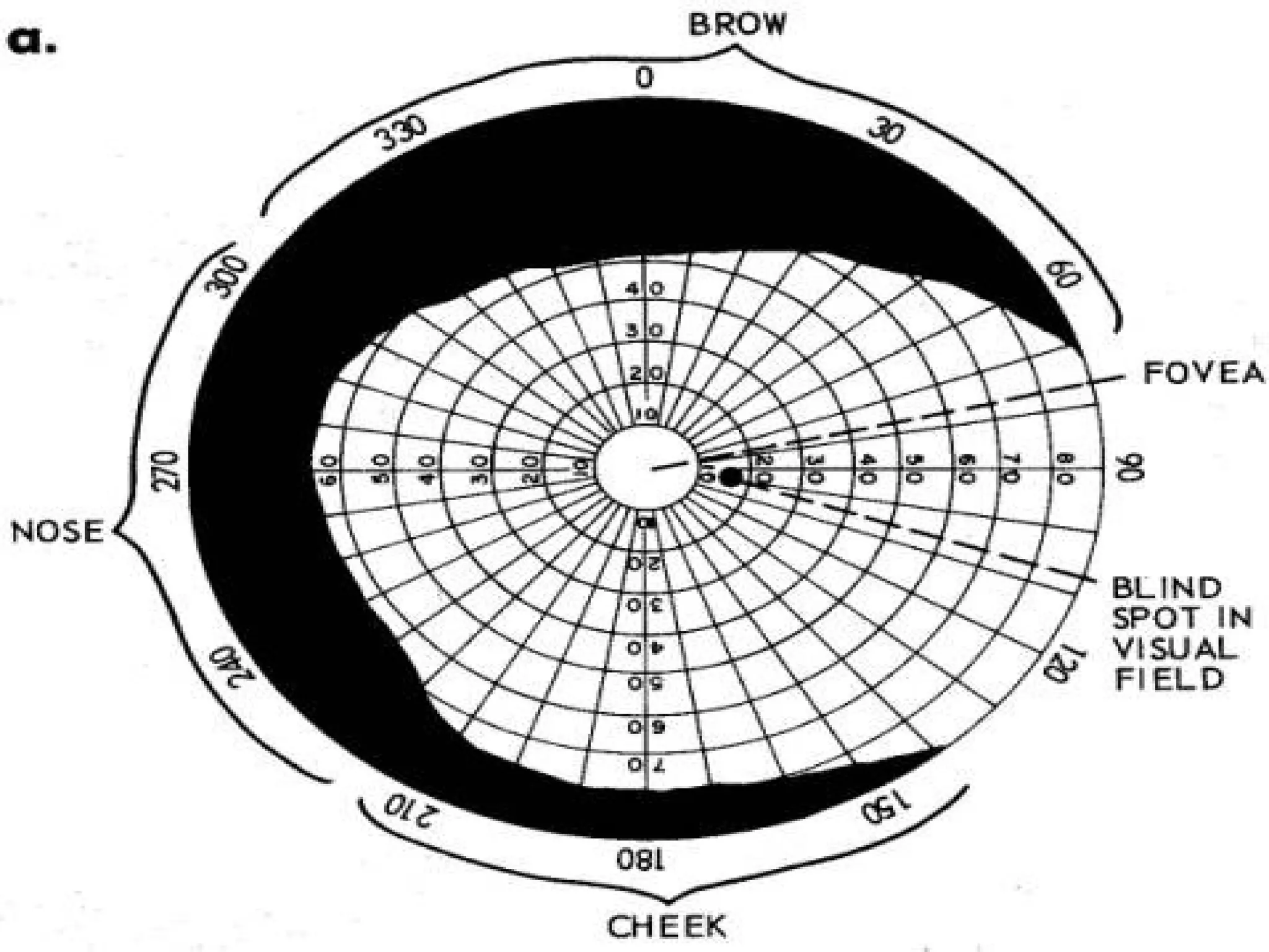
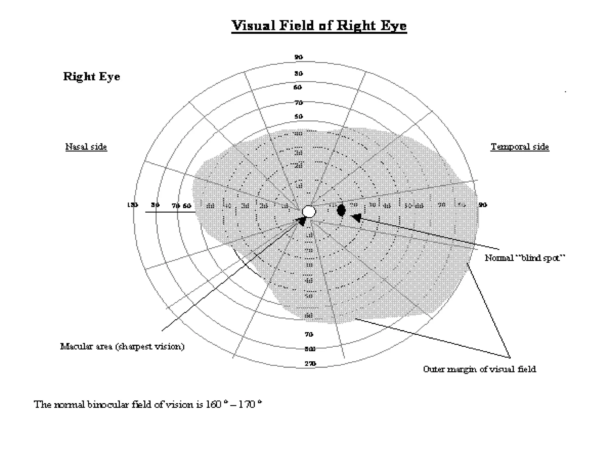
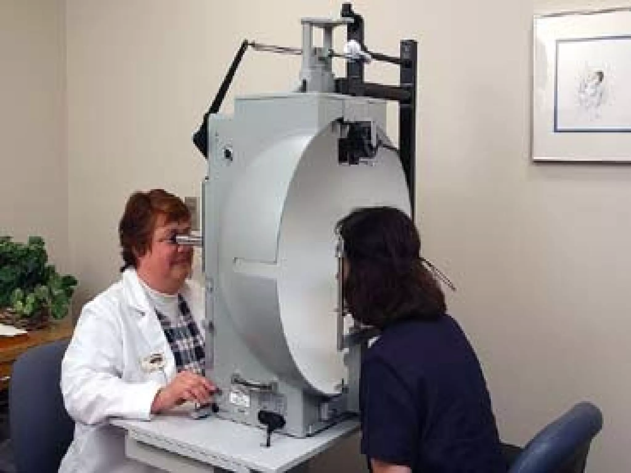
contents
Definition , normal limits .
Visual (optic nerve) pathway.
Examination of visual field .
Visual field defect .
References .
 The visual field refers to the total area
in which objects can be seen in the side
(peripheral) vision while you focus your
eyes on a central point .
 or it is the portion of the subject's
surroundings that can be seen at one
time.
Definition
 60°superiorly .
 60°nasally .
 75°inferiorly .
 100° temporally
* The macula corresponds to the central 13
degrees of the visual field; the fovea to the central 3
degrees.
** the visual field is limited by size of retina and
margins of the orbit .
Normal limits
The neurons from each eye form the two optic nerves
that transmit visual information to the brain.after leaving
the back of the eye it travels to the optic chiasm, located
just below and in front of the pituitary gland. In the optic
chiasm, the optic nerve fibers emanating from the nasal
half of each retina cross over to the other side; but the
nerve fibers originating in the temporal retina do not cross
over.
From there, the nerve fibers become the optic tract which
continues to the lateral geniculate nucleus . from the
lateral geniculate body, fibers of the optic radiation pass
to the visual cortex in the occipital lobe of the brain . The
visual cortex ultimately interprets the electrical signals
produced by light stimulation of the retina, via the optic
nerve, as visual images .
Visual (optic nerve) pathway.
 Confrontation visual field testing
* Sit or stand about 50 cms away from the patient ask
the patient to cover or close one eye and you close
the one opposite ask the subject to stare at your
eye or nose and you do the same .
Examination of the visual field
A visual field examination can detect dysfunction in
central and peripheral vision which may be caused
by various medical conditions such as glaucoma,
stroke, brain tumours or other neurological defects.
Confrontation visual field testing (continued
(
* Starting at the top outer quadrant, move your
fingers (or the pin( in from the side, wiggling
them as they go, and ask the patient to tell you
when he first see your fingers and, as you move
towards the centre, whether they disappear.
* Repeat the process in each quadrant and for
each eye separately.If you detect a defect, re-
examine that area and define it further.
* This technique compares the patient's visual field
with yours, so it does assume normal examiner
visual fields!
 Amsler grid testing
* This assesses the central 10° the visual
field. It detects central and paracentral
scotomas.
* In the test, the person looks with each eye
separately at the small dot in the center of
the grid. Patients with macular disease
may see wavy lines or some lines may be
missing.
Perimetry ( Static and kinetic(
* Static Perimetry :- points of light are
flashed onto a white screen and the
observer is asked to press a button if he or
she sees it. The most common perimeter
used is the automated Humphrey Field
Analyzer. This can assess various
amounts of the visual field (10° to full
field(.
Perimetry ( Static and kinetic
(
* Kinetic perimetry :- Here the patient is
asked to sit 1 meter from a screen with a target
on the center. The eye that isn't tested is
covered during the exam. While the patient
stares at the target the examiner will move an
object toward the patient's visual field. The
patient signals the examiner when the object
comes into view. This exam allows the patient's
visual field to be mapped.
• The most commonly used kinetic test is
Goldmann perimetry.
Visual field defects
* Visual field defect :- a portion of visual field missing. This
may be central or peripheral .
There are many causes of visual field loss :-
 Central field loss occurs with:
 Optic neuropathy
 Macular degeneration
 Macular holes
 Cone dystrophies
 Peripheral field loss occurs with:
 Retinitis pigmentosa
 Chorioretinitis
 Glaucoma
 Retinal detachment
 Lead poisining
Visual field defect
types of visual field defects:-
• Scotoma :- It is a blind spots surrounded
by normal visual field. Types :-
 Central scotoma :- Visual field loss
appears in the central vision .
 Peripheral scotoma :- Visual field
loss appears in the peripheral
region.
Visual field defect
• Hemianopia :- binocular visual defect in each eye's
hemifield.
 Bitemporal hemianopia :- the two halves lost are on the
outside of each eye's peripheral vision, effectively
creating a central visual tunnel.
 Homonymous hemianopia :- the two halves lost are on
the corresponding area of visual field in both eyes, i.e.
either the left or the right half of the visual field.
 Altitudinal hemianopia :- upper or lower halves of visual
field are lost; may be unilateral or bilateral.
 Quadrantanopia :- ( incomplete hemianopia) Visual field
loss in a quarter of the visual field of the eye. The defect
is usually bilateral.
References
 www.medlineplus .com
 Smythies J (1996). "A note on the concept of
the visual field in neurology, psychology, and
visual neuroscience".
 Jay WM (1981). "Visual field defects".
American Family Physician 24 (2): 138–42.
PMID 7258077.
Names
•
‫مهدي‬ ‫صل ح‬ ‫علي‬
‫مهدي‬ ‫صل ح‬ ‫علي‬
•
‫مرزوك‬ ‫حاكم‬ ‫ا‬ ‫عبد‬
•
‫كريم‬ ‫هادي‬ ‫رند‬
‫كريم‬ ‫هادي‬ ‫رند‬
Thank you

visualfield-140206115003-phpapp02.pdf

  • 2.
    contents Definition , normallimits . Visual (optic nerve) pathway. Examination of visual field . Visual field defect . References .
  • 3.
     The visualfield refers to the total area in which objects can be seen in the side (peripheral) vision while you focus your eyes on a central point .  or it is the portion of the subject's surroundings that can be seen at one time. Definition
  • 4.
     60°superiorly . 60°nasally .  75°inferiorly .  100° temporally * The macula corresponds to the central 13 degrees of the visual field; the fovea to the central 3 degrees. ** the visual field is limited by size of retina and margins of the orbit . Normal limits
  • 6.
    The neurons fromeach eye form the two optic nerves that transmit visual information to the brain.after leaving the back of the eye it travels to the optic chiasm, located just below and in front of the pituitary gland. In the optic chiasm, the optic nerve fibers emanating from the nasal half of each retina cross over to the other side; but the nerve fibers originating in the temporal retina do not cross over. From there, the nerve fibers become the optic tract which continues to the lateral geniculate nucleus . from the lateral geniculate body, fibers of the optic radiation pass to the visual cortex in the occipital lobe of the brain . The visual cortex ultimately interprets the electrical signals produced by light stimulation of the retina, via the optic nerve, as visual images . Visual (optic nerve) pathway.
  • 9.
     Confrontation visualfield testing * Sit or stand about 50 cms away from the patient ask the patient to cover or close one eye and you close the one opposite ask the subject to stare at your eye or nose and you do the same . Examination of the visual field A visual field examination can detect dysfunction in central and peripheral vision which may be caused by various medical conditions such as glaucoma, stroke, brain tumours or other neurological defects.
  • 10.
    Confrontation visual fieldtesting (continued ( * Starting at the top outer quadrant, move your fingers (or the pin( in from the side, wiggling them as they go, and ask the patient to tell you when he first see your fingers and, as you move towards the centre, whether they disappear. * Repeat the process in each quadrant and for each eye separately.If you detect a defect, re- examine that area and define it further. * This technique compares the patient's visual field with yours, so it does assume normal examiner visual fields!
  • 12.
     Amsler gridtesting * This assesses the central 10° the visual field. It detects central and paracentral scotomas. * In the test, the person looks with each eye separately at the small dot in the center of the grid. Patients with macular disease may see wavy lines or some lines may be missing.
  • 14.
    Perimetry ( Staticand kinetic( * Static Perimetry :- points of light are flashed onto a white screen and the observer is asked to press a button if he or she sees it. The most common perimeter used is the automated Humphrey Field Analyzer. This can assess various amounts of the visual field (10° to full field(.
  • 16.
    Perimetry ( Staticand kinetic ( * Kinetic perimetry :- Here the patient is asked to sit 1 meter from a screen with a target on the center. The eye that isn't tested is covered during the exam. While the patient stares at the target the examiner will move an object toward the patient's visual field. The patient signals the examiner when the object comes into view. This exam allows the patient's visual field to be mapped. • The most commonly used kinetic test is Goldmann perimetry.
  • 20.
    Visual field defects *Visual field defect :- a portion of visual field missing. This may be central or peripheral . There are many causes of visual field loss :-  Central field loss occurs with:  Optic neuropathy  Macular degeneration  Macular holes  Cone dystrophies  Peripheral field loss occurs with:  Retinitis pigmentosa  Chorioretinitis  Glaucoma  Retinal detachment  Lead poisining
  • 21.
    Visual field defect typesof visual field defects:- • Scotoma :- It is a blind spots surrounded by normal visual field. Types :-  Central scotoma :- Visual field loss appears in the central vision .  Peripheral scotoma :- Visual field loss appears in the peripheral region.
  • 26.
    Visual field defect •Hemianopia :- binocular visual defect in each eye's hemifield.  Bitemporal hemianopia :- the two halves lost are on the outside of each eye's peripheral vision, effectively creating a central visual tunnel.  Homonymous hemianopia :- the two halves lost are on the corresponding area of visual field in both eyes, i.e. either the left or the right half of the visual field.  Altitudinal hemianopia :- upper or lower halves of visual field are lost; may be unilateral or bilateral.  Quadrantanopia :- ( incomplete hemianopia) Visual field loss in a quarter of the visual field of the eye. The defect is usually bilateral.
  • 30.
    References  www.medlineplus .com Smythies J (1996). "A note on the concept of the visual field in neurology, psychology, and visual neuroscience".  Jay WM (1981). "Visual field defects". American Family Physician 24 (2): 138–42. PMID 7258077.
  • 31.
    Names • ‫مهدي‬ ‫صل ح‬‫علي‬ ‫مهدي‬ ‫صل ح‬ ‫علي‬ • ‫مرزوك‬ ‫حاكم‬ ‫ا‬ ‫عبد‬ ‫مرزوك‬ ‫حاكم‬ ‫ا‬ ‫عبد‬ • ‫كريم‬ ‫هادي‬ ‫رند‬ ‫كريم‬ ‫هادي‬ ‫رند‬
  • 32.