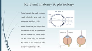
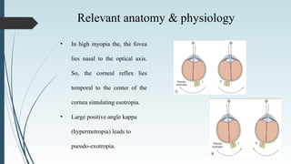
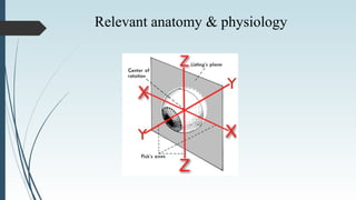
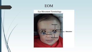
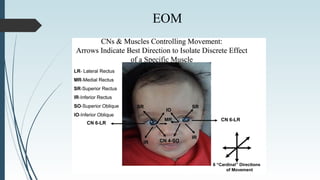
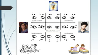
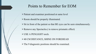
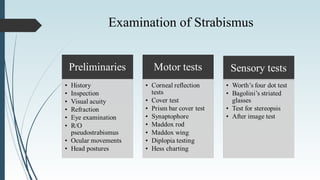
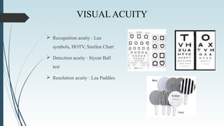
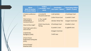
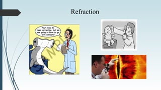
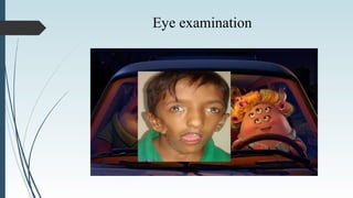
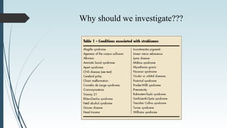
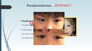
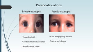
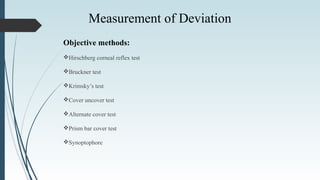
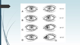
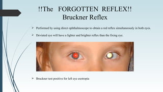
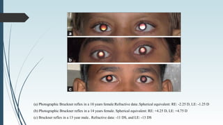
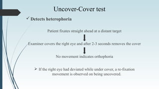
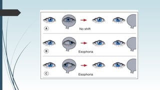
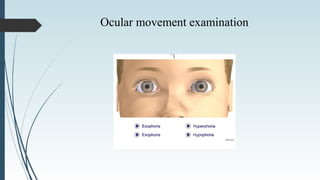
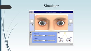
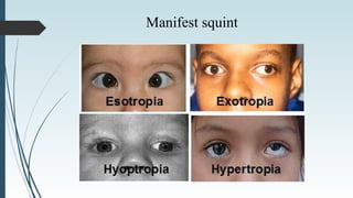
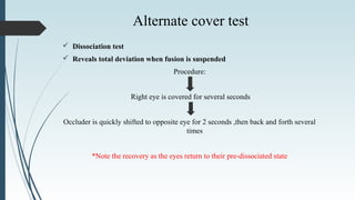
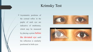
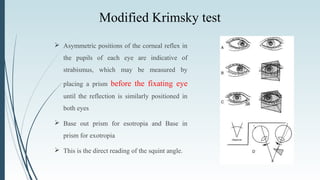
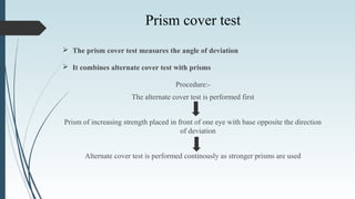
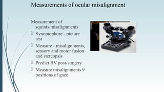
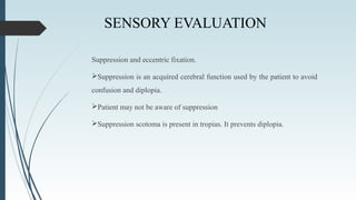
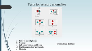
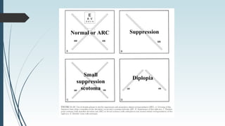
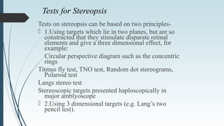
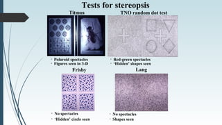
DIAGNOSTIC EVALUATION
FOR STRABISMUS
By
Dr Indeevar V Mishra
Disclaimer..
“The opinions expressed in this presentation and on the following
slides are solely those of the presenter and not necessarily those of
anyone else. The presentor to an extent guarantees the accuracy or
reliability of the information provided herein.”
Axes of the eye
 Visual axis:
Line passes from the fovea to the point of fixation (object of regard). The
normal visual axes (from both eyes) intersects at the point of fixation.
 Optical axis:
It is the line passing through the centre of the cornea and meets the retina
on the nasal side of the fovea
 Fixation axis:
It is the line joining the fixation point and the centre of rotation
Relevant anatomy & physiology
--
• Angle kappa is the angle between
visual (0ptical) axis and the
anatomical (pupillary) axis.
• As the fovea lies just temporal to
the anatomical axis, a light shown
into the cornea will cause reflex
(on the visual axis) just nasal to
the center of the cornea in both
eyes (+ve angle kappa = 5°).
• In high myopia the, the fovea
lies nasal to the optical axis.
So, the corneal reflex lies
temporal to the center of the
cornea simulating esotropia.
• Large positive angle kappa
(hypermetropia) leads to
pseudo-exotropia.
Relevant anatomy & physiology
EOM
EOM
Patient and examiner positioned at same level
Room should be properly illuminated.
Sit in front of the patient so that BE eyes can be seen simultaneously.
Remove any Spectacles.( to remove prismatic effect)
USE A PENLIGHT torch.
40 CM DISTANCE, SHINE ON FOREHEAD
The 9 diagnostic positions should be examined.
Points to Remember for EOM
Examination of Strabismus
HISTORY
Complaint & HOPC
•Age of onset of deviation…..
•Is the deviation constant or intermittent?
•Is the deviation present for distance, near or both?
•Is it unilateral or alternating?
•Is it present only when the patient is inattentive or fatigued?
•Is it associated with trauma or physical stress?
Birth history
o Regarding Pregnancy & Delivery
o Developmental Milestones(Delay)
Family History
o Squint/ Refractive Error / Lazy eye
Personal History: Are there any other medical problems?
o DM /HTN / Thyroid
Treatment History
o Glasses /Occlusion Therapy /Orthoptic Exercise / Prev. Surgery
o Is there a history of toxin or medication exposure?
Old photographs..
INSPECTION
 Lid Fissure:-
• Ptosis
• Mongoloid/Anti mongoloid
• Exophthalmos / enophthalmos
• Nasal Bridge
• Closure of one eye in bright light
• Epicanthal folds
 Head Posture (AHP)
• Face Turn (Right/Left)
• Head Tilt (Right/Left Shoulder)
• Chin (Elevation/Depression)
 Facial Asymmetry
 Fixation Preference
 Nystagmus
Head posture
VISUAL ACUITY
VISUAL ACUITY
 Recognition acuity : Lea
symbols, HOTV, Snellen Chart
 Detection acuity : Stycar Ball
test
 Resolution acuity : Lea Paddles
Refraction
Refraction why is it so important???
Eye examination
Why should we investigate???
Before Jumping to
conclusions!!!
Pseudostrabismus…BEWARE!!
What is it???
• Pseudoesotropia as a result of
a broad bridge of the nose.
• Telecanthus or hypertelorism
can also result in mis-diagnosis
of strabismus.
Pseudo-deviations
Pseudo-esotropia Pseudo-exotropia
•Epicanthic folds
•Short interpupillary distance
•Negative angle kappa
•Wide interpupillary distance
•Positive angle kappa
Objective methods:
Hirschberg corneal reflex test
Bruckner test
Krimsky’s test
Cover uncover test
Alternate cover test
Prism bar cover test
Synoptophore
Measurement of Deviation
Subjective methods:
Maddox rod test
Maddox wing test
Maddox tangent
Double maddox rod
Maddox double prism
Hess screen
Red green glass test
Diplopia field
Measurement of Deviation
Motor evaluation
Two principle methods of evaluating ocular motility
are:
1.Observation of ocular ductions, which are the actual monocular movements of the
eye.
2.Observation of binocular ocular alignment, using cover/uncover and alternate
cover testing.
Reflex at border of pupil = 15°° Reflex at limbus = 45°°
Hirschberg‘s test
 Used as an initial screening test for strabismus.
 Amount of deviation: 1mm = 7° or 15Δ
Hirschberg Simulator
 Performed by using direct ophthalmoscope to obtain a red reflex simultaneously in both eyes.
 Deviated eye will have a lighter and brighter reflex than the fixing eye.
 Bruckner test positive for left eye esotropia
!!The FORGOTTEN REFLEX!!
Bruckner Reflex
(a) Photographic Bruckner reflex in a 10 years female:Refractive data:.Spherical equivalent: RE: -2.25 D, LE: -1.25 D
(b) Photographic Bruckner reflex in a 14 years female. Spherical equivalent: RE: +4.25 D, LE: +4.75 D
(c) Bruckner reflex in a 13 year male.. Refractive data: -11 DS, and LE: -13 DS
Pre requisites for cover test
 Fixation must be adequate
 Must be done for distance & near
 With & without glasses
 Palm of hand or occluder for cover
 Spielman transluscent occluder
Detects heterophoria
Patient fixates straight ahead at a distant target
Examiner covers the right eye and after 2-3 seconds removes the cover
No movement indicates orthophoria
 If the right eye had deviated while under cover, a re-fixation
movement is observed on being uncovered.
Uncover-Cover test
Ocular movement examination
Simulator
Presence of deviation: Phoria or tropia
 Type of deviation Eso or Exo
 Eccentric fixation
 Amblyopia
 Degree of alternation
 Pseudoptosis
 Latent nystagmus
 Measurement of deviation
Inference of cover test
Drawbacks of cover test..
Misses the following….or may miss…
1.Small heterophoria
2.Small angle esotropia
3.Microtropia
4.Monofixation syndrome
5.Cyclodeviation
Manifest squint
 Dissociation test
 Reveals total deviation when fusion is suspended
Procedure:
Right eye is covered for several seconds
Occluder is quickly shifted to opposite eye for 2 seconds ,then back and forth several
times
*Note the recovery as the eyes return to their pre-dissociated state
Alternate cover test
Simulator
Krimsky Test
 Asymmetric positions of
the corneal reflex in the
pupils of each eye are
indicative of strabismus,
which may be measured
by placing a prism before
the deviated eye until
the reflection is similarly
positioned in both eyes
Modified Krimsky test
 Asymmetric positions of the corneal reflex in
the pupils of each eye are indicative of
strabismus, which may be measured by
placing a prism before the fixating eye
until the reflection is similarly positioned in
both eyes
 Base out prism for esotropia and Base in
prism for exotropia
 This is the direct reading of the squint angle.
 The prism cover test measures the angle of deviation
 It combines alternate cover test with prisms
Procedure:-
The alternate cover test is performed first
Prism of increasing strength placed in front of one eye with base opposite the direction
of deviation
Alternate cover test is performed continously as stronger prisms are used
Prism cover test
Amplitude of refixation gradually decreases
End point reached when no movement is seen
To ensure maximum angle is found , prism strength is increased until movement is
observed in opposite direction
Then reduced again to find neutral value
Angle of deviation equals the strength of the prism
Prism Cover Test
 Measure
squint/misalignment
 Single prism/prism bar
 Primary position or in all
positions of gaze
Dissimilar image tests
Maddox wing
Maddox rod
•Dissociates eyes for near
fixation (1/3 m)
•Measures heterophoria
•White spot converted into red streak
•Cannot differentiate tropia from phoria
Measurements of ocular misalignment
Measurement of
squints/misalignments
 Synoptophore - picture
test
 Measure - misalignments,
sensory and motor fusion
and stereopsis
 Predict BV post-surgery
 Measure misalignments 9
positions of gaze
SENSORY EVALUATION
Suppression and eccentric fixation.
Suppression is an acquired cerebral function used by the patient to avoid
confusion and diplopia.
Patient may not be aware of suppression
Suppression scotoma is present in tropias. It prevents diplopia.
Tests for sensory anomalies
Worth four-dot test
a - Prior to use of glasses
b - Normal
c - Left suppression/ amblyopia
d - Right suppression/ amblyopia
e - Diplopia
Bagolini striated glasses
Normal or ARC
Diplopia
Suppression
Small
suppression
scotoma
Tests for Stereopsis
Tests on stereopsis can be based on two principles-
 1.Using targets which lie in two planes, but are so
constructed that they stimulate disparate retinal
elements and give a three dimensional effect, for
example:
Circular perspective diagram such as the concentric
rings
Titmus fly test, TNO test, Random dot stereograms,
Polaroid test
Langs stereo test
Stereoscopic targets presented haploscopically in
major amblyoscope
 2.Using 3 dimensional targets (e.g. Lang’s two
pencil test).
 Qualitative tests for
Stereopsis:
 Lang’s 2 pencil test
 Synoptophore
 Quantitative tests for
Stereopsis:
 Random dot test
 TNO Test
 Lang’s stereo test
Tests for stereopsis
Titmus
• Red-green spectacles
TNO random dot test
• ‘Hidden’ shapes seen
• Polaroid spectacles
• Figures seen in 3-D
Lang
• No spectacles
Frisby
• ‘Hidden’ circle seen
• No spectacles
• Shapes seen
Thank you!!!

Evaluation of squint - The Basics

  • 1.
  • 2.
    Disclaimer.. “The opinions expressedin this presentation and on the following slides are solely those of the presenter and not necessarily those of anyone else. The presentor to an extent guarantees the accuracy or reliability of the information provided herein.”
  • 3.
    Axes of theeye  Visual axis: Line passes from the fovea to the point of fixation (object of regard). The normal visual axes (from both eyes) intersects at the point of fixation.  Optical axis: It is the line passing through the centre of the cornea and meets the retina on the nasal side of the fovea  Fixation axis: It is the line joining the fixation point and the centre of rotation
  • 4.
    Relevant anatomy &physiology -- • Angle kappa is the angle between visual (0ptical) axis and the anatomical (pupillary) axis. • As the fovea lies just temporal to the anatomical axis, a light shown into the cornea will cause reflex (on the visual axis) just nasal to the center of the cornea in both eyes (+ve angle kappa = 5°).
  • 5.
    • In highmyopia the, the fovea lies nasal to the optical axis. So, the corneal reflex lies temporal to the center of the cornea simulating esotropia. • Large positive angle kappa (hypermetropia) leads to pseudo-exotropia. Relevant anatomy & physiology
  • 6.
  • 7.
  • 8.
  • 10.
    Patient and examinerpositioned at same level Room should be properly illuminated. Sit in front of the patient so that BE eyes can be seen simultaneously. Remove any Spectacles.( to remove prismatic effect) USE A PENLIGHT torch. 40 CM DISTANCE, SHINE ON FOREHEAD The 9 diagnostic positions should be examined. Points to Remember for EOM
  • 11.
  • 12.
    HISTORY Complaint & HOPC •Ageof onset of deviation….. •Is the deviation constant or intermittent? •Is the deviation present for distance, near or both? •Is it unilateral or alternating? •Is it present only when the patient is inattentive or fatigued? •Is it associated with trauma or physical stress?
  • 13.
    Birth history o RegardingPregnancy & Delivery o Developmental Milestones(Delay) Family History o Squint/ Refractive Error / Lazy eye Personal History: Are there any other medical problems? o DM /HTN / Thyroid Treatment History o Glasses /Occlusion Therapy /Orthoptic Exercise / Prev. Surgery o Is there a history of toxin or medication exposure? Old photographs..
  • 14.
    INSPECTION  Lid Fissure:- •Ptosis • Mongoloid/Anti mongoloid • Exophthalmos / enophthalmos • Nasal Bridge • Closure of one eye in bright light • Epicanthal folds  Head Posture (AHP) • Face Turn (Right/Left) • Head Tilt (Right/Left Shoulder) • Chin (Elevation/Depression)  Facial Asymmetry  Fixation Preference  Nystagmus
  • 15.
  • 16.
  • 17.
    VISUAL ACUITY  Recognitionacuity : Lea symbols, HOTV, Snellen Chart  Detection acuity : Stycar Ball test  Resolution acuity : Lea Paddles
  • 19.
  • 20.
    Refraction why isit so important???
  • 21.
  • 22.
    Why should weinvestigate???
  • 23.
  • 24.
    Pseudostrabismus…BEWARE!! What is it??? •Pseudoesotropia as a result of a broad bridge of the nose. • Telecanthus or hypertelorism can also result in mis-diagnosis of strabismus.
  • 25.
    Pseudo-deviations Pseudo-esotropia Pseudo-exotropia •Epicanthic folds •Shortinterpupillary distance •Negative angle kappa •Wide interpupillary distance •Positive angle kappa
  • 26.
    Objective methods: Hirschberg cornealreflex test Bruckner test Krimsky’s test Cover uncover test Alternate cover test Prism bar cover test Synoptophore Measurement of Deviation
  • 27.
    Subjective methods: Maddox rodtest Maddox wing test Maddox tangent Double maddox rod Maddox double prism Hess screen Red green glass test Diplopia field Measurement of Deviation
  • 28.
    Motor evaluation Two principlemethods of evaluating ocular motility are: 1.Observation of ocular ductions, which are the actual monocular movements of the eye. 2.Observation of binocular ocular alignment, using cover/uncover and alternate cover testing.
  • 29.
    Reflex at borderof pupil = 15°° Reflex at limbus = 45°° Hirschberg‘s test  Used as an initial screening test for strabismus.  Amount of deviation: 1mm = 7° or 15Δ
  • 30.
  • 32.
     Performed byusing direct ophthalmoscope to obtain a red reflex simultaneously in both eyes.  Deviated eye will have a lighter and brighter reflex than the fixing eye.  Bruckner test positive for left eye esotropia !!The FORGOTTEN REFLEX!! Bruckner Reflex
  • 33.
    (a) Photographic Brucknerreflex in a 10 years female:Refractive data:.Spherical equivalent: RE: -2.25 D, LE: -1.25 D (b) Photographic Bruckner reflex in a 14 years female. Spherical equivalent: RE: +4.25 D, LE: +4.75 D (c) Bruckner reflex in a 13 year male.. Refractive data: -11 DS, and LE: -13 DS
  • 34.
    Pre requisites forcover test  Fixation must be adequate  Must be done for distance & near  With & without glasses  Palm of hand or occluder for cover  Spielman transluscent occluder
  • 35.
    Detects heterophoria Patient fixatesstraight ahead at a distant target Examiner covers the right eye and after 2-3 seconds removes the cover No movement indicates orthophoria  If the right eye had deviated while under cover, a re-fixation movement is observed on being uncovered. Uncover-Cover test
  • 37.
  • 38.
  • 39.
    Presence of deviation:Phoria or tropia  Type of deviation Eso or Exo  Eccentric fixation  Amblyopia  Degree of alternation  Pseudoptosis  Latent nystagmus  Measurement of deviation Inference of cover test
  • 40.
    Drawbacks of covertest.. Misses the following….or may miss… 1.Small heterophoria 2.Small angle esotropia 3.Microtropia 4.Monofixation syndrome 5.Cyclodeviation
  • 41.
  • 42.
     Dissociation test Reveals total deviation when fusion is suspended Procedure: Right eye is covered for several seconds Occluder is quickly shifted to opposite eye for 2 seconds ,then back and forth several times *Note the recovery as the eyes return to their pre-dissociated state Alternate cover test
  • 43.
  • 44.
    Krimsky Test  Asymmetricpositions of the corneal reflex in the pupils of each eye are indicative of strabismus, which may be measured by placing a prism before the deviated eye until the reflection is similarly positioned in both eyes
  • 45.
    Modified Krimsky test Asymmetric positions of the corneal reflex in the pupils of each eye are indicative of strabismus, which may be measured by placing a prism before the fixating eye until the reflection is similarly positioned in both eyes  Base out prism for esotropia and Base in prism for exotropia  This is the direct reading of the squint angle.
  • 46.
     The prismcover test measures the angle of deviation  It combines alternate cover test with prisms Procedure:- The alternate cover test is performed first Prism of increasing strength placed in front of one eye with base opposite the direction of deviation Alternate cover test is performed continously as stronger prisms are used Prism cover test
  • 47.
    Amplitude of refixationgradually decreases End point reached when no movement is seen To ensure maximum angle is found , prism strength is increased until movement is observed in opposite direction Then reduced again to find neutral value Angle of deviation equals the strength of the prism
  • 49.
    Prism Cover Test Measure squint/misalignment  Single prism/prism bar  Primary position or in all positions of gaze
  • 51.
    Dissimilar image tests Maddoxwing Maddox rod •Dissociates eyes for near fixation (1/3 m) •Measures heterophoria •White spot converted into red streak •Cannot differentiate tropia from phoria
  • 52.
    Measurements of ocularmisalignment Measurement of squints/misalignments  Synoptophore - picture test  Measure - misalignments, sensory and motor fusion and stereopsis  Predict BV post-surgery  Measure misalignments 9 positions of gaze
  • 53.
    SENSORY EVALUATION Suppression andeccentric fixation. Suppression is an acquired cerebral function used by the patient to avoid confusion and diplopia. Patient may not be aware of suppression Suppression scotoma is present in tropias. It prevents diplopia.
  • 54.
    Tests for sensoryanomalies Worth four-dot test a - Prior to use of glasses b - Normal c - Left suppression/ amblyopia d - Right suppression/ amblyopia e - Diplopia
  • 55.
  • 56.
  • 57.
    Tests for Stereopsis Testson stereopsis can be based on two principles-  1.Using targets which lie in two planes, but are so constructed that they stimulate disparate retinal elements and give a three dimensional effect, for example: Circular perspective diagram such as the concentric rings Titmus fly test, TNO test, Random dot stereograms, Polaroid test Langs stereo test Stereoscopic targets presented haploscopically in major amblyoscope  2.Using 3 dimensional targets (e.g. Lang’s two pencil test).
  • 58.
     Qualitative testsfor Stereopsis:  Lang’s 2 pencil test  Synoptophore  Quantitative tests for Stereopsis:  Random dot test  TNO Test  Lang’s stereo test
  • 59.
    Tests for stereopsis Titmus •Red-green spectacles TNO random dot test • ‘Hidden’ shapes seen • Polaroid spectacles • Figures seen in 3-D Lang • No spectacles Frisby • ‘Hidden’ circle seen • No spectacles • Shapes seen
  • 60.