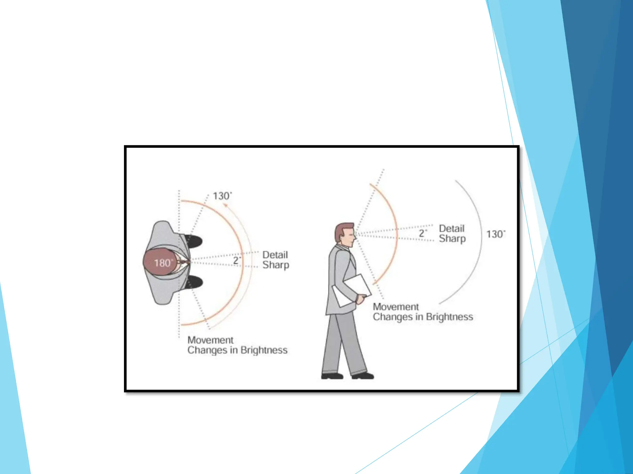
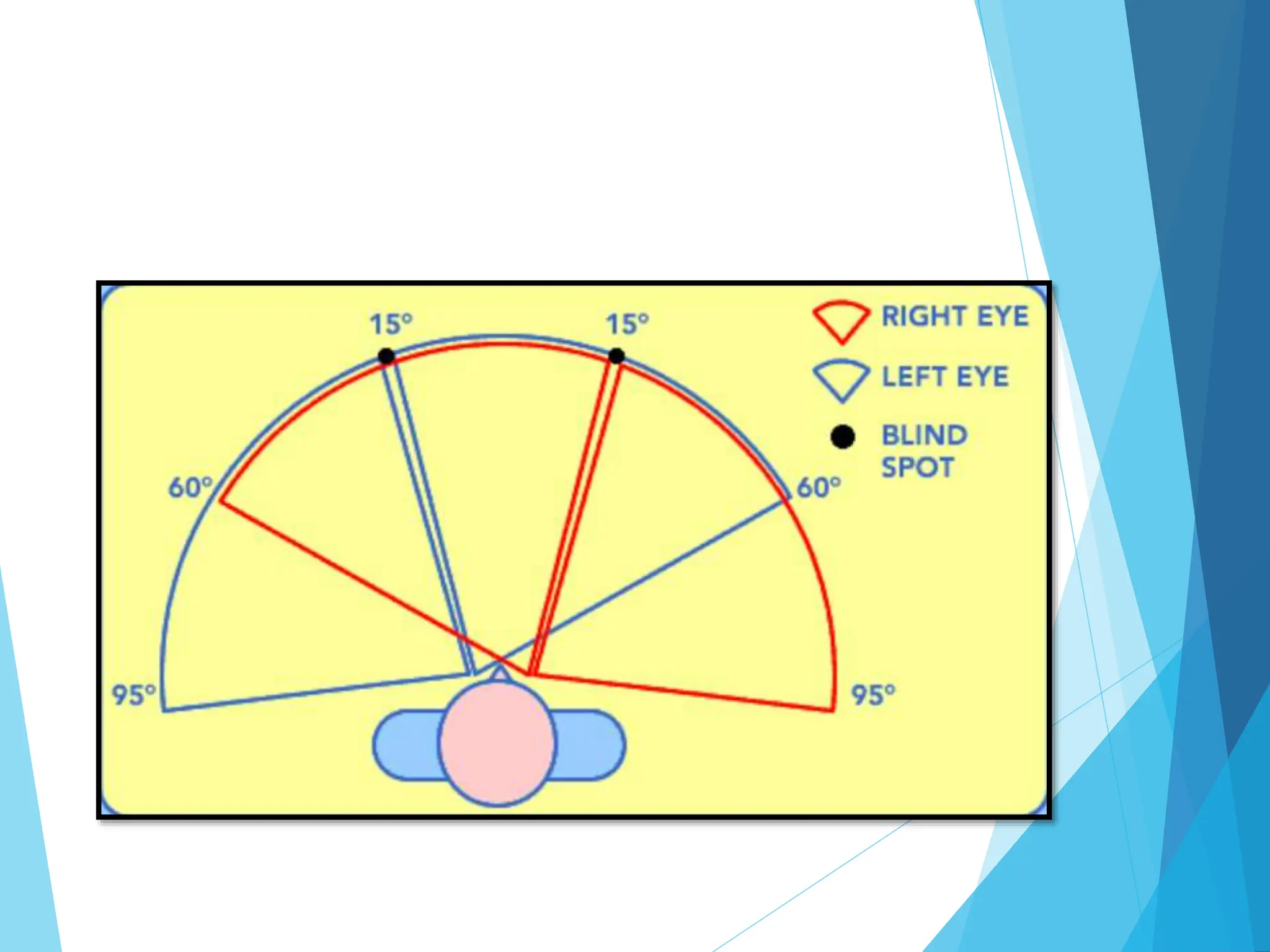
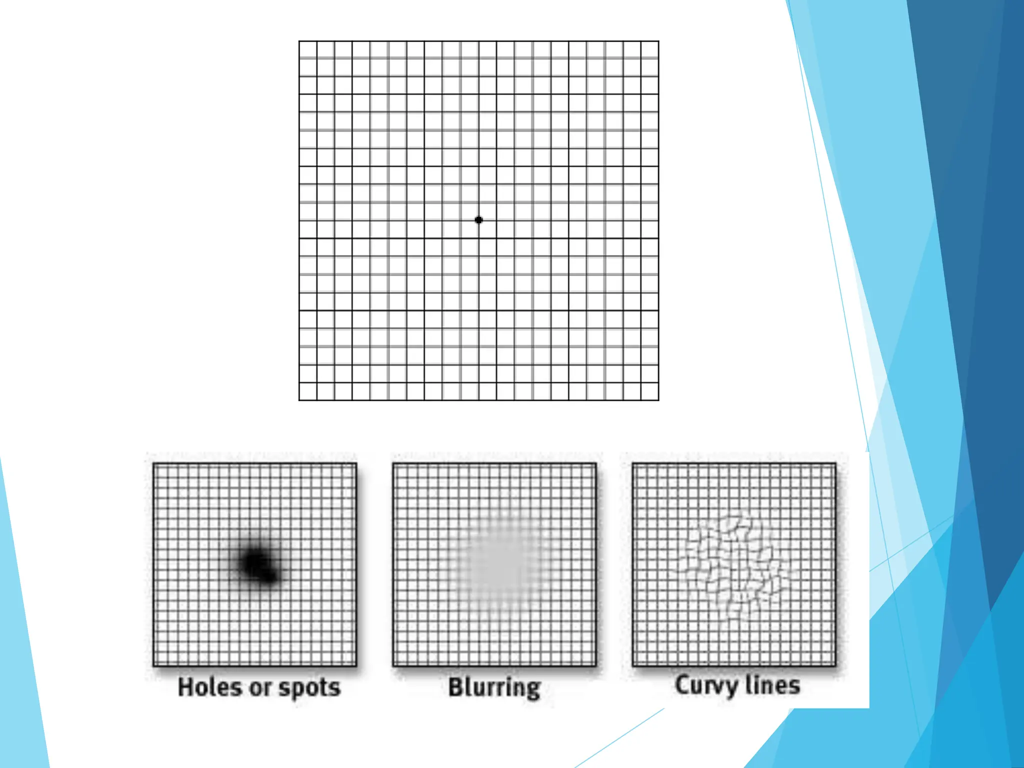
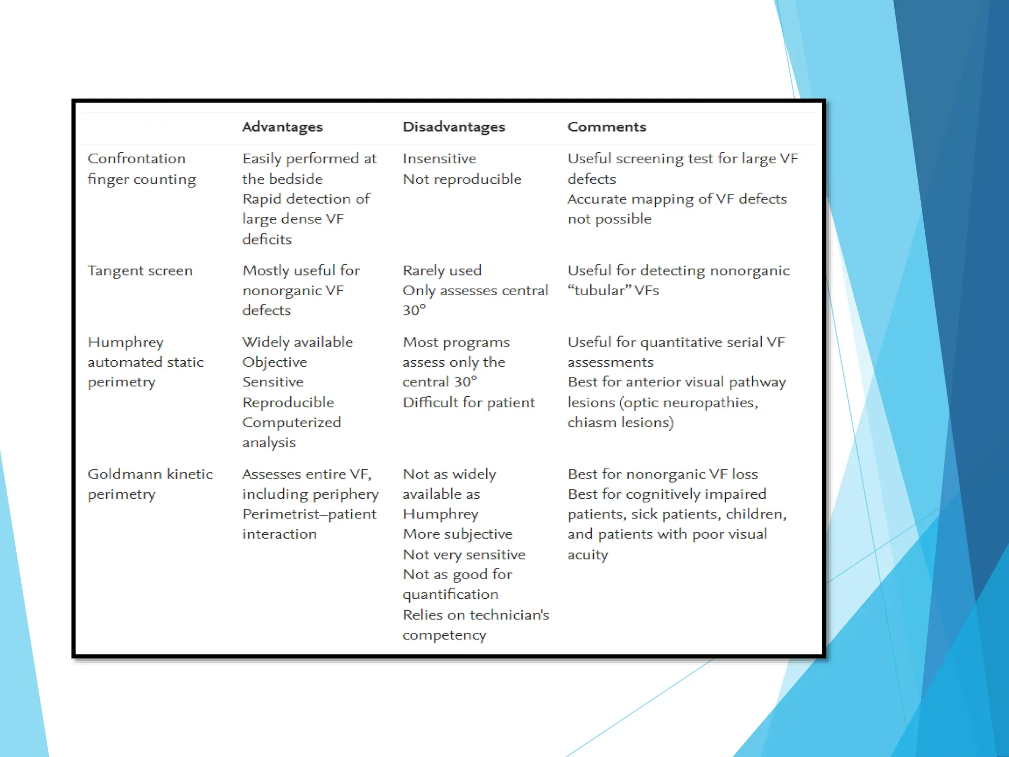
VISUALFIELD
EXAMINATION
INTRODUCTION
 Visual field is the visual area that is perceived
simultaneously by a fixating eye
 Visual function assessment is integral to the
evaluation and management of glaucoma.2
 Visual field testing can be performed by various
methods, including confrontation technique, amsler
grid, tangent screen, kinetic perimetry, or static
perimetry.2
 Defined visual field as island of vision in the sea of darkness.
 Hill of vision is a 3D representation of the retinal light sensitivity
 Sea represents the areas of no light perception
TRAQUAIR FIELD OF VISION
 Visual acuity is sharpest at the very top
of hill (fovea) and then declines
progressively towards the periphery, the
nasal slope being steeper than the
temporal.
 The ‘bottomless pit’ of blind spot is
located temporally 15˚, slightly below
the horizontal meridian 1.5˚, diameter
7.5˚
NORMAL LIMITS OF VISUAL
FIELD
PHYSIOLOGICAL BLIND
SPOT
 Every eye has a blind spot, where the nerve exits the eye and
blood vessels enter and exit
 There are no retinal photoreceptors in this area, therefore, no
vision
 It corresponds to the location of the optic nerve head in the
retina, it should show up on any visual field test.
 The blind spot is centered slightly below the horizontal midline
and 15.5 degrees temporal relative to fixation (macula),
measuring 5.5 degrees wide and 7.5 degrees high
SIGNIFICANCE OF VISUAL
FIELD TESTING
Find out the extent of VF
To diagnose and detect diseases as well
as extent of damage caused in VF by
the disease
To locate possible lesion in
neurological disorder
To find out the progression of diseases
CONFRONTATION TEST3
 The visual fields of both eyes overlap; therefore
each eye is tested independently
 The patient should cover their right eye with their
right hand (vice versa when testing the opposite
eye).
 With the examiner seated directly across from the
patient, the patient should direct their gaze to the
corresponding eye of the examiner.
 The testing itself can be performed using stationary
or moving targets (disk mounted on a stick or
examiner's fingers).
 A moving target should start outside the usual 180º
visual field, then move slowly to a more central position
until the patient confirms visualization of the target.
 To perform stationary testing, the examiner holds up a
certain number of fingers peripherally, equidistant
between the examiner and the patient.
 The patient is asked to correctly identify the number of
fingers.
 All four quadrants (upper and lower, temporal and nasal)
should be tested.
DISADVANTAGE
 With this approach alone, early (or even moderate)
visual field defect often go unnoticed, particularly
if one eye is affected
 It is a subjective test and sometimes
misinterpretations of report may be presented if the
patient has poor compliance, usually pediatric and
geratric patients
 It is the screening test and the diagnosis can’t be
made alone with this test
AMSLER GRID2
 A basic visual field test for central vision
 It is a pattern of straight lines that makes a grid of
many equal squares
 Patient look at a dot in the middle of the grid and
describe any areas that may appear wavy, blurry or
blank
 The Amsler grid is commonly used at home by
people with AMD
 This test only measures the middle of the visual
field, but is a simple yet helpful tool for monitoring
vision changes
TANGENT SCREEN
(BJERRUM)
 A type of kinetic perimetry used to measure
central 30 degree visual field
 Testing distance is 1 meter
 Consists of flat black surface made up of black
felt material and stitched with radial lines at 15
degree interval and circles at 5 degrees interval
 Done under subdued lighting
PROCEDURES
Before Test:
 Patient must be able to sit up, able to follow instruction
 Patient should be wearing their glasses for refractive error.
 Non tested eye must be occluded.
 Test stimulus is used 3 to 10mm.
During Test:
 Patient should sit at 1m from tangent screen and is asked to fixate the central
fixation spot.
 Target should be brought from non seeing to seeing part of the field.
 The patient signals the examiner when the object comes into view.
 Map out patient’s blind spot.
 To detect scotomas with central field test, object is moved in radial direction at
interval of 5 degree per sec, from all direction.
ADVANTAGES DISADVANTAGES
• Inexpensive
• Simple procedure
• Ability to control all
aspects of examination
from test distance, to
screen illumination, to
mode of stimulus
• Test distance magnifies
visual-field defects
• Difficult to achieve
consistent test
conditions and testing
process
• Static techniques are
not possible
PERIMETRY
• Systematic measurement of VF by the
use of a perimeter
• Modern perimeter
• Consist of A bowl positioned at A fixed
distance from the eye
• Enable the controlled presentation of stimuli
within the bowl
• Enables assessment of the visual function
through out the visual field
• Detection & quantification of damage to the
visual field
• Monitoring the change over time
PERIMETRY TYPES
KINETIC STATIC
• Measures extent of visual field
by plotting isopters (locus of
retinal points having same
sensitivity)
• Stimulus moves from non-
seeing to seeing area.
• Result depends upon the
experience of the operator.
• Goldman perimetry,
confrontation, Tangent screen
• Measures the sensitivity of each
retinal points
• The stimulus is stationary but
increases in luminance
• Mostly automatic, very little
role of the operator
• Automated perimetry, Goldman
perimetry
GOLDMAN PERIMETRY
•The most widely used instrument for
manual perimetry
•Has a calibrated bowl projection instrument
•With a background intensity of 31.5 apostilbs
(asb)
•Test targets: dots
•Varying size and illumination
 The visual field is considered abnormal if:
 The threshold values are significantly
brighter log units or more than the expected
values AND/OR
 Scotomas or depressions are present
AUTOMATED PERIMETRY
(STATIC)
 Machine constructed along the basic lines of a goldman
perimeter + sophisticated software programs
 Key reason for increased interest in automated perimetry has
been due to the standardization automated perimetry allows
 Visual threshold is measured at a series of fixed points in the
visual field
 The brightness of the test spot is varied, but not its location
 Threshold is usually plotted relative to normal fields, to reveal
defects
• HUMPHREY:
• OCTOPUS:
FACTORS AFFECTING
AUTOMATED PERIMETRY
•Background luminance
•Stimulus size
•Fixation control
•Refractive errors
•Cataracts and other
media opacities
•Miosis
•Facial structure
•Fatigue
•Experience of a
perimeter
THANK YOU
REFERENCES
1. https://www.ophthalmologyreview.org/articles/visual-
fields-introduction
2. Standard Automated Perimetry. A ref AA, December
25, 2022. Eyewiki AAO.
https://eyewiki.aao.org/Standard_Automated_Perimet
ry
3. https://emedicine.medscape.com/article/2094663-
technique
4. Neurology of Vision and Visual Disorders. Michael Wall,
in Handbook of Clinical Neurology, 2021.

VISUAL FIELD.pptx

  • 1.
  • 2.
    INTRODUCTION  Visual fieldis the visual area that is perceived simultaneously by a fixating eye  Visual function assessment is integral to the evaluation and management of glaucoma.2  Visual field testing can be performed by various methods, including confrontation technique, amsler grid, tangent screen, kinetic perimetry, or static perimetry.2
  • 3.
     Defined visualfield as island of vision in the sea of darkness.  Hill of vision is a 3D representation of the retinal light sensitivity  Sea represents the areas of no light perception TRAQUAIR FIELD OF VISION
  • 4.
    TRAQUAIR FIELD OFVISION  Visual acuity is sharpest at the very top of hill (fovea) and then declines progressively towards the periphery, the nasal slope being steeper than the temporal.  The ‘bottomless pit’ of blind spot is located temporally 15˚, slightly below the horizontal meridian 1.5˚, diameter 7.5˚
  • 5.
    NORMAL LIMITS OFVISUAL FIELD
  • 7.
    PHYSIOLOGICAL BLIND SPOT  Everyeye has a blind spot, where the nerve exits the eye and blood vessels enter and exit  There are no retinal photoreceptors in this area, therefore, no vision  It corresponds to the location of the optic nerve head in the retina, it should show up on any visual field test.  The blind spot is centered slightly below the horizontal midline and 15.5 degrees temporal relative to fixation (macula), measuring 5.5 degrees wide and 7.5 degrees high
  • 9.
    SIGNIFICANCE OF VISUAL FIELDTESTING Find out the extent of VF To diagnose and detect diseases as well as extent of damage caused in VF by the disease To locate possible lesion in neurological disorder To find out the progression of diseases
  • 10.
    CONFRONTATION TEST3  Thevisual fields of both eyes overlap; therefore each eye is tested independently  The patient should cover their right eye with their right hand (vice versa when testing the opposite eye).  With the examiner seated directly across from the patient, the patient should direct their gaze to the corresponding eye of the examiner.  The testing itself can be performed using stationary or moving targets (disk mounted on a stick or examiner's fingers).
  • 11.
     A movingtarget should start outside the usual 180º visual field, then move slowly to a more central position until the patient confirms visualization of the target.  To perform stationary testing, the examiner holds up a certain number of fingers peripherally, equidistant between the examiner and the patient.  The patient is asked to correctly identify the number of fingers.  All four quadrants (upper and lower, temporal and nasal) should be tested.
  • 13.
    DISADVANTAGE  With thisapproach alone, early (or even moderate) visual field defect often go unnoticed, particularly if one eye is affected  It is a subjective test and sometimes misinterpretations of report may be presented if the patient has poor compliance, usually pediatric and geratric patients  It is the screening test and the diagnosis can’t be made alone with this test
  • 14.
    AMSLER GRID2  Abasic visual field test for central vision  It is a pattern of straight lines that makes a grid of many equal squares  Patient look at a dot in the middle of the grid and describe any areas that may appear wavy, blurry or blank  The Amsler grid is commonly used at home by people with AMD  This test only measures the middle of the visual field, but is a simple yet helpful tool for monitoring vision changes
  • 16.
    TANGENT SCREEN (BJERRUM)  Atype of kinetic perimetry used to measure central 30 degree visual field  Testing distance is 1 meter  Consists of flat black surface made up of black felt material and stitched with radial lines at 15 degree interval and circles at 5 degrees interval  Done under subdued lighting
  • 18.
    PROCEDURES Before Test:  Patientmust be able to sit up, able to follow instruction  Patient should be wearing their glasses for refractive error.  Non tested eye must be occluded.  Test stimulus is used 3 to 10mm. During Test:  Patient should sit at 1m from tangent screen and is asked to fixate the central fixation spot.  Target should be brought from non seeing to seeing part of the field.  The patient signals the examiner when the object comes into view.  Map out patient’s blind spot.  To detect scotomas with central field test, object is moved in radial direction at interval of 5 degree per sec, from all direction.
  • 19.
    ADVANTAGES DISADVANTAGES • Inexpensive •Simple procedure • Ability to control all aspects of examination from test distance, to screen illumination, to mode of stimulus • Test distance magnifies visual-field defects • Difficult to achieve consistent test conditions and testing process • Static techniques are not possible
  • 20.
    PERIMETRY • Systematic measurementof VF by the use of a perimeter • Modern perimeter • Consist of A bowl positioned at A fixed distance from the eye • Enable the controlled presentation of stimuli within the bowl • Enables assessment of the visual function through out the visual field • Detection & quantification of damage to the visual field • Monitoring the change over time
  • 21.
    PERIMETRY TYPES KINETIC STATIC •Measures extent of visual field by plotting isopters (locus of retinal points having same sensitivity) • Stimulus moves from non- seeing to seeing area. • Result depends upon the experience of the operator. • Goldman perimetry, confrontation, Tangent screen • Measures the sensitivity of each retinal points • The stimulus is stationary but increases in luminance • Mostly automatic, very little role of the operator • Automated perimetry, Goldman perimetry
  • 22.
    GOLDMAN PERIMETRY •The mostwidely used instrument for manual perimetry •Has a calibrated bowl projection instrument •With a background intensity of 31.5 apostilbs (asb) •Test targets: dots •Varying size and illumination
  • 24.
     The visualfield is considered abnormal if:  The threshold values are significantly brighter log units or more than the expected values AND/OR  Scotomas or depressions are present
  • 25.
    AUTOMATED PERIMETRY (STATIC)  Machineconstructed along the basic lines of a goldman perimeter + sophisticated software programs  Key reason for increased interest in automated perimetry has been due to the standardization automated perimetry allows  Visual threshold is measured at a series of fixed points in the visual field  The brightness of the test spot is varied, but not its location  Threshold is usually plotted relative to normal fields, to reveal defects
  • 26.
  • 27.
    FACTORS AFFECTING AUTOMATED PERIMETRY •Backgroundluminance •Stimulus size •Fixation control •Refractive errors •Cataracts and other media opacities •Miosis •Facial structure •Fatigue •Experience of a perimeter
  • 29.
  • 30.
    REFERENCES 1. https://www.ophthalmologyreview.org/articles/visual- fields-introduction 2. StandardAutomated Perimetry. A ref AA, December 25, 2022. Eyewiki AAO. https://eyewiki.aao.org/Standard_Automated_Perimet ry 3. https://emedicine.medscape.com/article/2094663- technique 4. Neurology of Vision and Visual Disorders. Michael Wall, in Handbook of Clinical Neurology, 2021.

Editor's Notes

  • #4 Visual field is an area within which stimuli will produce the sensation of sight with the eye in a straight ahead position.
  • #5 From the point of fixation, stimuli can typically be detected 60° superiorly, 60 ° nasally, 70° inferiorly 90 °-110 ° temporally. The blind spot is centered slightly below the horizontal midline and 15.5 degrees temporal relative to fixation (macula), measuring 5.5 degrees wide and 7.5 degrees high
  • #6 From the point of fixation, stimuli can typically be detected 60º superiorly, 70º inferiorly, 60º nasally, and 100 degrees temporally,[1] though the true extent of the visual field depends on several features of the stimulus (size, brightness, motion) as well as the background conditions.2 Monocular visual field is limited by facial anatomy
  • #7 In normal eye, VF is binocular which allow us to perceive stereo acuity and depth perception - can detect stimuli over a 120º range vertically and a nearly 160 degree range horizontally. Diagram of horizontal and vertical visual field extents. Image from the University of Edinburgh, Scottish Sensory Centre. From the point of fixation, stimuli can typically be detected 60º superiorly, 70º inferiorly, 60º nasally, and 100 degrees temporally,[1] though the true extent of the visual field depends on several features of the stimulus (size, brightness, motion) as well as the background conditions.2 Diagram of horizontal and vertical visual field extents. Image from the University of Edinburgh, Scottish Sensory Centre.
  • #8 If it does not show up on the test, the patient is not fixating
  • #9 The diagram above depicts the normal horizontal field of vision, including the location of the blind spots for both eyes. In a normal person, the field of vision should span a total width of 190 degrees binocularly. Monocular 160 degrees. The macula, which is the central part of the retina (and is therefore important for central vision), corresponds to the central 12 to 13 degrees of your visual field. The center of the macula, called the fovea, has the highest concentration of cone photoreceptors and corresponds to the central 3 degrees of your field of vision. The optic nerve has no photoreceptor cells. Therefore, light that falls on the optic nerve head (where the optic nerve is attached to the eye) will not be detected. This is why you get a blind spot in your field of vision. The blind spot is approximately 15 degrees towards your ear (temporally) from the center.
  • #12 The normal visual field reaches 180º in the horizontal plane (160º for monocular vision) and 135º in the vertical plane. Stationary targets are more precise because they present a finer stimulus to the retina and are less easily identifiable relative to a moving target. In addition, for unknown reasons, colored targets such as red or green discs are more sensitive in detecting deficits when compared to a white test object (cotton disc mounted on a stick).
  • #13 interpretations of confrontation test: A comparison of examiner and patient’s fields is made,the assumption being that you, as the examiner have normal visual field. If the patient cant see the object when it is visible to you then it is interpretated that patient may have visual field defect. If defect is detected ,re-examine that area and define it further. Any discrepancies between the examiner’s and patient’s visual field after repetations then should prompt further field examinations.
  • #15 Age-related macular degeneration (AMD) is a medical condition, which is characterized by the deterioration of the central region of the retina– known as the macula. In the dry form of AMD, yellow debris, called drusen, accumulate between the retinal pigment epithelium and the choroid layer. In a rarer wet form of AMD, abnormal blood vessels grow beneath the macula, which leak fluid and can also break and leak blood, causing partial loss of vision and metamorphopsia with possible progression and severe damage to the light-sensitive retinal cells and thus to partial blindness.
  • #16  The main cause of metamorphopsia has been suggested to be the displacement of the photoreceptors in the sensory retina due to retinal contraction of proliferating membranes and the detachment of the retinal pigment epithelium from Bruch's membrane of the choroid
  • #18 Image source4 Perimetry using a black felt tangent screen. The examiner is using stimuli of different sizes and intensities to map the field of vision. 4
  • #19 Mark the location of each point on the tangent screen by putting small pin on screen and connect these points on the diagram/chart, to find type of visual field defect. The smaller the target more sensitive is the test.
  • #20 Tangent screen fields are also useful for patients who cannot use a bowl perimeter for physical or other reasons.
  • #21 Perimetry (curved surface): Kinetic: Goldman bowl perimeter Static/automated: Humphrey visual field analyser (HVF)
  • #22 Static perimetry It assesses fields in which the location of a stimulus remains fixed, with intensity increased until it is seen by the subject (threshold is reached) or decreased until it is no longer detected. Usually automated. Eg Humphrey Kinetic (dynamic) perimetry A moving stimulus of known intensity from a non-seeing area to a seeing area at a standardized speed until it is perceived, and the point of perception is recorded on a chart. Can be performed by means of a manual (Goldmann) or an automated. Kinetic perimetry is typically performed manually by confrontation, on a tangent screen, or with a Goldmann perimeter. In kinetic perimetry, the stimulus usually is presented in the non-seeing periphery and moved at approximately 2° per second toward fixation until the patient first perceives it. The stimulus is subsequently moved to another meridian in the periphery out of view and advanced toward fixation again until the patient sees it. By repeating these maneuvers at approximately 15° intervals around 360° of the visual field, the examiner defines a series of points that can be connected to describe an isopter corresponding to the stimulus used  Isopter - Lines that mark boundaries of visual field areas of equal or greater sensitivity. Used only on manual (Goldmann ) Perimetry. Each isopter is labled with the size and intesity of the test object. Threshold - A given point as that stimulus intensity which has a 50% probability of being seen. This test compares what has the probability of being seen with the possible inaccuracy of the test subjects. This test takes longer as it is comparing results and data. This can only be done on an automated visual field machine. Apostilb (asb) - A unit of light per a unit of area.Intensity value of the stimulus. Used in static (automated) perimetry. Smaller asb = greater retinal sensitivity Decibel (dB) - Used on manual VF. Larger dB= greater retinal sensitivity
  • #24 With Goldmann kinetic perimetry, the locations where a subject can first detect a stimulus of a specific size and intensity are plotted and then joined by an interpolated line (connecting the dots). This is an isosensitivity ellipse, called an isopter. This is repeated with stimuli of different brightness and size to give a series of isopters, with fainter, smaller stimuli needing to be closer to the center of vision to be seen In kinetic perimetry points of light are moved inwards slowly one by one until you see them. The location where you see the points of light are marked on a piece of paper. The brightness and size of the point of light can be changed. So the larger and brighter the point of light, the easier it should be seen. Kinetic perimetry is not automated. As such, the accuracy of the kinetic perimeter (right) depends a lot on the person operating it.
  • #27 Computers and automation permit static testing to be performed in an objective and standardized fashion, which minimizes perimetrist bias. A quantitative representation of the visual field can be obtained more rapidly than with manual testing. The computer presents stimuli in a pseudorandom, unpredictable fashion; patients do not know where the next stimulus will appear, improving fixation and increasing reliability of the test. Random presentations also increase the speed with which perimetry can be performed by bypassing the problem of local retinal adaptation, which requires a 2-second interval between stimuli if adjacent locations are tested.
  • #30 Table source4