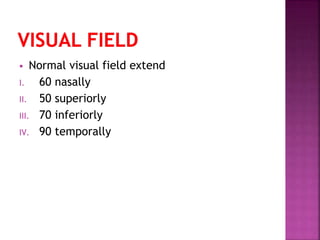
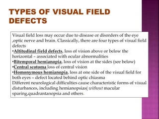
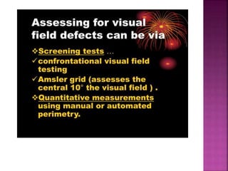
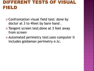
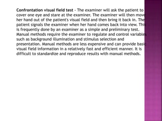
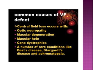
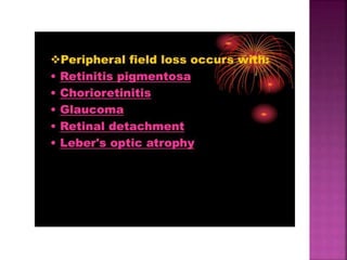
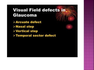
By SANA BUTT
What Is a Visual Field Test?
The visual field is the entire area (field of
vision) that can be seen when the eyes are
focused on a single point. In addition to what
can be seen straight ahead, the visual field
includes what can be seen above, below, and to
either side of the point the eyes are focused on.
Vision is typically the sharpest in the middle of
the visual field.
A visual field test is often given as part of an
eye exam. Visual field testing helps your doctor
to determine where your side vision (peripheral
vision) begins and ends and how well you can
see objects in your peripheral vision.
 Normal visual field extend
I. 60 nasally
II. 50 superiorly
III. 70 inferiorly
IV. 90 temporally
Visual field loss may occur due to disease or disorders of the eye
,optic nerve and brain. Classically, there are four types of visual field
defects
•Altitudinal field defects, loss of vision above or below the
horizontal – associated with ocular abnormalities
•Bitemporal hemianopia, loss of vision at the sides (see below)
•Central scotoma loss of central vision
•Homonymous hemianopia, loss at one side of the visual field for
both eyes – defect located behind optic chiasma
Different neurological difficulties cause characteristic forms of visual
disturbances, including hemianopsias( without macular
sparing,quadrantanopsia and others.
 Confrontation visual field test: done by
doctor at 3 to 4feet by bare hand .
 Tangent screen test;done at 3 feet away
from screen
 Automated perimetry test:uses computer it
includes goldaman perimetry e.tc.
Tangent Screen Visual Field Testing
The tangent screen is a relatively simple and
easy test to run and is much more sensitive than
confrontation fields. The tangent screen has a
black felt background with circular stitching
every five degrees and will test out to thirty(30)
degrees at one meter. It usually also has radial
stitching that starts at the 180 meridian running
through the fixation point every 22.5 degrees
The tangent screen targets are pigments.
Therefore, the test is more sensitive the dimmer
the lighting is on the screen (more difficult to
see). The light falling on the tangent screen
should be 7 foot candles
LCD and Heavy Duty Chin Rest
Small Size Projection Screen 120 cm
Tangent screen visual field or Goldmann field test - A simple tangent
screen is a relatively easy test to run and is much more sensitive than
confrontation fields. Here the patient is asked to sit approximately 3 feet
from a screen with a target on the center. The patient should be wearing
their Rx. If they are presbyopic they should have a +1.00 D lens placed in a
trial frame over their distant correction, assuming the tangent screen is at
one meter. Patients should never wear glasses with multifocal lenses when
being tested. The eye that isn't tested is covered during the test. While the
patient stares at the target the examiner will move an object toward the
patient's visual field. The patient signals the examiner when the object
comes into view. This test allows the patient's visual field to be mapped.
This method is still used because it is inexpensive, efficient, and it can
yield a fairly accurate measure of the central 30 degrees of the field. The
equipment needed is simple. The nice thing about the tangent screen
concept is that the procedure can be performed without any of the
standard equipment. All you really need is a blank wall and an improvised
stimulus. The fixation target can be a small dot of paper taped to the wall.
The tangent screen is also useful when measuring the effect that a ptosis
has on the superior visual field. Cost effective for special case fields or for
quick screening. Tangent Screen was the standard visual field test for many
years Declined use with advent of Goldmann and Automated perimeters.
Confrontation visual field test - The examiner will ask the patient to
cover one eye and stare at the examiner. The examiner will then move
her hand out of the patient's visual field and then bring it back in. The
patient signals the examiner when her hand comes back into view. This
is frequently done by an examiner as a simple and preliminary test.
Manual methods require the examiner to regulate and control variables
such as background illumination and stimulus selection and
presentation. Manual methods are less expensive and can provide basic
visual field information in a relatively fast and efficient manner. It is
difficult to standardize and reproduce results with manual methods.
Errors in Visual Field Testing
INADEQUATE PATIENT INSTRUCTION
INADEQUATE PATIENT SUPERVISION INATTENTIVE OR
UNCOOPERATIVE PATIENT (LEADING TO FIXATION LOSSES AND
UNRELIABLE RESPONSES)
FELLOW EYE NOT PATCHED OR PARTIALLY PATCHED
IMPROPER STIMULUS SELECTION (SIZE, BRIGHTNESS)
IMPROPER FIXATION TARGET (E.G., LARGE TARGET FOR LOW
VISION)
INCORRECT LENS POSITION,
OBSTRUCTING LENS RIM, A DROOPY EYELID OR PROMINENT BROW
OBSTRUCTS FIELD AND CAUSE WHAT APPEARS TO BE A SIGNIFICANT
FIELD DEFECT. T THIS IS TERMED ARTIFACTUAL FIELD LOSS,
MEANING IT IS DUE TO A "MECHANICAL" OBSTRUCTION AND IS NOT
DUE TO DECREASE RETINAL SENSITIVITY.
PUPIL TOO SMALL (LESS THAN 2MM) AND IMPROPER LENS
CORRECTIONS CAN PRODUCE ARTIFACTUAL TEST RESULTS THAT
SOMETIMES MIMIC PATHOLOGICAL SENSITIVITY CHANGES. SMALL
PUPILS (LESS THAN 2 MM IN DIAMETER) CAN GREATLY RESTRICT THE
AMOUNT OF LIGHT REACHING THE RETINA.

Tangent screen

  • 1.
  • 2.
    What Is aVisual Field Test? The visual field is the entire area (field of vision) that can be seen when the eyes are focused on a single point. In addition to what can be seen straight ahead, the visual field includes what can be seen above, below, and to either side of the point the eyes are focused on. Vision is typically the sharpest in the middle of the visual field. A visual field test is often given as part of an eye exam. Visual field testing helps your doctor to determine where your side vision (peripheral vision) begins and ends and how well you can see objects in your peripheral vision.
  • 3.
     Normal visualfield extend I. 60 nasally II. 50 superiorly III. 70 inferiorly IV. 90 temporally
  • 4.
    Visual field lossmay occur due to disease or disorders of the eye ,optic nerve and brain. Classically, there are four types of visual field defects •Altitudinal field defects, loss of vision above or below the horizontal – associated with ocular abnormalities •Bitemporal hemianopia, loss of vision at the sides (see below) •Central scotoma loss of central vision •Homonymous hemianopia, loss at one side of the visual field for both eyes – defect located behind optic chiasma Different neurological difficulties cause characteristic forms of visual disturbances, including hemianopsias( without macular sparing,quadrantanopsia and others.
  • 6.
     Confrontation visualfield test: done by doctor at 3 to 4feet by bare hand .  Tangent screen test;done at 3 feet away from screen  Automated perimetry test:uses computer it includes goldaman perimetry e.tc.
  • 7.
    Tangent Screen VisualField Testing The tangent screen is a relatively simple and easy test to run and is much more sensitive than confrontation fields. The tangent screen has a black felt background with circular stitching every five degrees and will test out to thirty(30) degrees at one meter. It usually also has radial stitching that starts at the 180 meridian running through the fixation point every 22.5 degrees The tangent screen targets are pigments. Therefore, the test is more sensitive the dimmer the lighting is on the screen (more difficult to see). The light falling on the tangent screen should be 7 foot candles
  • 9.
    LCD and HeavyDuty Chin Rest
  • 10.
    Small Size ProjectionScreen 120 cm
  • 11.
    Tangent screen visualfield or Goldmann field test - A simple tangent screen is a relatively easy test to run and is much more sensitive than confrontation fields. Here the patient is asked to sit approximately 3 feet from a screen with a target on the center. The patient should be wearing their Rx. If they are presbyopic they should have a +1.00 D lens placed in a trial frame over their distant correction, assuming the tangent screen is at one meter. Patients should never wear glasses with multifocal lenses when being tested. The eye that isn't tested is covered during the test. While the patient stares at the target the examiner will move an object toward the patient's visual field. The patient signals the examiner when the object comes into view. This test allows the patient's visual field to be mapped. This method is still used because it is inexpensive, efficient, and it can yield a fairly accurate measure of the central 30 degrees of the field. The equipment needed is simple. The nice thing about the tangent screen concept is that the procedure can be performed without any of the standard equipment. All you really need is a blank wall and an improvised stimulus. The fixation target can be a small dot of paper taped to the wall. The tangent screen is also useful when measuring the effect that a ptosis has on the superior visual field. Cost effective for special case fields or for quick screening. Tangent Screen was the standard visual field test for many years Declined use with advent of Goldmann and Automated perimeters.
  • 12.
    Confrontation visual fieldtest - The examiner will ask the patient to cover one eye and stare at the examiner. The examiner will then move her hand out of the patient's visual field and then bring it back in. The patient signals the examiner when her hand comes back into view. This is frequently done by an examiner as a simple and preliminary test. Manual methods require the examiner to regulate and control variables such as background illumination and stimulus selection and presentation. Manual methods are less expensive and can provide basic visual field information in a relatively fast and efficient manner. It is difficult to standardize and reproduce results with manual methods.
  • 16.
    Errors in VisualField Testing INADEQUATE PATIENT INSTRUCTION INADEQUATE PATIENT SUPERVISION INATTENTIVE OR UNCOOPERATIVE PATIENT (LEADING TO FIXATION LOSSES AND UNRELIABLE RESPONSES) FELLOW EYE NOT PATCHED OR PARTIALLY PATCHED IMPROPER STIMULUS SELECTION (SIZE, BRIGHTNESS) IMPROPER FIXATION TARGET (E.G., LARGE TARGET FOR LOW VISION) INCORRECT LENS POSITION, OBSTRUCTING LENS RIM, A DROOPY EYELID OR PROMINENT BROW OBSTRUCTS FIELD AND CAUSE WHAT APPEARS TO BE A SIGNIFICANT FIELD DEFECT. T THIS IS TERMED ARTIFACTUAL FIELD LOSS, MEANING IT IS DUE TO A "MECHANICAL" OBSTRUCTION AND IS NOT DUE TO DECREASE RETINAL SENSITIVITY. PUPIL TOO SMALL (LESS THAN 2MM) AND IMPROPER LENS CORRECTIONS CAN PRODUCE ARTIFACTUAL TEST RESULTS THAT SOMETIMES MIMIC PATHOLOGICAL SENSITIVITY CHANGES. SMALL PUPILS (LESS THAN 2 MM IN DIAMETER) CAN GREATLY RESTRICT THE AMOUNT OF LIGHT REACHING THE RETINA.