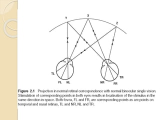
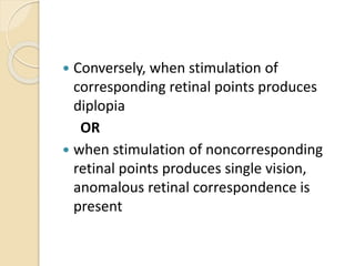
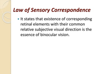
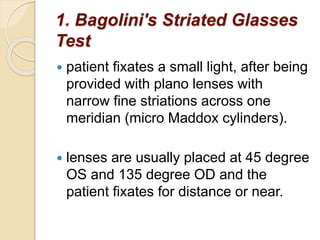
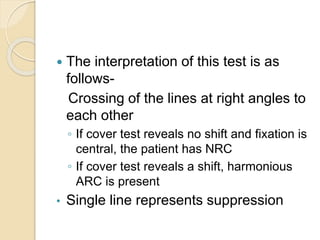
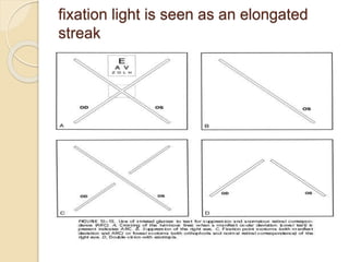
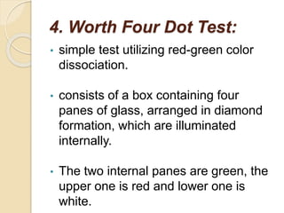
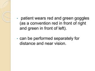
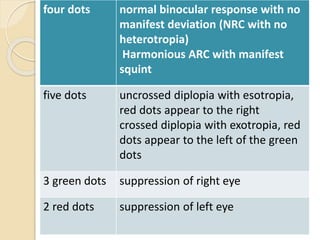
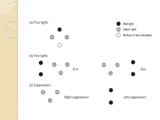
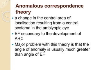
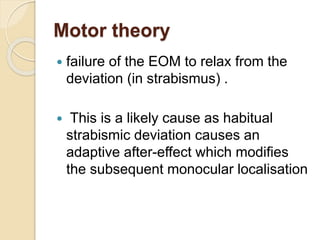
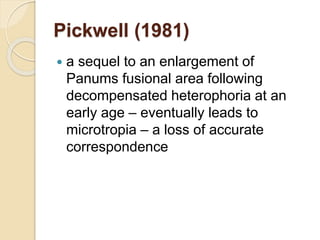
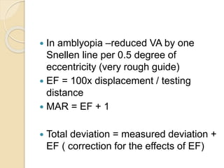
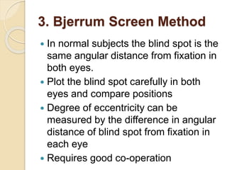
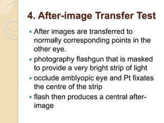
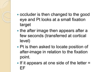
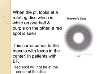
SlideShare a Scribd company logo
Abnormal retinal
correspondence and eccentric
fixation
Rajeshwori Ngakhushi
Retinal Correspondence
 Retinal elements of the two eyes that
share a common subjective visual
direction.
 All other retinal elements are non-
corresponding or disparate with respect
to a given retinal element in the fellow
eye for a particular visual direction.
 Retinal correspondence is the
inherent relationship between paired
retinal visual cells in the two eyes.
 Images from one object stimulate
both cells, which transmit the
information to the brain, permitting
a single visual impression localized in
the same direction in space.
 Specifically, the right nasal retina contains
points that correspond to their
counterpart in the left temporal retina,
and vice versa.
 When stimulation of corresponding
retinal points or areas produces single
vision, normal retinal correspondence is
said to be present.
 Conversely, when stimulation of
corresponding retinal points produces
diplopia
OR
 when stimulation of noncorresponding
retinal points produces single vision,
anomalous retinal correspondence is
present
Law of Sensory Correspondence
 It states that existence of corresponding
retinal elements with their common
relative subjective visual direction is the
essence of binocular vision.
Retinal Correspondence can be of two
types:
1. Normal Retinal Correspondence
2. Abnormal Retinal Correspondence
Normal Retinal
Correspondence
 Retinal correspondence is called
normal when both the fovea have a
common visual direction and the
retinal elements nasal to the fovea in
one eye corresponds to the retinal
elements temporal to the fovea in the
other eye.
Abnormal Retinal
Correspondence
• when the fovea of one eye has a
common visual direction with an
extrafoveal area in the other eye
• angle of squint is small and the
extrafoveal point is close to the fovea - to
regain the binocular advantage, although
anomalous
 This results in the eyes seeing
binocularly single inspite of a manifest
squint.
 under binocular conditions the fovea and
the extafoveal point share the common
subjective visual direction
 when the normal eye is closed the
extrafoveal element loses any advantage
over the fovea of that eye, which retains
its primary visual direction.
Concept of a Horopter
 From the Greek words horos
(boundary) and opter (observer).
 The term Horopter (the horizon of
vision) was introduced in 1613 by
Aguilonius.
 locus of all object points that are
imaged on corresponding retinal
elements at a given fixation distance
Geometric Vieth Muller horopter
 According to this model of horopter if
corresponding points have a regular
horizontal distance from the retina the
horopter would be a circle passing
through the center of rotation of the
two eyes and the fixation point.
Empirical Horopter Curve
 slightly flatter than vieth muller
geometric horopter
 Hering-Hillebrand deviation - due to
both neural and optical factors - nasal
hemi-retina at any given eccentricity
contains more photoreceptors per unit
area than the temporal hemi retina
producing a deviation in the horopter
mapping in the visual cortex.
Panum’s Fusion Area
 Region in front and back of the
horopter in which single vision is
present
 diplopia elicited by object points off the
horopter - Physiological diplopia
 narrowest at the fixation point and
becomes broader in the periphery at a
rate of 1-2 arc min per degree of
visual field eccentricity.
 expands and contracts depending on the
size, sharpness and speed of the stimuli
 Panum’s area for the stimuli that are fuzzy
and slow moving is 20 times wider than it
is for stimuli that are sharply focused and
rapidly moving.
Fusion
Fusion is defined as the unification of
visual excitations from the
corresponding retinal images into a
single visual percept.
 Sensory Fusion
It is the ability to appreciate two
similar images, one with each eye and
interpret them as one. Single visual
image is the hallmark of retinal
Motor Fusion
• It is the ability to align the eyes in such
a manner that sensory fusion can be
maintained.
• The stimulus for these fusional eye
movements is retinal disparity outside
panum’s area and the eyes moving in
opposite direction (vergence).
Diplopia
 simultaneous stimulation of non-
corresponding or disparate retinal
elements by an object point causes
this point to be localized in two
different subjective visual directions.
 Double vision is the hallmark of retinal
disparity.
Retinal Rivalry / Binocular
Rivalry
 When dissimilar
contours are
presented to
corresponding retinal
areas fusion
becomes impossible
and retinal rivalry
may be observed.
 Simultaneous excitation of
corresponding retinal areas by
dissimilar objects does not permit
fusion and leads to confusion
Test for Retinal
Correspondence
 Clinically the tests used can be based on
either of the two principles:
A) Assesment of relationship between the
fovea of the fixing eye and the retinal
area stimulated in the squinting eye. This
includes:
1. Bagolini's striated glasses test
2. red filter test
3. Synaptophore using SMP slides for
measuring the objective and subjective
angles
4. Worth's 4 dot test
B) Assessment of the visual directions
of the two foveas. Included in this are:
1. After image test (Hering
Bielschowsky)
2. Cuppers binocular visuoscopy test
(foveo-foveal test of Cuppers)
1. Bagolini's Striated Glasses
Test
 patient fixates a small light, after being
provided with plano lenses with
narrow fine striations across one
meridian (micro Maddox cylinders).
 lenses are usually placed at 45 degree
OS and 135 degree OD and the
patient fixates for distance or near.
 The interpretation of this test is as
follows-
Crossing of the lines at right angles to
each other
◦ If cover test reveals no shift and fixation is
central, the patient has NRC
◦ If cover test reveals a shift, harmonious
ARC is present
• Single line represents suppression
fixation light is seen as an elongated
streak
2. Red Filter Test
 place a red filter in front of the habitually
fixating eye while the patient is looking at
a small light source
 number of different responses can be
elicited.
 patient may report that two lights are
seen, a red one and a white one.
 In esotropia the
images appear in
homonymous
(uncrossed)
diplopia,
 In exotropia the images
appear in
heteronymous
(crossed) diplopia
 This represents NRC.
 patient may report
that only one pinkish
light in the position
of the white fixation
light is seen
 This is clearly an
abnormal response
in presence of
heterophoria. This is
termed Harmonious
ARC.
 may report that the
measured distance
between the double
images proves to be
smaller than
expected from the
magnitude of
deviation
 This represents
unharmonious ARC.
3. Measurement of Angle of
Anomaly:
 The angle of anomaly denotes the
degree of shift in visual direction.
 It is determined by calculating the
difference between the objective and
subjective angles of deviation.
subjective angle of anomaly is the angle at which the
visual targets are superimposed.
no further fixation movement of the patient’s eye
reading of both the arms is noted
sum total of the reading of both the arms
gives the objective angle of anomaly
SMP slides is made
arms of the synaptophore are set at zero
moved by the examiner while alternately
flashing the light
 The interpretation of this test is as follows-
 Angle of Anomaly = Objective Angle –
Subjective Angle
 If Subjective Angle = Objective Angle → NRC
 If Subjective Angle < Objective Angle → ARC
 If Angle of Anomaly = Objective Angle
→Harmonious ARC (full sensory adaptation)
 If Angle of Anomaly < Objective Angle
→Unharmonious ARC
4. Worth Four Dot Test:
• simple test utilizing red-green color
dissociation.
• consists of a box containing four
panes of glass, arranged in diamond
formation, which are illuminated
internally.
• The two internal panes are green, the
upper one is red and lower one is
white.
• patient wears red and green goggles
(as a convention red in front of right
and green in front of left).
• can be performed separately for
distance and near vision.
The interpretation of this test is
as follows-
four dots normal binocular response with no
manifest deviation (NRC with no
heterotropia)
Harmonious ARC with manifest
squint
five dots uncrossed diplopia with esotropia,
red dots appear to the right
crossed diplopia with exotropia, red
dots appear to the left of the green
dots
3 green dots suppression of right eye
2 red dots suppression of left eye
5. Hering Bielschowsky After-
Image Test:
 highly dissociating orthoptic test in
which battery- powered camera flash
is used to produce a vertical after
image in one eye and a horizontal
after image in the other eye.
 center of flash is covered with a black
mark (serves as a point of fixation and
protects the fovea).
Procedure
• Each eye fixates on the center black mark
of a glowing filament horizontally to the
better eye vertically to the poorer eye
for 20 sec in a darkened room patient
indicates the relative position of the two
gaps in the center of each afterimage
gaps correspond to the visual direction of
each fovea if central fixation is present.
Interpretation of results -
• Cross response: A symmetrical cross
with the central gaps superimposed
indicates a normal bifoveal
correspondence.
• Asymmetrical crossing: In case of ARC
the horizontal and vertical lines have
their center separated, the amount of
separation dependent on the angle of
anomaly.
• Single line with a gap: A single line with
a gap indicates suppression in the fellow
 Displacement between the centres of
the two after images is proportional to
the angle of anomaly
 Tan Ø = dispalcement / distance of
testing
6. Foveo-Foveal Test of
Cuppers:
• Cuppers test for retinal
correspondence determines whether
the two foveas have common or
different visual directions.
• It permits quantitative analysis of the
angle of anomaly when eccentric
fixation is present.
Procedure -
 patient fixates with the normal eye on the
central light of a Maddox scale via a plano
mirror
 the amblyopic eye looks straight ahead
 visuoscope asterisk is projected onto the
fovea of the amblyopic eye
 figure of the Maddox scale on which the
patient sees the asterisk indicated the
angle of anomaly.
Modification -
• To determine which parts of the
peripheral retina in the deviating eye
have acquired a common visual direction
with the fovea of the fixating eye
• the patient is asked to guide the
Visuoscope until he sees the asterisk
superimposed on the central light of the
Maddox
Abnormal retinal
correspondence
 Abnormal retinal correspondence is a
physiological part of normal binocular
vision in many persons and is a natural
accompaniment of the binocular
adjustment when the visual axes are not
parallel in all positions.
 It is a binocular condition and not a
uniocular adaptation of projection.
 Anomalous retinal correspondence
(ARC) is a neural adaptation to eye
misalignment in which non-
corresponding retinal points are linked
in the visual cortex to provide
binocular fusion.
Variety of descriptive terms
 Anomalous correspondence
 Anomalous projection
 False associated fixation
 Positive secondary retinal
correspondence
 False projection
 Retinal incongruity
 False macula
 Anomalous binocular junction
Requirements for ARC
 Early onset squint
 Constant angle of deviation
 Small esodeviations
Classification
(1) Harmonious ARC
 angle of anomaly is equal to the objective
angle of deviation ( subjective angle = o)
 This indicates that the
ARC fully corresponds to the strabismus.
 provides a compensation for the angle of
squint
 Eliminate the awareness of diplopia and
confusion
(2) Unharmonious ARC
angle of anomaly is less than the objective
angle of deviation
subjective angle ≠ 0
(3)Paradoxical, when the angle of anomaly is
greater than the objective angle of deviation
Theory of ARC
 Linksz returned to the original rigid
theory (Mu¨ller and von Graefe) that
normal correspondence is a strictly
anatomical fact based on an
immutable connection between
distinct retinal and cortical areas
 ARC has been put forward as cause
of strabismus
 Morgan proposed that some ocular
movements are ‘‘registered’’ in
coordinating centers and some are
‘‘not registered,’’ depending on
whether they affect egocentric
localization.
 He used this concept to explain not
only anomalous correspondence but
also monocular diplopia.
Advantages
 Avoids the necessity for dense
amblyopia ( HRC )
 Permits anomalous fusion range and
some degree of stereopsis
 Prevents consecutive divergence
 Allows a form of binocular convergence
to take place
Disadvantages
 Prevents the development of normal
retinal correspondence, true fusion
and normal stereopsis
 Represents an abnormal reflex
development
Treatment
 Occlusion Preventive as well as
curative measure
 Prolonged alternating occlusion is not
recommended - prevent further
development of ARC as well as
normal correspondence
 Exercises with major amblyoscope
Eccentric fixation
 A uniocular condition in which some
part of retina other than fovea is used
for fixation, but in which the fovea
retains its normal straight ahead
projection
 It may be associated with a defect of
central vision caused by a stuctural
lesion of fovea.
 Eccentric fixation can be present
during both monocular and binocular
viewing conditions, but it is best
diagnosed under monocular viewing
conditions.
 This is important to keep in mind to
avoid confusion with anomalous
correspondence. Anomalous
correspondence is relevant only
during binocular fusion
Four Theories as to the cause of
Eccentric
1. Fixation Suppression Theory (Worth,
1906, Bangerter,1953)
2. Anomalous correspondence theory
(Chavasse, 1939, Cuppers, 1956)
3. Motor theory (Schor, 1978)
4. Pickwell (1981)
Suppression Theory:
 occurs when central acuity has dropped
to a level below that of the surrounding
area, so that better acuity results
 now thought to be unlikely as foveal VA
still seems to be better than in the rest of
the retina
 Strabismic patients suppress to get rid
of double vision
Anomalous correspondence
theory
 a change in the central area of
localisation resulting from a central
scotoma in the amblyopic eye
 EF secondary to the development of
ARC
 Major problem with this theory is that the
angle of anomaly is usually much greater
than angle of EF
Motor theory
 failure of the EOM to relax from the
deviation (in strabismus) .
 This is a likely cause as habitual
strabismic deviation causes an
adaptive after-effect which modifies
the subsequent monocular localisation
Pickwell (1981)
 a sequel to an enlargement of
Panums fusional area following
decompensated heterophoria at an
early age – eventually leads to
microtropia – a loss of accurate
correspondence
 In amblyopia –reduced VA by one
Snellen line per 0.5 degree of
eccentricity (very rough guide)
 EF = 100x displacement / testing
distance
 MAR = EF + 1
 Total deviation = measured deviation +
EF ( correction for the effects of EF)
Diagnostic testing methods
1. Corneal reflex test
2. Ophthalmoscopic
methods/Visuscope
3. Bjerrum Screen Method
4. After-image Transfer Test
5. Haidingers Brushes
6. Maxwell’s spots
7. Projectoscope
8. Euthyscope
9. Past pointing test
10. Angle kappa
11. Neutral density filter
12. Speed of accommodation
1. Corneal Reflex Test:
 comparing the position of corneal
reflection of pentorch in amblyopic eye
with that of the fellow eye.
 Relative displacement of reflex by
1mm would indicate the eccentric
fixation of 11⁰(or20prism)
approximately
2. Ophthalmoscopic
methods/Visuscope:
 Project the ophthalmoscopic target
onto the patients retina
 Dilated pupil may be necessary
 With the fixating eye occluded the
examiner observes the amblyopic eye
noting the point or area of retina upon
which the star is projected when the
patient is attempting to look at the star
straight at it.
Visuoscopy Targets
A. Propper ophthalmoscope
B. Welch-Allyn ophthalmoscope
C. Keeler ophthalmoscope
D. Determining location & magnitude of EF using Welch-Allyn
ophthalmoscope
3. Bjerrum Screen Method
 In normal subjects the blind spot is the
same angular distance from fixation in
both eyes.
 Plot the blind spot carefully in both
eyes and compare positions
 Degree of eccentricity can be
measured by the difference in angular
distance of blind spot from fixation in
each eye
 Requires good co-operation
4. After-image Transfer Test
 After images are transferred to
normally corresponding points in the
other eye.
 photography flashgun that is masked
to provide a very bright strip of light
 occlude amblyopic eye and Pt fixates
the centre of the strip
 flash then produces a central after-
image
 occluder is then changed to the good
eye and Pt looks at a small fixation
target
 the after image then appears after a
few seconds (transferred at cortical
level)
 Pt is then asked to locate position of
after-image in relation to the fixation
point.
 If it appears at one side of the letter =
EF
5. Haidingers Brushes
 an entoptic phenomenon due to
characteristics of the central fovea
area
 seen with a brightly illuminated blue
polarised field when the direction of
the polarisation is rotated
 looks like two darkened and opposing
sections rotating in the central field
 The direction and magnitude of EF can be
assessed by asking the px to report the
location of Propeller in relation to fixation
point
 Practitioner can measure distance between
these two points and convert mm
dispalcement to PD.eg at 1 meter,10 mm=1
PD,at 40 cm,4mm=1 PD.
6. Maxwell’s spot
 Entopic phenomenon caused by the
radial orientation of the yellow pigment
xanthophyll in the macula
 Orientation of the macular pigment acts
like a polarizing filter
 It absorbs blue light & transmits red &
yellow light
When the pt. looks at a
rotating disc which is
white on one half &
purple on the other, a red
spot is seen
This corresponds to the
macula with fovea in the
center. In patients with
EF,
Red spot will not be at the
center of the disc
7. Projectoscope
 Modified keeler opthalmoscope which
may be used for diagnosis and
treatment.
 The Nut Auto-disc is provided with
three graticules.
 Stage 1: Localisation of the fovea
With the Auto-disc position 1 The
linksz star in green light may be
focused on the retina and accurately
placed on the patient’s fovea.
 Stage 2: Dazziling the extra macular
retina With the Auto-disc position 2 By
pressing the trigger on the
projectoscope the second graticule
consisting of a 3ᴼ or 5ᴼ black spot
 Stage 3: Foveal stimulation by
flashing light With the Auto-disc
position 3 The 3ᴼ black spot is
automatically replaced by a disc of
white light, whilst the extra macular
retina is projected by a green filtered
light
8. Euthyscope:
 modified opthalmoscope by cuppers
 beam of light illuminates an area of
approximately 30ᴼ at the posterior
pole of the eye.
 green filter is incorporated and also
two black discs of 5ᴼ and 3ᴼ diameter
which can be placed in the centre of
the beam of the light.
 light allowed to stimulate the circular
paramacular zone for 20 to 30
seconds.
 instructed to look through one of the
instrument incorporating haidingers
brushes encouraged to find the
brushes within the clear centre of the
after image.
9. Past Pointing Test
 Related to localisation
 Occlude amblyopic eye, hold pen 25cm in
front and ask patient to touch pen with
the tip of their finger
 If finger goes a few cm to the side then
past pointing has been demonstrated
 This result indicates that fixation does not
coincide with the centre of localisation
10. Angle kappa
 Angle Kappa- Worth Test
 Compare angle lambda of each eye
under monocular conditions
 Any difference associated with reduced
acuity indicate EF
◦ 1 mm of displacement = 22 pd
11. Neutral density Filters
 If a ND filter is added and no reduction in
VA occurs then EF is likely to be present
12. Speed of Accommodation
 Much slower in EF (?also in other
amblyopes)
Treatment
 The principles of treatment were those
used to restore the normal sensory
relationship of the two retinae.
 encourage foveal fixation
 Refractive Error Correction, especially in
high anisometropia.
Inverse occlusion
 Infants with eccentric fixation who are
too young for treatment with
Haidinger’s brushes
 Continued as long as improvement is
made
 Rarely suitable in adult – eccentric
fixation is too firmly established
Red filter
 600 – 640 nm
 Only stimulate cones
 Patient will attempt to fix with area of
the retina which contains maximum no
of cones
Pleoptics Therapy
 Pleos=full,optikos=sight
 Aim is to actively stimulate macula in
dense amblyopia with eccentric
fixation
 Two methods,either Bangerter or
Cupper
Bangerter’s method
 Bangerter dazzled the extramacular
retina including the eccentric point by
bright light protecting the macula by a
disc projected onto it.
 It was followed by intermittent
stimulation of macula with flashes of
light
 Performed by modified Gullstrand’s
Ophthalmoscope,called as
Pleoptophore.
Cupper’s method
 Cupper used Euthyscope which had
discs of varying sizes to create a
central after image apart from dazzling
the eccentric point
 He used the alternate flashing of room
illumination(Alternascope) to
perpetuate the after images(forming
negative after image in light and
positive after image in dark)
 patient perceives an afterimage
resembling the white doughnut
 Treat underlying amblyopia by
occlusion of non-squinting eye (if
patient <6 years old).
 In patients >6 years – correct
refractive error, otherwise do not treat
the microtropia.
 Surgery is not appropriate
Refrences
 Lyle and Jackson’s Practical orthoptics
in the treatment of squint
 Strabismus simplified, Pradip Sharma
 Binocular single vision and ocular
motility, Von Noorden
 Previous presentations
 internet
anomalous retinal correspondence

More Related Content

What's hot

Laws of ocular motility 2
Laws of ocular motility 2Laws of ocular motility 2
Laws of ocular motility 2
suchismita Rout
 
binocular single vision
binocular single visionbinocular single vision
binocular single vision
DrShrey Maheshwari
 
Detail of suppression and AC
Detail of suppression and ACDetail of suppression and AC
Detail of suppression and AC
Raju Kaiti
 
Binocular Single Vision Tests
Binocular Single Vision TestsBinocular Single Vision Tests
Binocular Single Vision Tests
Rabia Ammer
 
Optics of contact lens
Optics of contact lensOptics of contact lens
Optics of contact lens
Aayush Chandan
 
Synaptophore
SynaptophoreSynaptophore
Synaptophore
Manjusha Lakshmi
 
Ac/a ratio
Ac/a ratio Ac/a ratio
Ac/a ratio
anupama manoharan
 
Amblyopia : classification & Occlusion therapy
Amblyopia : classification & Occlusion therapy Amblyopia : classification & Occlusion therapy
Amblyopia : classification & Occlusion therapy
Aayush Chandan
 
Binocular refraction techniques, binocular balancing
Binocular refraction techniques, binocular balancing Binocular refraction techniques, binocular balancing
Binocular refraction techniques, binocular balancing Mohammad Arman Bin Aziz
 
Multifocal contact lens
Multifocal contact lensMultifocal contact lens
Multifocal contact lens
OPTOM FASLU MUHAMMED
 
JCC -Jackson Cross Cylinder
JCC -Jackson Cross CylinderJCC -Jackson Cross Cylinder
JCC -Jackson Cross Cylinder
Indra Prasad Sharma
 
Vertex distance and power
Vertex distance and powerVertex distance and power
History taking of low vision
History taking of low visionHistory taking of low vision
History taking of low vision
sagarkalamkar05
 
Aniseikonia
Aniseikonia Aniseikonia
Aniseikonia
Azizul Islam
 
Measuring interpupillary distance
Measuring interpupillary distanceMeasuring interpupillary distance
Measuring interpupillary distance
Indra Prasad Sharma
 
Introduction to binocular single vision (BSV)
Introduction to binocular single vision (BSV)Introduction to binocular single vision (BSV)
Introduction to binocular single vision (BSV)
Anis Suzanna Mohamad
 
Bifocal lenses: types and principles
Bifocal lenses: types and principlesBifocal lenses: types and principles
Bifocal lenses: types and principles
Garima Poudel
 
Test for stereopsis
Test for stereopsisTest for stereopsis
Test for stereopsis
Aliasger Fakhruddin
 
Hvid
Hvid Hvid

What's hot (20)

Laws of ocular motility 2
Laws of ocular motility 2Laws of ocular motility 2
Laws of ocular motility 2
 
binocular single vision
binocular single visionbinocular single vision
binocular single vision
 
Detail of suppression and AC
Detail of suppression and ACDetail of suppression and AC
Detail of suppression and AC
 
Binocular Single Vision Tests
Binocular Single Vision TestsBinocular Single Vision Tests
Binocular Single Vision Tests
 
Optics of contact lens
Optics of contact lensOptics of contact lens
Optics of contact lens
 
Synaptophore
SynaptophoreSynaptophore
Synaptophore
 
Ac/a ratio
Ac/a ratio Ac/a ratio
Ac/a ratio
 
Amblyopia : classification & Occlusion therapy
Amblyopia : classification & Occlusion therapy Amblyopia : classification & Occlusion therapy
Amblyopia : classification & Occlusion therapy
 
Binocular refraction techniques, binocular balancing
Binocular refraction techniques, binocular balancing Binocular refraction techniques, binocular balancing
Binocular refraction techniques, binocular balancing
 
Multifocal contact lens
Multifocal contact lensMultifocal contact lens
Multifocal contact lens
 
Low vision chart
Low vision chart Low vision chart
Low vision chart
 
JCC -Jackson Cross Cylinder
JCC -Jackson Cross CylinderJCC -Jackson Cross Cylinder
JCC -Jackson Cross Cylinder
 
Vertex distance and power
Vertex distance and powerVertex distance and power
Vertex distance and power
 
History taking of low vision
History taking of low visionHistory taking of low vision
History taking of low vision
 
Aniseikonia
Aniseikonia Aniseikonia
Aniseikonia
 
Measuring interpupillary distance
Measuring interpupillary distanceMeasuring interpupillary distance
Measuring interpupillary distance
 
Introduction to binocular single vision (BSV)
Introduction to binocular single vision (BSV)Introduction to binocular single vision (BSV)
Introduction to binocular single vision (BSV)
 
Bifocal lenses: types and principles
Bifocal lenses: types and principlesBifocal lenses: types and principles
Bifocal lenses: types and principles
 
Test for stereopsis
Test for stereopsisTest for stereopsis
Test for stereopsis
 
Hvid
Hvid Hvid
Hvid
 

Viewers also liked

Stereopsis
Stereopsis  Stereopsis
stereopsis and types of stereopsis tests
stereopsis and types of stereopsis testsstereopsis and types of stereopsis tests
stereopsis and types of stereopsis tests
Sac Hin
 
Bifocal Spectacles..
Bifocal Spectacles..Bifocal Spectacles..
Bifocal Spectacles..Vishakh Nair
 
Relative and Atribute Risk
Relative and Atribute RiskRelative and Atribute Risk
Relative and Atribute RiskTauseef Jawaid
 
Sensory Adaptation to Strabismus
Sensory Adaptation to StrabismusSensory Adaptation to Strabismus
Sensory Adaptation to StrabismusHossein Mirzaie
 
Progressive addition lens1
Progressive addition lens1Progressive addition lens1
Progressive addition lens1GauriSShrestha
 
2016 I. Binocular Vision & Retinal Correspondence; II Amblyopia
2016 I. Binocular Vision & Retinal Correspondence; II Amblyopia2016 I. Binocular Vision & Retinal Correspondence; II Amblyopia
2016 I. Binocular Vision & Retinal Correspondence; II Amblyopia
Alvina Pauline Santiago, MD
 
Binomial probability distributions ppt
Binomial probability distributions pptBinomial probability distributions ppt
Binomial probability distributions pptTayab Ali
 
Association and causation
Association and causationAssociation and causation
Association and causationdrravimr
 
descriptive and inferential statistics
descriptive and inferential statisticsdescriptive and inferential statistics
descriptive and inferential statisticsMona Sajid
 

Viewers also liked (13)

Eccentric Fixation
Eccentric FixationEccentric Fixation
Eccentric Fixation
 
Stereopsis
Stereopsis  Stereopsis
Stereopsis
 
stereopsis and types of stereopsis tests
stereopsis and types of stereopsis testsstereopsis and types of stereopsis tests
stereopsis and types of stereopsis tests
 
Assesment of streopsis
Assesment of streopsisAssesment of streopsis
Assesment of streopsis
 
Bifocal Spectacles..
Bifocal Spectacles..Bifocal Spectacles..
Bifocal Spectacles..
 
Stereoscopic Vision
Stereoscopic VisionStereoscopic Vision
Stereoscopic Vision
 
Relative and Atribute Risk
Relative and Atribute RiskRelative and Atribute Risk
Relative and Atribute Risk
 
Sensory Adaptation to Strabismus
Sensory Adaptation to StrabismusSensory Adaptation to Strabismus
Sensory Adaptation to Strabismus
 
Progressive addition lens1
Progressive addition lens1Progressive addition lens1
Progressive addition lens1
 
2016 I. Binocular Vision & Retinal Correspondence; II Amblyopia
2016 I. Binocular Vision & Retinal Correspondence; II Amblyopia2016 I. Binocular Vision & Retinal Correspondence; II Amblyopia
2016 I. Binocular Vision & Retinal Correspondence; II Amblyopia
 
Binomial probability distributions ppt
Binomial probability distributions pptBinomial probability distributions ppt
Binomial probability distributions ppt
 
Association and causation
Association and causationAssociation and causation
Association and causation
 
descriptive and inferential statistics
descriptive and inferential statisticsdescriptive and inferential statistics
descriptive and inferential statistics
 

Similar to anomalous retinal correspondence

ARC: abnormal retinal correspondence, eccentric fixation
ARC: abnormal retinal correspondence, eccentric fixationARC: abnormal retinal correspondence, eccentric fixation
ARC: abnormal retinal correspondence, eccentric fixation
aditi Jain
 
Binocular Single Vision Ophthalmology .pptx
Binocular Single Vision Ophthalmology .pptxBinocular Single Vision Ophthalmology .pptx
Binocular Single Vision Ophthalmology .pptx
Vishnu645963
 
MECHANISMS AND TERMINOLOGIES IN BINOCULAR VISION.pptx
MECHANISMS AND TERMINOLOGIES IN BINOCULAR VISION.pptxMECHANISMS AND TERMINOLOGIES IN BINOCULAR VISION.pptx
MECHANISMS AND TERMINOLOGIES IN BINOCULAR VISION.pptx
kajal bhagat
 
Tests of binocularity
Tests of binocularityTests of binocularity
Tests of binocularity
Anuradha Chandra
 
Binocular vision
Binocular visionBinocular vision
Binocular vision
Yesenia Castillo Salinas
 
Binocular vision basics
Binocular vision basicsBinocular vision basics
Binocular vision basics
Isha Gupta
 
Sensory evaluation of strabismus
Sensory evaluation of strabismusSensory evaluation of strabismus
Sensory evaluation of strabismusGauriSShrestha
 
Sensory evaluation of strabismus
Sensory evaluation of strabismusSensory evaluation of strabismus
Sensory evaluation of strabismus
GauriSShrestha
 
Binocular vision
Binocular visionBinocular vision
Binocular vision
Sujay Chauhan
 
evaluation of strabismus
evaluation of strabismusevaluation of strabismus
evaluation of strabismus
Sudheer Kumar
 
Heterophoria investigation and management
Heterophoria investigation and managementHeterophoria investigation and management
Heterophoria investigation and management
Ananta poudel
 
pupil examination step by step for ophthalmic exam.pptx
pupil examination step by step for ophthalmic exam.pptxpupil examination step by step for ophthalmic exam.pptx
pupil examination step by step for ophthalmic exam.pptx
AhmadElSayed52
 
Simultaneous Macular Perception -Nayana
Simultaneous Macular Perception -NayanaSimultaneous Macular Perception -Nayana
Simultaneous Macular Perception -Nayana
NayanaKT
 
Binocular vision and stereopsis
Binocular vision and stereopsisBinocular vision and stereopsis
Binocular vision and stereopsis
SAMEEKSHA AGRAWAL
 
Synoptophore.pptx
Synoptophore.pptxSynoptophore.pptx
Synoptophore.pptx
Anisha Heka
 
BSV.pptx
BSV.pptxBSV.pptx
Binocular vision
Binocular visionBinocular vision
Binocular vision
Saransh Jain
 
Binocular vision and space perception
Binocular vision and space perceptionBinocular vision and space perception
Binocular vision and space perception
SimiAfroz2
 
Binocular vision final
Binocular vision finalBinocular vision final
Binocular vision final
Desta Genete
 

Similar to anomalous retinal correspondence (20)

ARC: abnormal retinal correspondence, eccentric fixation
ARC: abnormal retinal correspondence, eccentric fixationARC: abnormal retinal correspondence, eccentric fixation
ARC: abnormal retinal correspondence, eccentric fixation
 
Binocular Single Vision Ophthalmology .pptx
Binocular Single Vision Ophthalmology .pptxBinocular Single Vision Ophthalmology .pptx
Binocular Single Vision Ophthalmology .pptx
 
MECHANISMS AND TERMINOLOGIES IN BINOCULAR VISION.pptx
MECHANISMS AND TERMINOLOGIES IN BINOCULAR VISION.pptxMECHANISMS AND TERMINOLOGIES IN BINOCULAR VISION.pptx
MECHANISMS AND TERMINOLOGIES IN BINOCULAR VISION.pptx
 
Tests of binocularity
Tests of binocularityTests of binocularity
Tests of binocularity
 
Diplopia
DiplopiaDiplopia
Diplopia
 
Binocular vision
Binocular visionBinocular vision
Binocular vision
 
Binocular vision basics
Binocular vision basicsBinocular vision basics
Binocular vision basics
 
Sensory evaluation of strabismus
Sensory evaluation of strabismusSensory evaluation of strabismus
Sensory evaluation of strabismus
 
Sensory evaluation of strabismus
Sensory evaluation of strabismusSensory evaluation of strabismus
Sensory evaluation of strabismus
 
Binocular vision
Binocular visionBinocular vision
Binocular vision
 
evaluation of strabismus
evaluation of strabismusevaluation of strabismus
evaluation of strabismus
 
Heterophoria investigation and management
Heterophoria investigation and managementHeterophoria investigation and management
Heterophoria investigation and management
 
pupil examination step by step for ophthalmic exam.pptx
pupil examination step by step for ophthalmic exam.pptxpupil examination step by step for ophthalmic exam.pptx
pupil examination step by step for ophthalmic exam.pptx
 
Simultaneous Macular Perception -Nayana
Simultaneous Macular Perception -NayanaSimultaneous Macular Perception -Nayana
Simultaneous Macular Perception -Nayana
 
Binocular vision and stereopsis
Binocular vision and stereopsisBinocular vision and stereopsis
Binocular vision and stereopsis
 
Synoptophore.pptx
Synoptophore.pptxSynoptophore.pptx
Synoptophore.pptx
 
BSV.pptx
BSV.pptxBSV.pptx
BSV.pptx
 
Binocular vision
Binocular visionBinocular vision
Binocular vision
 
Binocular vision and space perception
Binocular vision and space perceptionBinocular vision and space perception
Binocular vision and space perception
 
Binocular vision final
Binocular vision finalBinocular vision final
Binocular vision final
 

Recently uploaded

Maxilla, Mandible & Hyoid Bone & Clinical Correlations by Dr. RIG.pptx
Maxilla, Mandible & Hyoid Bone & Clinical Correlations by Dr. RIG.pptxMaxilla, Mandible & Hyoid Bone & Clinical Correlations by Dr. RIG.pptx
Maxilla, Mandible & Hyoid Bone & Clinical Correlations by Dr. RIG.pptx
Dr. Rabia Inam Gandapore
 
Ozempic: Preoperative Management of Patients on GLP-1 Receptor Agonists
Ozempic: Preoperative Management of Patients on GLP-1 Receptor Agonists  Ozempic: Preoperative Management of Patients on GLP-1 Receptor Agonists
Ozempic: Preoperative Management of Patients on GLP-1 Receptor Agonists
Saeid Safari
 
Alcohol_Dr. Jeenal Mistry MD Pharmacology.pdf
Alcohol_Dr. Jeenal Mistry MD Pharmacology.pdfAlcohol_Dr. Jeenal Mistry MD Pharmacology.pdf
Alcohol_Dr. Jeenal Mistry MD Pharmacology.pdf
Dr Jeenal Mistry
 
Ophthalmology Clinical Tests for OSCE exam
Ophthalmology Clinical Tests for OSCE examOphthalmology Clinical Tests for OSCE exam
Ophthalmology Clinical Tests for OSCE exam
KafrELShiekh University
 
Flu Vaccine Alert in Bangalore Karnataka
Flu Vaccine Alert in Bangalore KarnatakaFlu Vaccine Alert in Bangalore Karnataka
Flu Vaccine Alert in Bangalore Karnataka
addon Scans
 
The POPPY STUDY (Preconception to post-partum cardiovascular function in prim...
The POPPY STUDY (Preconception to post-partum cardiovascular function in prim...The POPPY STUDY (Preconception to post-partum cardiovascular function in prim...
The POPPY STUDY (Preconception to post-partum cardiovascular function in prim...
Catherine Liao
 
Lung Cancer: Artificial Intelligence, Synergetics, Complex System Analysis, S...
Lung Cancer: Artificial Intelligence, Synergetics, Complex System Analysis, S...Lung Cancer: Artificial Intelligence, Synergetics, Complex System Analysis, S...
Lung Cancer: Artificial Intelligence, Synergetics, Complex System Analysis, S...
Oleg Kshivets
 
ARTHROLOGY PPT NCISM SYLLABUS AYURVEDA STUDENTS
ARTHROLOGY PPT NCISM SYLLABUS AYURVEDA STUDENTSARTHROLOGY PPT NCISM SYLLABUS AYURVEDA STUDENTS
ARTHROLOGY PPT NCISM SYLLABUS AYURVEDA STUDENTS
Dr. Vinay Pareek
 
Prix Galien International 2024 Forum Program
Prix Galien International 2024 Forum ProgramPrix Galien International 2024 Forum Program
Prix Galien International 2024 Forum Program
Levi Shapiro
 
Non-respiratory Functions of the Lungs.pdf
Non-respiratory Functions of the Lungs.pdfNon-respiratory Functions of the Lungs.pdf
Non-respiratory Functions of the Lungs.pdf
MedicoseAcademics
 
For Better Surat #ℂall #Girl Service ❤85270-49040❤ Surat #ℂall #Girls
For Better Surat #ℂall #Girl Service ❤85270-49040❤ Surat #ℂall #GirlsFor Better Surat #ℂall #Girl Service ❤85270-49040❤ Surat #ℂall #Girls
For Better Surat #ℂall #Girl Service ❤85270-49040❤ Surat #ℂall #Girls
Savita Shen $i11
 
basicmodesofventilation2022-220313203758.pdf
basicmodesofventilation2022-220313203758.pdfbasicmodesofventilation2022-220313203758.pdf
basicmodesofventilation2022-220313203758.pdf
aljamhori teaching hospital
 
Tom Selleck Health: A Comprehensive Look at the Iconic Actor’s Wellness Journey
Tom Selleck Health: A Comprehensive Look at the Iconic Actor’s Wellness JourneyTom Selleck Health: A Comprehensive Look at the Iconic Actor’s Wellness Journey
Tom Selleck Health: A Comprehensive Look at the Iconic Actor’s Wellness Journey
greendigital
 
BENIGN PROSTATIC HYPERPLASIA.BPH. BPHpdf
BENIGN PROSTATIC HYPERPLASIA.BPH. BPHpdfBENIGN PROSTATIC HYPERPLASIA.BPH. BPHpdf
BENIGN PROSTATIC HYPERPLASIA.BPH. BPHpdf
DR SETH JOTHAM
 
Evaluation of antidepressant activity of clitoris ternatea in animals
Evaluation of antidepressant activity of clitoris ternatea in animalsEvaluation of antidepressant activity of clitoris ternatea in animals
Evaluation of antidepressant activity of clitoris ternatea in animals
Shweta
 
ARTIFICIAL INTELLIGENCE IN HEALTHCARE.pdf
ARTIFICIAL INTELLIGENCE IN  HEALTHCARE.pdfARTIFICIAL INTELLIGENCE IN  HEALTHCARE.pdf
ARTIFICIAL INTELLIGENCE IN HEALTHCARE.pdf
Anujkumaranit
 
Triangles of Neck and Clinical Correlation by Dr. RIG.pptx
Triangles of Neck and Clinical Correlation by Dr. RIG.pptxTriangles of Neck and Clinical Correlation by Dr. RIG.pptx
Triangles of Neck and Clinical Correlation by Dr. RIG.pptx
Dr. Rabia Inam Gandapore
 
Surat @ℂall @Girls ꧁❤8527049040❤꧂@ℂall @Girls Service Vip Top Model Safe
Surat @ℂall @Girls ꧁❤8527049040❤꧂@ℂall @Girls Service Vip Top Model SafeSurat @ℂall @Girls ꧁❤8527049040❤꧂@ℂall @Girls Service Vip Top Model Safe
Surat @ℂall @Girls ꧁❤8527049040❤꧂@ℂall @Girls Service Vip Top Model Safe
Savita Shen $i11
 
Pulmonary Thromboembolism - etilogy, types, medical- Surgical and nursing man...
Pulmonary Thromboembolism - etilogy, types, medical- Surgical and nursing man...Pulmonary Thromboembolism - etilogy, types, medical- Surgical and nursing man...
Pulmonary Thromboembolism - etilogy, types, medical- Surgical and nursing man...
VarunMahajani
 
KDIGO 2024 guidelines for diabetologists
KDIGO 2024 guidelines for diabetologistsKDIGO 2024 guidelines for diabetologists
KDIGO 2024 guidelines for diabetologists
د.محمود نجيب
 

Recently uploaded (20)

Maxilla, Mandible & Hyoid Bone & Clinical Correlations by Dr. RIG.pptx
Maxilla, Mandible & Hyoid Bone & Clinical Correlations by Dr. RIG.pptxMaxilla, Mandible & Hyoid Bone & Clinical Correlations by Dr. RIG.pptx
Maxilla, Mandible & Hyoid Bone & Clinical Correlations by Dr. RIG.pptx
 
Ozempic: Preoperative Management of Patients on GLP-1 Receptor Agonists
Ozempic: Preoperative Management of Patients on GLP-1 Receptor Agonists  Ozempic: Preoperative Management of Patients on GLP-1 Receptor Agonists
Ozempic: Preoperative Management of Patients on GLP-1 Receptor Agonists
 
Alcohol_Dr. Jeenal Mistry MD Pharmacology.pdf
Alcohol_Dr. Jeenal Mistry MD Pharmacology.pdfAlcohol_Dr. Jeenal Mistry MD Pharmacology.pdf
Alcohol_Dr. Jeenal Mistry MD Pharmacology.pdf
 
Ophthalmology Clinical Tests for OSCE exam
Ophthalmology Clinical Tests for OSCE examOphthalmology Clinical Tests for OSCE exam
Ophthalmology Clinical Tests for OSCE exam
 
Flu Vaccine Alert in Bangalore Karnataka
Flu Vaccine Alert in Bangalore KarnatakaFlu Vaccine Alert in Bangalore Karnataka
Flu Vaccine Alert in Bangalore Karnataka
 
The POPPY STUDY (Preconception to post-partum cardiovascular function in prim...
The POPPY STUDY (Preconception to post-partum cardiovascular function in prim...The POPPY STUDY (Preconception to post-partum cardiovascular function in prim...
The POPPY STUDY (Preconception to post-partum cardiovascular function in prim...
 
Lung Cancer: Artificial Intelligence, Synergetics, Complex System Analysis, S...
Lung Cancer: Artificial Intelligence, Synergetics, Complex System Analysis, S...Lung Cancer: Artificial Intelligence, Synergetics, Complex System Analysis, S...
Lung Cancer: Artificial Intelligence, Synergetics, Complex System Analysis, S...
 
ARTHROLOGY PPT NCISM SYLLABUS AYURVEDA STUDENTS
ARTHROLOGY PPT NCISM SYLLABUS AYURVEDA STUDENTSARTHROLOGY PPT NCISM SYLLABUS AYURVEDA STUDENTS
ARTHROLOGY PPT NCISM SYLLABUS AYURVEDA STUDENTS
 
Prix Galien International 2024 Forum Program
Prix Galien International 2024 Forum ProgramPrix Galien International 2024 Forum Program
Prix Galien International 2024 Forum Program
 
Non-respiratory Functions of the Lungs.pdf
Non-respiratory Functions of the Lungs.pdfNon-respiratory Functions of the Lungs.pdf
Non-respiratory Functions of the Lungs.pdf
 
For Better Surat #ℂall #Girl Service ❤85270-49040❤ Surat #ℂall #Girls
For Better Surat #ℂall #Girl Service ❤85270-49040❤ Surat #ℂall #GirlsFor Better Surat #ℂall #Girl Service ❤85270-49040❤ Surat #ℂall #Girls
For Better Surat #ℂall #Girl Service ❤85270-49040❤ Surat #ℂall #Girls
 
basicmodesofventilation2022-220313203758.pdf
basicmodesofventilation2022-220313203758.pdfbasicmodesofventilation2022-220313203758.pdf
basicmodesofventilation2022-220313203758.pdf
 
Tom Selleck Health: A Comprehensive Look at the Iconic Actor’s Wellness Journey
Tom Selleck Health: A Comprehensive Look at the Iconic Actor’s Wellness JourneyTom Selleck Health: A Comprehensive Look at the Iconic Actor’s Wellness Journey
Tom Selleck Health: A Comprehensive Look at the Iconic Actor’s Wellness Journey
 
BENIGN PROSTATIC HYPERPLASIA.BPH. BPHpdf
BENIGN PROSTATIC HYPERPLASIA.BPH. BPHpdfBENIGN PROSTATIC HYPERPLASIA.BPH. BPHpdf
BENIGN PROSTATIC HYPERPLASIA.BPH. BPHpdf
 
Evaluation of antidepressant activity of clitoris ternatea in animals
Evaluation of antidepressant activity of clitoris ternatea in animalsEvaluation of antidepressant activity of clitoris ternatea in animals
Evaluation of antidepressant activity of clitoris ternatea in animals
 
ARTIFICIAL INTELLIGENCE IN HEALTHCARE.pdf
ARTIFICIAL INTELLIGENCE IN  HEALTHCARE.pdfARTIFICIAL INTELLIGENCE IN  HEALTHCARE.pdf
ARTIFICIAL INTELLIGENCE IN HEALTHCARE.pdf
 
Triangles of Neck and Clinical Correlation by Dr. RIG.pptx
Triangles of Neck and Clinical Correlation by Dr. RIG.pptxTriangles of Neck and Clinical Correlation by Dr. RIG.pptx
Triangles of Neck and Clinical Correlation by Dr. RIG.pptx
 
Surat @ℂall @Girls ꧁❤8527049040❤꧂@ℂall @Girls Service Vip Top Model Safe
Surat @ℂall @Girls ꧁❤8527049040❤꧂@ℂall @Girls Service Vip Top Model SafeSurat @ℂall @Girls ꧁❤8527049040❤꧂@ℂall @Girls Service Vip Top Model Safe
Surat @ℂall @Girls ꧁❤8527049040❤꧂@ℂall @Girls Service Vip Top Model Safe
 
Pulmonary Thromboembolism - etilogy, types, medical- Surgical and nursing man...
Pulmonary Thromboembolism - etilogy, types, medical- Surgical and nursing man...Pulmonary Thromboembolism - etilogy, types, medical- Surgical and nursing man...
Pulmonary Thromboembolism - etilogy, types, medical- Surgical and nursing man...
 
KDIGO 2024 guidelines for diabetologists
KDIGO 2024 guidelines for diabetologistsKDIGO 2024 guidelines for diabetologists
KDIGO 2024 guidelines for diabetologists
 

anomalous retinal correspondence

  • 1. Abnormal retinal correspondence and eccentric fixation Rajeshwori Ngakhushi
  • 2. Retinal Correspondence  Retinal elements of the two eyes that share a common subjective visual direction.  All other retinal elements are non- corresponding or disparate with respect to a given retinal element in the fellow eye for a particular visual direction.
  • 3.  Retinal correspondence is the inherent relationship between paired retinal visual cells in the two eyes.  Images from one object stimulate both cells, which transmit the information to the brain, permitting a single visual impression localized in the same direction in space.
  • 4.  Specifically, the right nasal retina contains points that correspond to their counterpart in the left temporal retina, and vice versa.  When stimulation of corresponding retinal points or areas produces single vision, normal retinal correspondence is said to be present.
  • 5.
  • 6.  Conversely, when stimulation of corresponding retinal points produces diplopia OR  when stimulation of noncorresponding retinal points produces single vision, anomalous retinal correspondence is present
  • 7. Law of Sensory Correspondence  It states that existence of corresponding retinal elements with their common relative subjective visual direction is the essence of binocular vision.
  • 8. Retinal Correspondence can be of two types: 1. Normal Retinal Correspondence 2. Abnormal Retinal Correspondence
  • 9. Normal Retinal Correspondence  Retinal correspondence is called normal when both the fovea have a common visual direction and the retinal elements nasal to the fovea in one eye corresponds to the retinal elements temporal to the fovea in the other eye.
  • 10. Abnormal Retinal Correspondence • when the fovea of one eye has a common visual direction with an extrafoveal area in the other eye • angle of squint is small and the extrafoveal point is close to the fovea - to regain the binocular advantage, although anomalous
  • 11.  This results in the eyes seeing binocularly single inspite of a manifest squint.  under binocular conditions the fovea and the extafoveal point share the common subjective visual direction  when the normal eye is closed the extrafoveal element loses any advantage over the fovea of that eye, which retains its primary visual direction.
  • 12. Concept of a Horopter  From the Greek words horos (boundary) and opter (observer).  The term Horopter (the horizon of vision) was introduced in 1613 by Aguilonius.  locus of all object points that are imaged on corresponding retinal elements at a given fixation distance
  • 13. Geometric Vieth Muller horopter  According to this model of horopter if corresponding points have a regular horizontal distance from the retina the horopter would be a circle passing through the center of rotation of the two eyes and the fixation point.
  • 14.
  • 15. Empirical Horopter Curve  slightly flatter than vieth muller geometric horopter  Hering-Hillebrand deviation - due to both neural and optical factors - nasal hemi-retina at any given eccentricity contains more photoreceptors per unit area than the temporal hemi retina producing a deviation in the horopter mapping in the visual cortex.
  • 16. Panum’s Fusion Area  Region in front and back of the horopter in which single vision is present  diplopia elicited by object points off the horopter - Physiological diplopia
  • 17.  narrowest at the fixation point and becomes broader in the periphery at a rate of 1-2 arc min per degree of visual field eccentricity.
  • 18.  expands and contracts depending on the size, sharpness and speed of the stimuli  Panum’s area for the stimuli that are fuzzy and slow moving is 20 times wider than it is for stimuli that are sharply focused and rapidly moving.
  • 19. Fusion Fusion is defined as the unification of visual excitations from the corresponding retinal images into a single visual percept.  Sensory Fusion It is the ability to appreciate two similar images, one with each eye and interpret them as one. Single visual image is the hallmark of retinal
  • 20. Motor Fusion • It is the ability to align the eyes in such a manner that sensory fusion can be maintained. • The stimulus for these fusional eye movements is retinal disparity outside panum’s area and the eyes moving in opposite direction (vergence).
  • 21. Diplopia  simultaneous stimulation of non- corresponding or disparate retinal elements by an object point causes this point to be localized in two different subjective visual directions.  Double vision is the hallmark of retinal disparity.
  • 22. Retinal Rivalry / Binocular Rivalry  When dissimilar contours are presented to corresponding retinal areas fusion becomes impossible and retinal rivalry may be observed.
  • 23.  Simultaneous excitation of corresponding retinal areas by dissimilar objects does not permit fusion and leads to confusion
  • 24. Test for Retinal Correspondence  Clinically the tests used can be based on either of the two principles: A) Assesment of relationship between the fovea of the fixing eye and the retinal area stimulated in the squinting eye. This includes: 1. Bagolini's striated glasses test 2. red filter test 3. Synaptophore using SMP slides for measuring the objective and subjective angles 4. Worth's 4 dot test
  • 25. B) Assessment of the visual directions of the two foveas. Included in this are: 1. After image test (Hering Bielschowsky) 2. Cuppers binocular visuoscopy test (foveo-foveal test of Cuppers)
  • 26. 1. Bagolini's Striated Glasses Test  patient fixates a small light, after being provided with plano lenses with narrow fine striations across one meridian (micro Maddox cylinders).  lenses are usually placed at 45 degree OS and 135 degree OD and the patient fixates for distance or near.
  • 27.  The interpretation of this test is as follows- Crossing of the lines at right angles to each other ◦ If cover test reveals no shift and fixation is central, the patient has NRC ◦ If cover test reveals a shift, harmonious ARC is present • Single line represents suppression
  • 28. fixation light is seen as an elongated streak
  • 29. 2. Red Filter Test  place a red filter in front of the habitually fixating eye while the patient is looking at a small light source  number of different responses can be elicited.  patient may report that two lights are seen, a red one and a white one.
  • 30.  In esotropia the images appear in homonymous (uncrossed) diplopia,
  • 31.  In exotropia the images appear in heteronymous (crossed) diplopia  This represents NRC.
  • 32.  patient may report that only one pinkish light in the position of the white fixation light is seen  This is clearly an abnormal response in presence of heterophoria. This is termed Harmonious ARC.
  • 33.  may report that the measured distance between the double images proves to be smaller than expected from the magnitude of deviation  This represents unharmonious ARC.
  • 34. 3. Measurement of Angle of Anomaly:  The angle of anomaly denotes the degree of shift in visual direction.  It is determined by calculating the difference between the objective and subjective angles of deviation.
  • 35. subjective angle of anomaly is the angle at which the visual targets are superimposed. no further fixation movement of the patient’s eye reading of both the arms is noted sum total of the reading of both the arms gives the objective angle of anomaly SMP slides is made arms of the synaptophore are set at zero moved by the examiner while alternately flashing the light
  • 36.  The interpretation of this test is as follows-  Angle of Anomaly = Objective Angle – Subjective Angle  If Subjective Angle = Objective Angle → NRC  If Subjective Angle < Objective Angle → ARC  If Angle of Anomaly = Objective Angle →Harmonious ARC (full sensory adaptation)  If Angle of Anomaly < Objective Angle →Unharmonious ARC
  • 37. 4. Worth Four Dot Test: • simple test utilizing red-green color dissociation. • consists of a box containing four panes of glass, arranged in diamond formation, which are illuminated internally. • The two internal panes are green, the upper one is red and lower one is white.
  • 38. • patient wears red and green goggles (as a convention red in front of right and green in front of left). • can be performed separately for distance and near vision.
  • 39. The interpretation of this test is as follows- four dots normal binocular response with no manifest deviation (NRC with no heterotropia) Harmonious ARC with manifest squint five dots uncrossed diplopia with esotropia, red dots appear to the right crossed diplopia with exotropia, red dots appear to the left of the green dots 3 green dots suppression of right eye 2 red dots suppression of left eye
  • 40.
  • 41. 5. Hering Bielschowsky After- Image Test:  highly dissociating orthoptic test in which battery- powered camera flash is used to produce a vertical after image in one eye and a horizontal after image in the other eye.  center of flash is covered with a black mark (serves as a point of fixation and protects the fovea).
  • 42. Procedure • Each eye fixates on the center black mark of a glowing filament horizontally to the better eye vertically to the poorer eye for 20 sec in a darkened room patient indicates the relative position of the two gaps in the center of each afterimage gaps correspond to the visual direction of each fovea if central fixation is present.
  • 43. Interpretation of results - • Cross response: A symmetrical cross with the central gaps superimposed indicates a normal bifoveal correspondence. • Asymmetrical crossing: In case of ARC the horizontal and vertical lines have their center separated, the amount of separation dependent on the angle of anomaly. • Single line with a gap: A single line with a gap indicates suppression in the fellow
  • 44.
  • 45.  Displacement between the centres of the two after images is proportional to the angle of anomaly  Tan Ø = dispalcement / distance of testing
  • 46. 6. Foveo-Foveal Test of Cuppers: • Cuppers test for retinal correspondence determines whether the two foveas have common or different visual directions. • It permits quantitative analysis of the angle of anomaly when eccentric fixation is present.
  • 47. Procedure -  patient fixates with the normal eye on the central light of a Maddox scale via a plano mirror  the amblyopic eye looks straight ahead  visuoscope asterisk is projected onto the fovea of the amblyopic eye  figure of the Maddox scale on which the patient sees the asterisk indicated the angle of anomaly.
  • 48.
  • 49. Modification - • To determine which parts of the peripheral retina in the deviating eye have acquired a common visual direction with the fovea of the fixating eye • the patient is asked to guide the Visuoscope until he sees the asterisk superimposed on the central light of the Maddox
  • 50. Abnormal retinal correspondence  Abnormal retinal correspondence is a physiological part of normal binocular vision in many persons and is a natural accompaniment of the binocular adjustment when the visual axes are not parallel in all positions.  It is a binocular condition and not a uniocular adaptation of projection.
  • 51.  Anomalous retinal correspondence (ARC) is a neural adaptation to eye misalignment in which non- corresponding retinal points are linked in the visual cortex to provide binocular fusion.
  • 52. Variety of descriptive terms  Anomalous correspondence  Anomalous projection  False associated fixation  Positive secondary retinal correspondence  False projection  Retinal incongruity  False macula  Anomalous binocular junction
  • 53. Requirements for ARC  Early onset squint  Constant angle of deviation  Small esodeviations
  • 54. Classification (1) Harmonious ARC  angle of anomaly is equal to the objective angle of deviation ( subjective angle = o)  This indicates that the ARC fully corresponds to the strabismus.  provides a compensation for the angle of squint  Eliminate the awareness of diplopia and confusion
  • 55. (2) Unharmonious ARC angle of anomaly is less than the objective angle of deviation subjective angle ≠ 0 (3)Paradoxical, when the angle of anomaly is greater than the objective angle of deviation
  • 56. Theory of ARC  Linksz returned to the original rigid theory (Mu¨ller and von Graefe) that normal correspondence is a strictly anatomical fact based on an immutable connection between distinct retinal and cortical areas  ARC has been put forward as cause of strabismus
  • 57.  Morgan proposed that some ocular movements are ‘‘registered’’ in coordinating centers and some are ‘‘not registered,’’ depending on whether they affect egocentric localization.  He used this concept to explain not only anomalous correspondence but also monocular diplopia.
  • 58. Advantages  Avoids the necessity for dense amblyopia ( HRC )  Permits anomalous fusion range and some degree of stereopsis  Prevents consecutive divergence  Allows a form of binocular convergence to take place
  • 59. Disadvantages  Prevents the development of normal retinal correspondence, true fusion and normal stereopsis  Represents an abnormal reflex development
  • 60. Treatment  Occlusion Preventive as well as curative measure  Prolonged alternating occlusion is not recommended - prevent further development of ARC as well as normal correspondence  Exercises with major amblyoscope
  • 61. Eccentric fixation  A uniocular condition in which some part of retina other than fovea is used for fixation, but in which the fovea retains its normal straight ahead projection  It may be associated with a defect of central vision caused by a stuctural lesion of fovea.
  • 62.  Eccentric fixation can be present during both monocular and binocular viewing conditions, but it is best diagnosed under monocular viewing conditions.  This is important to keep in mind to avoid confusion with anomalous correspondence. Anomalous correspondence is relevant only during binocular fusion
  • 63. Four Theories as to the cause of Eccentric 1. Fixation Suppression Theory (Worth, 1906, Bangerter,1953) 2. Anomalous correspondence theory (Chavasse, 1939, Cuppers, 1956) 3. Motor theory (Schor, 1978) 4. Pickwell (1981)
  • 64. Suppression Theory:  occurs when central acuity has dropped to a level below that of the surrounding area, so that better acuity results  now thought to be unlikely as foveal VA still seems to be better than in the rest of the retina  Strabismic patients suppress to get rid of double vision
  • 65. Anomalous correspondence theory  a change in the central area of localisation resulting from a central scotoma in the amblyopic eye  EF secondary to the development of ARC  Major problem with this theory is that the angle of anomaly is usually much greater than angle of EF
  • 66. Motor theory  failure of the EOM to relax from the deviation (in strabismus) .  This is a likely cause as habitual strabismic deviation causes an adaptive after-effect which modifies the subsequent monocular localisation
  • 67. Pickwell (1981)  a sequel to an enlargement of Panums fusional area following decompensated heterophoria at an early age – eventually leads to microtropia – a loss of accurate correspondence
  • 68.  In amblyopia –reduced VA by one Snellen line per 0.5 degree of eccentricity (very rough guide)  EF = 100x displacement / testing distance  MAR = EF + 1  Total deviation = measured deviation + EF ( correction for the effects of EF)
  • 69. Diagnostic testing methods 1. Corneal reflex test 2. Ophthalmoscopic methods/Visuscope 3. Bjerrum Screen Method 4. After-image Transfer Test 5. Haidingers Brushes 6. Maxwell’s spots 7. Projectoscope 8. Euthyscope
  • 70. 9. Past pointing test 10. Angle kappa 11. Neutral density filter 12. Speed of accommodation
  • 71. 1. Corneal Reflex Test:  comparing the position of corneal reflection of pentorch in amblyopic eye with that of the fellow eye.  Relative displacement of reflex by 1mm would indicate the eccentric fixation of 11⁰(or20prism) approximately
  • 72. 2. Ophthalmoscopic methods/Visuscope:  Project the ophthalmoscopic target onto the patients retina  Dilated pupil may be necessary  With the fixating eye occluded the examiner observes the amblyopic eye noting the point or area of retina upon which the star is projected when the patient is attempting to look at the star straight at it.
  • 73. Visuoscopy Targets A. Propper ophthalmoscope B. Welch-Allyn ophthalmoscope C. Keeler ophthalmoscope D. Determining location & magnitude of EF using Welch-Allyn ophthalmoscope
  • 74. 3. Bjerrum Screen Method  In normal subjects the blind spot is the same angular distance from fixation in both eyes.  Plot the blind spot carefully in both eyes and compare positions  Degree of eccentricity can be measured by the difference in angular distance of blind spot from fixation in each eye  Requires good co-operation
  • 75. 4. After-image Transfer Test  After images are transferred to normally corresponding points in the other eye.  photography flashgun that is masked to provide a very bright strip of light  occlude amblyopic eye and Pt fixates the centre of the strip  flash then produces a central after- image
  • 76.  occluder is then changed to the good eye and Pt looks at a small fixation target  the after image then appears after a few seconds (transferred at cortical level)  Pt is then asked to locate position of after-image in relation to the fixation point.  If it appears at one side of the letter = EF
  • 77. 5. Haidingers Brushes  an entoptic phenomenon due to characteristics of the central fovea area  seen with a brightly illuminated blue polarised field when the direction of the polarisation is rotated  looks like two darkened and opposing sections rotating in the central field
  • 78.  The direction and magnitude of EF can be assessed by asking the px to report the location of Propeller in relation to fixation point  Practitioner can measure distance between these two points and convert mm dispalcement to PD.eg at 1 meter,10 mm=1 PD,at 40 cm,4mm=1 PD.
  • 79.
  • 80. 6. Maxwell’s spot  Entopic phenomenon caused by the radial orientation of the yellow pigment xanthophyll in the macula  Orientation of the macular pigment acts like a polarizing filter  It absorbs blue light & transmits red & yellow light
  • 81. When the pt. looks at a rotating disc which is white on one half & purple on the other, a red spot is seen This corresponds to the macula with fovea in the center. In patients with EF, Red spot will not be at the center of the disc
  • 82. 7. Projectoscope  Modified keeler opthalmoscope which may be used for diagnosis and treatment.  The Nut Auto-disc is provided with three graticules.  Stage 1: Localisation of the fovea With the Auto-disc position 1 The linksz star in green light may be focused on the retina and accurately placed on the patient’s fovea.
  • 83.  Stage 2: Dazziling the extra macular retina With the Auto-disc position 2 By pressing the trigger on the projectoscope the second graticule consisting of a 3ᴼ or 5ᴼ black spot
  • 84.  Stage 3: Foveal stimulation by flashing light With the Auto-disc position 3 The 3ᴼ black spot is automatically replaced by a disc of white light, whilst the extra macular retina is projected by a green filtered light
  • 85. 8. Euthyscope:  modified opthalmoscope by cuppers  beam of light illuminates an area of approximately 30ᴼ at the posterior pole of the eye.  green filter is incorporated and also two black discs of 5ᴼ and 3ᴼ diameter which can be placed in the centre of the beam of the light.
  • 86.  light allowed to stimulate the circular paramacular zone for 20 to 30 seconds.  instructed to look through one of the instrument incorporating haidingers brushes encouraged to find the brushes within the clear centre of the after image.
  • 87. 9. Past Pointing Test  Related to localisation  Occlude amblyopic eye, hold pen 25cm in front and ask patient to touch pen with the tip of their finger  If finger goes a few cm to the side then past pointing has been demonstrated  This result indicates that fixation does not coincide with the centre of localisation
  • 88. 10. Angle kappa  Angle Kappa- Worth Test  Compare angle lambda of each eye under monocular conditions  Any difference associated with reduced acuity indicate EF ◦ 1 mm of displacement = 22 pd
  • 89. 11. Neutral density Filters  If a ND filter is added and no reduction in VA occurs then EF is likely to be present 12. Speed of Accommodation  Much slower in EF (?also in other amblyopes)
  • 90.
  • 91.
  • 92. Treatment  The principles of treatment were those used to restore the normal sensory relationship of the two retinae.  encourage foveal fixation  Refractive Error Correction, especially in high anisometropia.
  • 93. Inverse occlusion  Infants with eccentric fixation who are too young for treatment with Haidinger’s brushes  Continued as long as improvement is made  Rarely suitable in adult – eccentric fixation is too firmly established
  • 94. Red filter  600 – 640 nm  Only stimulate cones  Patient will attempt to fix with area of the retina which contains maximum no of cones
  • 95. Pleoptics Therapy  Pleos=full,optikos=sight  Aim is to actively stimulate macula in dense amblyopia with eccentric fixation  Two methods,either Bangerter or Cupper
  • 96. Bangerter’s method  Bangerter dazzled the extramacular retina including the eccentric point by bright light protecting the macula by a disc projected onto it.  It was followed by intermittent stimulation of macula with flashes of light  Performed by modified Gullstrand’s Ophthalmoscope,called as Pleoptophore.
  • 97. Cupper’s method  Cupper used Euthyscope which had discs of varying sizes to create a central after image apart from dazzling the eccentric point  He used the alternate flashing of room illumination(Alternascope) to perpetuate the after images(forming negative after image in light and positive after image in dark)  patient perceives an afterimage resembling the white doughnut
  • 98.  Treat underlying amblyopia by occlusion of non-squinting eye (if patient <6 years old).  In patients >6 years – correct refractive error, otherwise do not treat the microtropia.  Surgery is not appropriate
  • 99. Refrences  Lyle and Jackson’s Practical orthoptics in the treatment of squint  Strabismus simplified, Pradip Sharma  Binocular single vision and ocular motility, Von Noorden  Previous presentations  internet