Downloaded 285 times


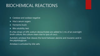


This document discusses Streptococcus pneumoniae, a common bacterium that can cause pneumonia, meningitis, and other infections. It describes the morphology and cultural characteristics of S. pneumoniae, including that it appears as paired diplococci and forms alpha-hemolytic colonies on blood agar. It also covers the antigenic properties, virulence factors, pathogenicity, epidemiology, laboratory diagnosis, prophylaxis, and treatment of infections caused by S. pneumoniae.


