SlideShare a Scribd company logo
1 of 9
Download to read offline
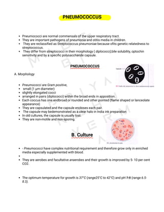
PNEUMOCOCCUS
Pneumococci are normal commensals of the upper respiratory tract. 
They are important pathogens of pneumonia and otitis media in children.
 They are reclassified as Streptococcus pneumoniae because ofits genetic relatedness to
streptococcus.
 They differ from streptococci in their morphology ( diplococci),bile solubility, optochin
sensitivity and by a specific polysaccharide capsule.
       PNEUMOCOCCUS
A. Morphology
Pneumococci are Gram positive,
 small (1 µm diameter)
slightly elongated cocci 
arranged in pairs (diplococci) within the broad ends in apposition. 
Each coccus has one endbroad or rounded and other pointed (flame shaped or lanceolate
appearance)
They are capsulated and the capsule encloses each pair.
 The capsule may bedemonstrated as a clear halo in India ink preparation
In old cultures, the capsule is usually lost.
They are non-motile and non-sporing.
B. Culture
- Pneumococci have complex nutritional requirement and therefore grow only in enriched
media especially supplemented with blood. 
They are aerobes and facultative anaerobes and their growth is improved by 5- 10 per cent
CO2. 
The optimum temperature for growth is 37°C (range25°C to 42°C) and pH 7.8 (range 6.5-
8.3).
On blood agar, after incubation for 18 hours, the colonies are usually small (0.5- 1 mm),
dome shaped, withan area of greenish discolouration (alpha haemolysis) around them. 
On prolonged incubation, the coloniesbecome flat, with raised edges and central
umbonation(due to autolysis occuring at centre) which creates adraughtsman
appearance (concentric rings are seen whenviewed from above).
 Some strains such as those of serotype 3 and 7 form large mucoid colonies due to
productionof abundant capsular polysaccharides. 
Pneumococci are typically alpha haemolytic but under anaerobic conditions colonies
show beta haemolysis due to liberation of oxygen labile pneumolysin O by these bacteria.
In liquid medium such as glucose broth, pneumococci produce uniform turbidity. 
These cocci readily undergoautolysis in cultures due to the action of
intracellularenzymes. 
Autolysis is enhanced by bile salts andother surface active agents. 
This property is helpful to differentiate it from other streptococci.
C. Biochemical Reactions
Pneumococci ferment several sugars with production of acid only.
 Fermentation is tested in Hiss's serum water.
Fermentation of inslin by pneumococci is of great valueto differentiate them from
streptococci.
Pneumococci are soluble in bile. When 2% sodiumdeoxycholate solution is added to a
broth culture atneutral pH, the culture clears due to the lysisof the cocci.Alternatively, if a
loopful of 10% sodium deoxycholatesolution is placed on a pneumococcus colony, lysis
ofcolony occurs within a few minutes. 
Bile solubility test is an important diagnostic test to differentiate pneumococcusfrom
other streptococci.
 This test is based on the presenceof an autolytic enzyme amidase in the pneumococci. 
Thisenzyme cleaves the bond between alanine and muramicacid in the peptidoglycan.
 The amidase is activated bybile salts, resulting in lysis of the bacteria.Pneumococci are
catalase and oxidase negative.
D. Resistance
Pneumococci are delicate organisms and are destroyedat 52°C for 15 minutes.
 They are more sensitive to usual antiseptics.
They are sensitive to a wide range of antibiotics including penicillins, erythromycin,
tetracycline, chloramphenicol, vancomycin and clindamycin. 
Penicillin resistant strainsfirst appeared in 1967. It may be due to mutation orgene
transfer. 
 The drug resistance to penicillin is notdue to production of beta lactamase, but alteration
inthe penicillin binding proteins on the bacterial surface.
These strains are also resistant to multiple drugs
Pneumococci are sensitive to optochin (ethyl-hydrocuprein hydrochloride) in a
concentration of1/500,000. When a 6 mm optochin disc (5 µg) is applied ona blood agar
plate inoculated with pneumococci, a widezone (14 mm or more) of inhibition occurs on
incubation.
This is very useful test to differentiate pneumococci fromother streptococci which do not show
zone of inhibition by optochin disc.
                 E. Antigenic Structure: 
The most important antigen of the pneumococcus iscapsular polysaccharide.
 Other antigens are somatic M protein and a group specific cell wall carbohydrate.
1. Capsular Polysaccharide: 
Capsular polysaccharide is type specific. 
Since this polysaccharide is soluble in tissue and culture fluid, it diffusesinto the culture
medium or tissues and infective exudates,hence, it is also called the soluble specific
substance (SSS).
Pneumococci are classified into types based on the nature of the capsular
polysaccharide. 
More than 90 serotypesare recognised.
 These are named 1, 2, 3, and so on.Serological typing of pneumococcus is carried out by
three methods.
(i) Agglutination of organisms with type specific
antiserum.
(ii) Precipitation of capsular polysaccharide with
type specific antiserum.
(iii) Quellung reaction or capsule swelling reaction
was described by Neufeld (1902).
 In this reaction,a suspension of pneumococci is mixed on a slidewith a drop of specific
antiserum and a loopfulof methylene blue solution. In presence of thehomologous
antiserum, the capsule around pneumococci reveals an apparent swelling,sharply
delineated and refractile under the microscope.
 The Quellung test can be done eitherin sputum or in culture, and used to be a routine
bedside procedure in olden days.
2. M Protein
M protein is characteristic for each type of pneumococcus.
It is not associated with virulence and antibody to M protein is non-protective.
3. Cell Wall Carbohydrate
(C-Substance) Pneumococci contain a species specific carbohydrateantigen which is
named as C-substance.
It is present in all pneumococci.
The C-substance is precipitated by a abnormal protein (ß-globulin), that appears in the
acute phase sera of cases of pneumonia but disappears during convalescence.
 It is also detected in blood of patients with some other illnesses.
 This is known as the C-reactive protein (CRP).
 It is not an antibody of C substance. 
It is an 'acute phase' substance, produced in hepatocytes.
Its production is stimulated by bacterial infections,malignancies and tissue destruction.
 CRP is used as anindex of activity in rheumatic fever and certain otherconditions.
 CRP is routinely detected in serum by latex agglutination test.
F. Variation Pneumococcus is virulent in capsulated (smooth) form but non-capsulated
(rough) forms are avirulent. On repeatedsubculture, a smooth to rough (S-.R) variation
occurs.
R forms arise as spontaneous mutants and outgrow the parental 
S forms in artificial culture.
 Such R mutants are eliminated in tissues by phagocytosis.
Rough pneumococci of one serotype may be made to produce capsules of the same or
different serotypes,on treatment with DNA from the respective serotypes of
pneumococci. 
This transformation was discovered byGriffith (1928) as the first instance of genetic
exchange of information in bacteria.
G. Toxins and Other Virulence Factors
Pneumococci produce an oxygen labile intracellular
haemolysin and a leucocidin. 
The virulence of pneumococci is dependant upon its capsule which prevents or inhibits
phagocytosis. 
Non-capsulated strains are avirulent.
 The antibody to the capsular polysaccharide protects against infection. 
Due to the abdundance of capsular material, the virulence of type-3 pneumococcus is
enhanced.
Pneumolysin, a toxin produced by penumococci, is another virulence factor.
 It has cytotoxic and complement activating properties. 
It is immunogenic H. Pathogenesis Str. pneumoniae is one of the most common bacteria
causing pneumonia, both lobar and bronchopneumonia.
It is also responsible for acute tracheobronchitis and empyema.
1. Lobar Pneumonia - 
Pneumonia results only when the general resistance is lowered.
 Common infective types of Str. pneumoniae include types 1- 12 in adults 
and types 6, 14, 19 and 23 are responsible in children.
2. Bronchopneumonia- 
It is almost always a secondary infection following viralinfections of the respiratory tract. 
Any serotype of pneumococcus can produce bronchopneumonia. 
Other causative agents responsible for bronchopneumonia include Staph. aureus, K.
pneumoniae, Str. pyogenes,H. influenzae, Fusobacterium species and Bacteroides.
3. Meningitis
It is the most serious of pneumococcal infections. Str. pneumoniae is the second most
important cause of pyogenic meningitis after Nmeningitidis.
 This disease is common  in children. Pneumococcus spreads from the pharynx to the
meninges via blood stream. 
Other bacterialagents of pyogenic meningitis include N meningitidis,H. influenzae, Str.
agalactiae (group-B) and Listeria monocytogenes.
4.Other Infections 
Pneumococcus may also produce empyema, pericarditis,otitis media, sinusitis,
conjunctivitis, peritonitis andsuppurative arthritis, usually as complications of pneumoma.
      I. Epidemiology
The source of human infection is carrier and less often patient. 
Pneumococci are transmitted by inhalation of contaminated dust, droplets or droplet
nuclei.
 Infection usually leads to pharyngeal carriage. 
Disease results only when the host resistance is lowered by factors such asrespiratory
viral infections, malnutrition etc.
Lobar pneumonia is usually a sporadic disease but epidemics may occur.
 The incidence of bronchopneumonia is more when an epidemic of influenza or other viral
infections of the respiratory tract occurs.
J. Laboratory Diagnosis
1. Specimens:
Clinical samples, such as
 sputum cerebrospinal fluid (CSF)
pleural exudate 
 blood are collected according to the site of lesion.
 Blood culture is useful in pneumococcal septicaemia.
2. Collection and Transport
All the specimens should be collected in sterile containers
under all aseptic conditions. 
They should be processed immediately. 
In case of delay, CSF specimen should never be refrigerated but kept at 37°C (H.
influenzae, another causative agent of pyogenic meningitis may die at cold temperature).
3. Direct Microscopy and Antigen Detection :
Gram staining of smear reveals a large number of polymorphs and typical organism.
 In case of meningitis, presumptive diagnosis may be made by finding Gram positive
diplococci which may be intracellular as well as extracellular in CSP smear. 
Capsule swelling may beobserved under microscope, when pneumococci are mixed with
type specific antisera.
Capsular polysaccharide antigen can be demonstrated by counterimmunoelectrophoresis. 
This has been employed in blood, urine and cerebrospinal fluid. 
Antigen may also be detected by immunochromatographic assay, latex agglutination or
coagglutination.
4. Culture
Specimen is inoculated on blood agar and incubated at 37°C for 24 hours in the presence
of 5- 10% CO2.
Typical colonies develop with a- haemolysis.
Organisms from the isolated colony are identified by Gram staining and biochemical
reactions  
5. Colony Morphology and Staining
Colonies are usually small (0.5- 1 mm), with alpha haemolysis around them. 
On prolonged incubation, colonies have draughtsman appearance.
On Gram staining pneumococci are Gram positive, small (1 µm diameter), diplococci
They are flameshaped or lanceolate in appearance.
 The capsule may be demonstrated as a clear halo in India ink preparation 
6. Biochemical Reactions
Important biochemical tests are inulin fermentation and bile solubility tests.
 Another test which has a great value is optochin sensitivity test. 
7. Animal Pathogenicity Test
From specimens where organisms are expected to be scanty, intraperitoneal inoculation
in mice may be used.
Inoculated mice die in 24-48 hours.
 Heart blood and peritoneal exudate of the animal shows pneumococci.
The test may be negative with occasional strains which are avirulent for mice (type 14
strains).
8. Antibiotic Sensitivity Test
It is especially useful in strains which are resistant.
K. Treatment
The antibiotic of choice is parenteral penicillin. 
Cephalosporin is indicated in case of penicillin resistant strains.
L. Prophylaxis
Immunity is type specific and is associated with antibody to the capsular polysaccharide. 
The existence of more than 90 serotypes makes prophylactic immunisation impracticable.
But, as a limited number of serotypes cause most of the pneumonia cases, vaccination is
possible.
Three pneumococcal vaccines are available.
A polysaccharide vaccine containing prevalentserotypes (23 serotypes) is administered
by a single dose injection.
 Such vaccines are used only in those persons who are at enhanced risk of pneumococcal
infection.
These include persons with absent or dysfunctional spleen, nephrotic syndrome, sickle cell
anaemia, multiple myeloma, hepatic cirrhosis, diabetes mellitus and immunodeficiencies
including HIV infection. 
Vaccination is contraindicated in lymphomas.
The heptavalent pneumococcal conjugate vaccine (7 serotypes) is composed of purified
polysaccharides of
seven serotypes conjugated to a diphtheria protein is now available for use in children
from two months to two years.
More recently, a new 13-valent vaccine that contain six additional serotypes was
approved for young children.
However, protection would de
pend on the principle thatserotypes prevalent in the community and serotypes used in the
vaccine are the same.
Pdf made by : Priyanshu verma
Reference : CP BAVEJA MICROBIOLOGY
pdf made by : Priyanshu verma
Reference : CV BAVEJA MICROBIOLOGY

More Related Content

What's hot (20)

3. pneumococcus
3. pneumococcus3. pneumococcus
3. pneumococcus
 
Pseudomonas
PseudomonasPseudomonas
Pseudomonas
 
Clostridium tetani
Clostridium tetaniClostridium tetani
Clostridium tetani
 
PNEUMOCOCCUS
PNEUMOCOCCUSPNEUMOCOCCUS
PNEUMOCOCCUS
 
Lab diagnosis of viral infection
Lab diagnosis of viral infection Lab diagnosis of viral infection
Lab diagnosis of viral infection
 
Staphylococcus
StaphylococcusStaphylococcus
Staphylococcus
 
Staphylococcus
StaphylococcusStaphylococcus
Staphylococcus
 
Bacillus
BacillusBacillus
Bacillus
 
Clostridium
ClostridiumClostridium
Clostridium
 
Staphylococcus
StaphylococcusStaphylococcus
Staphylococcus
 
Streptococcus pneumoniae
Streptococcus pneumoniaeStreptococcus pneumoniae
Streptococcus pneumoniae
 
Neisseria Meningitidis
Neisseria MeningitidisNeisseria Meningitidis
Neisseria Meningitidis
 
CLS Mycetoma.pptx
CLS Mycetoma.pptxCLS Mycetoma.pptx
CLS Mycetoma.pptx
 
Corynebacterium diptheriae
Corynebacterium diptheriaeCorynebacterium diptheriae
Corynebacterium diptheriae
 
Streptococcus
Streptococcus Streptococcus
Streptococcus
 
PROTEUS
PROTEUS PROTEUS
PROTEUS
 
Neisseria
NeisseriaNeisseria
Neisseria
 
Mycobacteria
MycobacteriaMycobacteria
Mycobacteria
 
streptococci
streptococcistreptococci
streptococci
 
Gram Positive Cocci-Streptococcus
Gram Positive Cocci-StreptococcusGram Positive Cocci-Streptococcus
Gram Positive Cocci-Streptococcus
 

Similar to Pneumococcus (microbiology)

Streptococcus pneumoniae: Una revisión
Streptococcus pneumoniae: Una revisiónStreptococcus pneumoniae: Una revisión
Streptococcus pneumoniae: Una revisiónfranklinaranda
 
Streptococcus pneumoniae: Una revisión
Streptococcus pneumoniae: Una revisiónStreptococcus pneumoniae: Una revisión
Streptococcus pneumoniae: Una revisiónfranklinaranda
 
streptococcus pneumoniae: Revisión
streptococcus pneumoniae: Revisión streptococcus pneumoniae: Revisión
streptococcus pneumoniae: Revisión franklinaranda
 
Fungal lesions of the eye
Fungal lesions of the eyeFungal lesions of the eye
Fungal lesions of the eyevayyu
 
Pneumonia Disease Research Paper
Pneumonia Disease Research PaperPneumonia Disease Research Paper
Pneumonia Disease Research PaperHaley Johnson
 
Opportunistic Infections. Genera Proteus. Klebsiella & Pseudomonas. Hospital ...
Opportunistic Infections. Genera Proteus. Klebsiella & Pseudomonas. Hospital ...Opportunistic Infections. Genera Proteus. Klebsiella & Pseudomonas. Hospital ...
Opportunistic Infections. Genera Proteus. Klebsiella & Pseudomonas. Hospital ...Eneutron
 
Gram positive bacteria in ocular pathology
Gram positive bacteria in ocular pathologyGram positive bacteria in ocular pathology
Gram positive bacteria in ocular pathologySudheer Kumar
 
Bacterial Diseases of the Respiratory System.pptx
Bacterial Diseases of the Respiratory System.pptxBacterial Diseases of the Respiratory System.pptx
Bacterial Diseases of the Respiratory System.pptxTofikMohammed3
 
FUNGAL INFECTIONS OF ORAL CAVITY
FUNGAL INFECTIONS OF ORAL CAVITYFUNGAL INFECTIONS OF ORAL CAVITY
FUNGAL INFECTIONS OF ORAL CAVITYDRROSHINCN1
 
Sterptococcus pneumoniae
Sterptococcus pneumoniaeSterptococcus pneumoniae
Sterptococcus pneumoniaeBruno Mmassy
 
Bohomolets Microbiology Lecture #20
Bohomolets Microbiology Lecture #20Bohomolets Microbiology Lecture #20
Bohomolets Microbiology Lecture #20Dr. Rubz
 

Similar to Pneumococcus (microbiology) (20)

Pneumococci ppt mahadi
Pneumococci ppt mahadiPneumococci ppt mahadi
Pneumococci ppt mahadi
 
Pyogenic cocci.pptx
Pyogenic cocci.pptxPyogenic cocci.pptx
Pyogenic cocci.pptx
 
neisseria gonorrhoea
neisseria gonorrhoeaneisseria gonorrhoea
neisseria gonorrhoea
 
Streptococcus pneumoniae: Una revisión
Streptococcus pneumoniae: Una revisiónStreptococcus pneumoniae: Una revisión
Streptococcus pneumoniae: Una revisión
 
Streptococcus pneumoniae: Una revisión
Streptococcus pneumoniae: Una revisiónStreptococcus pneumoniae: Una revisión
Streptococcus pneumoniae: Una revisión
 
streptococcus pneumoniae: Revisión
streptococcus pneumoniae: Revisión streptococcus pneumoniae: Revisión
streptococcus pneumoniae: Revisión
 
Fungal lesions of the eye
Fungal lesions of the eyeFungal lesions of the eye
Fungal lesions of the eye
 
Mycobacterium .pptx
Mycobacterium .pptxMycobacterium .pptx
Mycobacterium .pptx
 
Pneumococcus
PneumococcusPneumococcus
Pneumococcus
 
Bacterial diseases
Bacterial diseasesBacterial diseases
Bacterial diseases
 
Pneumonia Disease Research Paper
Pneumonia Disease Research PaperPneumonia Disease Research Paper
Pneumonia Disease Research Paper
 
Pneumococcus
PneumococcusPneumococcus
Pneumococcus
 
Bacteriology
BacteriologyBacteriology
Bacteriology
 
Opportunistic Infections. Genera Proteus. Klebsiella & Pseudomonas. Hospital ...
Opportunistic Infections. Genera Proteus. Klebsiella & Pseudomonas. Hospital ...Opportunistic Infections. Genera Proteus. Klebsiella & Pseudomonas. Hospital ...
Opportunistic Infections. Genera Proteus. Klebsiella & Pseudomonas. Hospital ...
 
Gram positive bacteria in ocular pathology
Gram positive bacteria in ocular pathologyGram positive bacteria in ocular pathology
Gram positive bacteria in ocular pathology
 
Bacterial Diseases of the Respiratory System.pptx
Bacterial Diseases of the Respiratory System.pptxBacterial Diseases of the Respiratory System.pptx
Bacterial Diseases of the Respiratory System.pptx
 
FUNGAL INFECTIONS OF ORAL CAVITY
FUNGAL INFECTIONS OF ORAL CAVITYFUNGAL INFECTIONS OF ORAL CAVITY
FUNGAL INFECTIONS OF ORAL CAVITY
 
Sterptococcus pneumoniae
Sterptococcus pneumoniaeSterptococcus pneumoniae
Sterptococcus pneumoniae
 
تشخيص بكتريا.pptx
تشخيص بكتريا.pptxتشخيص بكتريا.pptx
تشخيص بكتريا.pptx
 
Bohomolets Microbiology Lecture #20
Bohomolets Microbiology Lecture #20Bohomolets Microbiology Lecture #20
Bohomolets Microbiology Lecture #20
 

More from MrVermaasahab

Gastroesophageal reflux diseases (GERD)
Gastroesophageal reflux diseases (GERD) Gastroesophageal reflux diseases (GERD)
Gastroesophageal reflux diseases (GERD) MrVermaasahab
 
Acute respiratory distress syndrome, msn
Acute respiratory distress syndrome, msnAcute respiratory distress syndrome, msn
Acute respiratory distress syndrome, msnMrVermaasahab
 
HYPERTENSION || Medical surgical nursing || msn
HYPERTENSION || Medical surgical nursing || msnHYPERTENSION || Medical surgical nursing || msn
HYPERTENSION || Medical surgical nursing || msnMrVermaasahab
 
CORYNEBACTERIUM (microbiology)
CORYNEBACTERIUM (microbiology)CORYNEBACTERIUM (microbiology)
CORYNEBACTERIUM (microbiology)MrVermaasahab
 

More from MrVermaasahab (6)

PDFPrint.pdf
PDFPrint.pdfPDFPrint.pdf
PDFPrint.pdf
 
PNEUMONIA.pdf
PNEUMONIA.pdfPNEUMONIA.pdf
PNEUMONIA.pdf
 
Gastroesophageal reflux diseases (GERD)
Gastroesophageal reflux diseases (GERD) Gastroesophageal reflux diseases (GERD)
Gastroesophageal reflux diseases (GERD)
 
Acute respiratory distress syndrome, msn
Acute respiratory distress syndrome, msnAcute respiratory distress syndrome, msn
Acute respiratory distress syndrome, msn
 
HYPERTENSION || Medical surgical nursing || msn
HYPERTENSION || Medical surgical nursing || msnHYPERTENSION || Medical surgical nursing || msn
HYPERTENSION || Medical surgical nursing || msn
 
CORYNEBACTERIUM (microbiology)
CORYNEBACTERIUM (microbiology)CORYNEBACTERIUM (microbiology)
CORYNEBACTERIUM (microbiology)
 

Recently uploaded

All-domain Anomaly Resolution Office U.S. Department of Defense (U) Case: “Eg...
All-domain Anomaly Resolution Office U.S. Department of Defense (U) Case: “Eg...All-domain Anomaly Resolution Office U.S. Department of Defense (U) Case: “Eg...
All-domain Anomaly Resolution Office U.S. Department of Defense (U) Case: “Eg...Sérgio Sacani
 
Behavioral Disorder: Schizophrenia & it's Case Study.pdf
Behavioral Disorder: Schizophrenia & it's Case Study.pdfBehavioral Disorder: Schizophrenia & it's Case Study.pdf
Behavioral Disorder: Schizophrenia & it's Case Study.pdfSELF-EXPLANATORY
 
PossibleEoarcheanRecordsoftheGeomagneticFieldPreservedintheIsuaSupracrustalBe...
PossibleEoarcheanRecordsoftheGeomagneticFieldPreservedintheIsuaSupracrustalBe...PossibleEoarcheanRecordsoftheGeomagneticFieldPreservedintheIsuaSupracrustalBe...
PossibleEoarcheanRecordsoftheGeomagneticFieldPreservedintheIsuaSupracrustalBe...Sérgio Sacani
 
Call Girls in Munirka Delhi 💯Call Us 🔝8264348440🔝
Call Girls in Munirka Delhi 💯Call Us 🔝8264348440🔝Call Girls in Munirka Delhi 💯Call Us 🔝8264348440🔝
Call Girls in Munirka Delhi 💯Call Us 🔝8264348440🔝soniya singh
 
A relative description on Sonoporation.pdf
A relative description on Sonoporation.pdfA relative description on Sonoporation.pdf
A relative description on Sonoporation.pdfnehabiju2046
 
SOLUBLE PATTERN RECOGNITION RECEPTORS.pptx
SOLUBLE PATTERN RECOGNITION RECEPTORS.pptxSOLUBLE PATTERN RECOGNITION RECEPTORS.pptx
SOLUBLE PATTERN RECOGNITION RECEPTORS.pptxkessiyaTpeter
 
Discovery of an Accretion Streamer and a Slow Wide-angle Outflow around FUOri...
Discovery of an Accretion Streamer and a Slow Wide-angle Outflow around FUOri...Discovery of an Accretion Streamer and a Slow Wide-angle Outflow around FUOri...
Discovery of an Accretion Streamer and a Slow Wide-angle Outflow around FUOri...Sérgio Sacani
 
Traditional Agroforestry System in India- Shifting Cultivation, Taungya, Home...
Traditional Agroforestry System in India- Shifting Cultivation, Taungya, Home...Traditional Agroforestry System in India- Shifting Cultivation, Taungya, Home...
Traditional Agroforestry System in India- Shifting Cultivation, Taungya, Home...jana861314
 
Labelling Requirements and Label Claims for Dietary Supplements and Recommend...
Labelling Requirements and Label Claims for Dietary Supplements and Recommend...Labelling Requirements and Label Claims for Dietary Supplements and Recommend...
Labelling Requirements and Label Claims for Dietary Supplements and Recommend...Lokesh Kothari
 
Scheme-of-Work-Science-Stage-4 cambridge science.docx
Scheme-of-Work-Science-Stage-4 cambridge science.docxScheme-of-Work-Science-Stage-4 cambridge science.docx
Scheme-of-Work-Science-Stage-4 cambridge science.docxyaramohamed343013
 
Call Girls in Mayapuri Delhi 💯Call Us 🔝9953322196🔝 💯Escort.
Call Girls in Mayapuri Delhi 💯Call Us 🔝9953322196🔝 💯Escort.Call Girls in Mayapuri Delhi 💯Call Us 🔝9953322196🔝 💯Escort.
Call Girls in Mayapuri Delhi 💯Call Us 🔝9953322196🔝 💯Escort.aasikanpl
 
Call Us ≽ 9953322196 ≼ Call Girls In Mukherjee Nagar(Delhi) |
Call Us ≽ 9953322196 ≼ Call Girls In Mukherjee Nagar(Delhi) |Call Us ≽ 9953322196 ≼ Call Girls In Mukherjee Nagar(Delhi) |
Call Us ≽ 9953322196 ≼ Call Girls In Mukherjee Nagar(Delhi) |aasikanpl
 
Biological Classification BioHack (3).pdf
Biological Classification BioHack (3).pdfBiological Classification BioHack (3).pdf
Biological Classification BioHack (3).pdfmuntazimhurra
 
Artificial Intelligence In Microbiology by Dr. Prince C P
Artificial Intelligence In Microbiology by Dr. Prince C PArtificial Intelligence In Microbiology by Dr. Prince C P
Artificial Intelligence In Microbiology by Dr. Prince C PPRINCE C P
 
Cultivation of KODO MILLET . made by Ghanshyam pptx
Cultivation of KODO MILLET . made by Ghanshyam pptxCultivation of KODO MILLET . made by Ghanshyam pptx
Cultivation of KODO MILLET . made by Ghanshyam pptxpradhanghanshyam7136
 
Nightside clouds and disequilibrium chemistry on the hot Jupiter WASP-43b
Nightside clouds and disequilibrium chemistry on the hot Jupiter WASP-43bNightside clouds and disequilibrium chemistry on the hot Jupiter WASP-43b
Nightside clouds and disequilibrium chemistry on the hot Jupiter WASP-43bSérgio Sacani
 
Grafana in space: Monitoring Japan's SLIM moon lander in real time
Grafana in space: Monitoring Japan's SLIM moon lander  in real timeGrafana in space: Monitoring Japan's SLIM moon lander  in real time
Grafana in space: Monitoring Japan's SLIM moon lander in real timeSatoshi NAKAHIRA
 
Is RISC-V ready for HPC workload? Maybe?
Is RISC-V ready for HPC workload? Maybe?Is RISC-V ready for HPC workload? Maybe?
Is RISC-V ready for HPC workload? Maybe?Patrick Diehl
 
Orientation, design and principles of polyhouse
Orientation, design and principles of polyhouseOrientation, design and principles of polyhouse
Orientation, design and principles of polyhousejana861314
 

Recently uploaded (20)

All-domain Anomaly Resolution Office U.S. Department of Defense (U) Case: “Eg...
All-domain Anomaly Resolution Office U.S. Department of Defense (U) Case: “Eg...All-domain Anomaly Resolution Office U.S. Department of Defense (U) Case: “Eg...
All-domain Anomaly Resolution Office U.S. Department of Defense (U) Case: “Eg...
 
Behavioral Disorder: Schizophrenia & it's Case Study.pdf
Behavioral Disorder: Schizophrenia & it's Case Study.pdfBehavioral Disorder: Schizophrenia & it's Case Study.pdf
Behavioral Disorder: Schizophrenia & it's Case Study.pdf
 
Engler and Prantl system of classification in plant taxonomy
Engler and Prantl system of classification in plant taxonomyEngler and Prantl system of classification in plant taxonomy
Engler and Prantl system of classification in plant taxonomy
 
PossibleEoarcheanRecordsoftheGeomagneticFieldPreservedintheIsuaSupracrustalBe...
PossibleEoarcheanRecordsoftheGeomagneticFieldPreservedintheIsuaSupracrustalBe...PossibleEoarcheanRecordsoftheGeomagneticFieldPreservedintheIsuaSupracrustalBe...
PossibleEoarcheanRecordsoftheGeomagneticFieldPreservedintheIsuaSupracrustalBe...
 
Call Girls in Munirka Delhi 💯Call Us 🔝8264348440🔝
Call Girls in Munirka Delhi 💯Call Us 🔝8264348440🔝Call Girls in Munirka Delhi 💯Call Us 🔝8264348440🔝
Call Girls in Munirka Delhi 💯Call Us 🔝8264348440🔝
 
A relative description on Sonoporation.pdf
A relative description on Sonoporation.pdfA relative description on Sonoporation.pdf
A relative description on Sonoporation.pdf
 
SOLUBLE PATTERN RECOGNITION RECEPTORS.pptx
SOLUBLE PATTERN RECOGNITION RECEPTORS.pptxSOLUBLE PATTERN RECOGNITION RECEPTORS.pptx
SOLUBLE PATTERN RECOGNITION RECEPTORS.pptx
 
Discovery of an Accretion Streamer and a Slow Wide-angle Outflow around FUOri...
Discovery of an Accretion Streamer and a Slow Wide-angle Outflow around FUOri...Discovery of an Accretion Streamer and a Slow Wide-angle Outflow around FUOri...
Discovery of an Accretion Streamer and a Slow Wide-angle Outflow around FUOri...
 
Traditional Agroforestry System in India- Shifting Cultivation, Taungya, Home...
Traditional Agroforestry System in India- Shifting Cultivation, Taungya, Home...Traditional Agroforestry System in India- Shifting Cultivation, Taungya, Home...
Traditional Agroforestry System in India- Shifting Cultivation, Taungya, Home...
 
Labelling Requirements and Label Claims for Dietary Supplements and Recommend...
Labelling Requirements and Label Claims for Dietary Supplements and Recommend...Labelling Requirements and Label Claims for Dietary Supplements and Recommend...
Labelling Requirements and Label Claims for Dietary Supplements and Recommend...
 
Scheme-of-Work-Science-Stage-4 cambridge science.docx
Scheme-of-Work-Science-Stage-4 cambridge science.docxScheme-of-Work-Science-Stage-4 cambridge science.docx
Scheme-of-Work-Science-Stage-4 cambridge science.docx
 
Call Girls in Mayapuri Delhi 💯Call Us 🔝9953322196🔝 💯Escort.
Call Girls in Mayapuri Delhi 💯Call Us 🔝9953322196🔝 💯Escort.Call Girls in Mayapuri Delhi 💯Call Us 🔝9953322196🔝 💯Escort.
Call Girls in Mayapuri Delhi 💯Call Us 🔝9953322196🔝 💯Escort.
 
Call Us ≽ 9953322196 ≼ Call Girls In Mukherjee Nagar(Delhi) |
Call Us ≽ 9953322196 ≼ Call Girls In Mukherjee Nagar(Delhi) |Call Us ≽ 9953322196 ≼ Call Girls In Mukherjee Nagar(Delhi) |
Call Us ≽ 9953322196 ≼ Call Girls In Mukherjee Nagar(Delhi) |
 
Biological Classification BioHack (3).pdf
Biological Classification BioHack (3).pdfBiological Classification BioHack (3).pdf
Biological Classification BioHack (3).pdf
 
Artificial Intelligence In Microbiology by Dr. Prince C P
Artificial Intelligence In Microbiology by Dr. Prince C PArtificial Intelligence In Microbiology by Dr. Prince C P
Artificial Intelligence In Microbiology by Dr. Prince C P
 
Cultivation of KODO MILLET . made by Ghanshyam pptx
Cultivation of KODO MILLET . made by Ghanshyam pptxCultivation of KODO MILLET . made by Ghanshyam pptx
Cultivation of KODO MILLET . made by Ghanshyam pptx
 
Nightside clouds and disequilibrium chemistry on the hot Jupiter WASP-43b
Nightside clouds and disequilibrium chemistry on the hot Jupiter WASP-43bNightside clouds and disequilibrium chemistry on the hot Jupiter WASP-43b
Nightside clouds and disequilibrium chemistry on the hot Jupiter WASP-43b
 
Grafana in space: Monitoring Japan's SLIM moon lander in real time
Grafana in space: Monitoring Japan's SLIM moon lander  in real timeGrafana in space: Monitoring Japan's SLIM moon lander  in real time
Grafana in space: Monitoring Japan's SLIM moon lander in real time
 
Is RISC-V ready for HPC workload? Maybe?
Is RISC-V ready for HPC workload? Maybe?Is RISC-V ready for HPC workload? Maybe?
Is RISC-V ready for HPC workload? Maybe?
 
Orientation, design and principles of polyhouse
Orientation, design and principles of polyhouseOrientation, design and principles of polyhouse
Orientation, design and principles of polyhouse
 

Pneumococcus (microbiology)

  • 1. PNEUMOCOCCUS Pneumococci are normal commensals of the upper respiratory tract.  They are important pathogens of pneumonia and otitis media in children.  They are reclassified as Streptococcus pneumoniae because ofits genetic relatedness to streptococcus.  They differ from streptococci in their morphology ( diplococci),bile solubility, optochin sensitivity and by a specific polysaccharide capsule.        PNEUMOCOCCUS A. Morphology Pneumococci are Gram positive,  small (1 µm diameter) slightly elongated cocci  arranged in pairs (diplococci) within the broad ends in apposition.  Each coccus has one endbroad or rounded and other pointed (flame shaped or lanceolate appearance) They are capsulated and the capsule encloses each pair.  The capsule may bedemonstrated as a clear halo in India ink preparation In old cultures, the capsule is usually lost. They are non-motile and non-sporing. B. Culture - Pneumococci have complex nutritional requirement and therefore grow only in enriched media especially supplemented with blood.  They are aerobes and facultative anaerobes and their growth is improved by 5- 10 per cent CO2.  The optimum temperature for growth is 37°C (range25°C to 42°C) and pH 7.8 (range 6.5- 8.3).
  • 2. On blood agar, after incubation for 18 hours, the colonies are usually small (0.5- 1 mm), dome shaped, withan area of greenish discolouration (alpha haemolysis) around them.  On prolonged incubation, the coloniesbecome flat, with raised edges and central umbonation(due to autolysis occuring at centre) which creates adraughtsman appearance (concentric rings are seen whenviewed from above).  Some strains such as those of serotype 3 and 7 form large mucoid colonies due to productionof abundant capsular polysaccharides.  Pneumococci are typically alpha haemolytic but under anaerobic conditions colonies show beta haemolysis due to liberation of oxygen labile pneumolysin O by these bacteria. In liquid medium such as glucose broth, pneumococci produce uniform turbidity.  These cocci readily undergoautolysis in cultures due to the action of intracellularenzymes.  Autolysis is enhanced by bile salts andother surface active agents.  This property is helpful to differentiate it from other streptococci. C. Biochemical Reactions Pneumococci ferment several sugars with production of acid only.  Fermentation is tested in Hiss's serum water. Fermentation of inslin by pneumococci is of great valueto differentiate them from streptococci.
  • 3. Pneumococci are soluble in bile. When 2% sodiumdeoxycholate solution is added to a broth culture atneutral pH, the culture clears due to the lysisof the cocci.Alternatively, if a loopful of 10% sodium deoxycholatesolution is placed on a pneumococcus colony, lysis ofcolony occurs within a few minutes.  Bile solubility test is an important diagnostic test to differentiate pneumococcusfrom other streptococci.  This test is based on the presenceof an autolytic enzyme amidase in the pneumococci.  Thisenzyme cleaves the bond between alanine and muramicacid in the peptidoglycan.  The amidase is activated bybile salts, resulting in lysis of the bacteria.Pneumococci are catalase and oxidase negative. D. Resistance Pneumococci are delicate organisms and are destroyedat 52°C for 15 minutes.  They are more sensitive to usual antiseptics. They are sensitive to a wide range of antibiotics including penicillins, erythromycin, tetracycline, chloramphenicol, vancomycin and clindamycin.  Penicillin resistant strainsfirst appeared in 1967. It may be due to mutation orgene transfer.   The drug resistance to penicillin is notdue to production of beta lactamase, but alteration inthe penicillin binding proteins on the bacterial surface. These strains are also resistant to multiple drugs Pneumococci are sensitive to optochin (ethyl-hydrocuprein hydrochloride) in a concentration of1/500,000. When a 6 mm optochin disc (5 µg) is applied ona blood agar plate inoculated with pneumococci, a widezone (14 mm or more) of inhibition occurs on incubation. This is very useful test to differentiate pneumococci fromother streptococci which do not show zone of inhibition by optochin disc.
  • 4.                  E. Antigenic Structure:  The most important antigen of the pneumococcus iscapsular polysaccharide.  Other antigens are somatic M protein and a group specific cell wall carbohydrate. 1. Capsular Polysaccharide:  Capsular polysaccharide is type specific.  Since this polysaccharide is soluble in tissue and culture fluid, it diffusesinto the culture medium or tissues and infective exudates,hence, it is also called the soluble specific substance (SSS). Pneumococci are classified into types based on the nature of the capsular polysaccharide.  More than 90 serotypesare recognised.  These are named 1, 2, 3, and so on.Serological typing of pneumococcus is carried out by three methods. (i) Agglutination of organisms with type specific antiserum. (ii) Precipitation of capsular polysaccharide with type specific antiserum. (iii) Quellung reaction or capsule swelling reaction was described by Neufeld (1902).  In this reaction,a suspension of pneumococci is mixed on a slidewith a drop of specific antiserum and a loopfulof methylene blue solution. In presence of thehomologous antiserum, the capsule around pneumococci reveals an apparent swelling,sharply delineated and refractile under the microscope.  The Quellung test can be done eitherin sputum or in culture, and used to be a routine bedside procedure in olden days. 2. M Protein M protein is characteristic for each type of pneumococcus. It is not associated with virulence and antibody to M protein is non-protective. 3. Cell Wall Carbohydrate
  • 5. (C-Substance) Pneumococci contain a species specific carbohydrateantigen which is named as C-substance. It is present in all pneumococci. The C-substance is precipitated by a abnormal protein (ß-globulin), that appears in the acute phase sera of cases of pneumonia but disappears during convalescence.  It is also detected in blood of patients with some other illnesses.  This is known as the C-reactive protein (CRP).  It is not an antibody of C substance.  It is an 'acute phase' substance, produced in hepatocytes. Its production is stimulated by bacterial infections,malignancies and tissue destruction.  CRP is used as anindex of activity in rheumatic fever and certain otherconditions.  CRP is routinely detected in serum by latex agglutination test. F. Variation Pneumococcus is virulent in capsulated (smooth) form but non-capsulated (rough) forms are avirulent. On repeatedsubculture, a smooth to rough (S-.R) variation occurs. R forms arise as spontaneous mutants and outgrow the parental  S forms in artificial culture.  Such R mutants are eliminated in tissues by phagocytosis. Rough pneumococci of one serotype may be made to produce capsules of the same or different serotypes,on treatment with DNA from the respective serotypes of pneumococci.  This transformation was discovered byGriffith (1928) as the first instance of genetic exchange of information in bacteria. G. Toxins and Other Virulence Factors Pneumococci produce an oxygen labile intracellular haemolysin and a leucocidin.  The virulence of pneumococci is dependant upon its capsule which prevents or inhibits phagocytosis.  Non-capsulated strains are avirulent.  The antibody to the capsular polysaccharide protects against infection.  Due to the abdundance of capsular material, the virulence of type-3 pneumococcus is enhanced. Pneumolysin, a toxin produced by penumococci, is another virulence factor.  It has cytotoxic and complement activating properties.  It is immunogenic H. Pathogenesis Str. pneumoniae is one of the most common bacteria causing pneumonia, both lobar and bronchopneumonia. It is also responsible for acute tracheobronchitis and empyema. 1. Lobar Pneumonia -  Pneumonia results only when the general resistance is lowered.
  • 6.  Common infective types of Str. pneumoniae include types 1- 12 in adults  and types 6, 14, 19 and 23 are responsible in children. 2. Bronchopneumonia-  It is almost always a secondary infection following viralinfections of the respiratory tract.  Any serotype of pneumococcus can produce bronchopneumonia.  Other causative agents responsible for bronchopneumonia include Staph. aureus, K. pneumoniae, Str. pyogenes,H. influenzae, Fusobacterium species and Bacteroides. 3. Meningitis It is the most serious of pneumococcal infections. Str. pneumoniae is the second most important cause of pyogenic meningitis after Nmeningitidis.  This disease is common  in children. Pneumococcus spreads from the pharynx to the meninges via blood stream.  Other bacterialagents of pyogenic meningitis include N meningitidis,H. influenzae, Str. agalactiae (group-B) and Listeria monocytogenes. 4.Other Infections  Pneumococcus may also produce empyema, pericarditis,otitis media, sinusitis, conjunctivitis, peritonitis andsuppurative arthritis, usually as complications of pneumoma.       I. Epidemiology The source of human infection is carrier and less often patient.  Pneumococci are transmitted by inhalation of contaminated dust, droplets or droplet nuclei.  Infection usually leads to pharyngeal carriage.  Disease results only when the host resistance is lowered by factors such asrespiratory viral infections, malnutrition etc. Lobar pneumonia is usually a sporadic disease but epidemics may occur.  The incidence of bronchopneumonia is more when an epidemic of influenza or other viral infections of the respiratory tract occurs. J. Laboratory Diagnosis 1. Specimens: Clinical samples, such as  sputum cerebrospinal fluid (CSF) pleural exudate   blood are collected according to the site of lesion.  Blood culture is useful in pneumococcal septicaemia.
  • 7. 2. Collection and Transport All the specimens should be collected in sterile containers under all aseptic conditions.  They should be processed immediately.  In case of delay, CSF specimen should never be refrigerated but kept at 37°C (H. influenzae, another causative agent of pyogenic meningitis may die at cold temperature). 3. Direct Microscopy and Antigen Detection : Gram staining of smear reveals a large number of polymorphs and typical organism.  In case of meningitis, presumptive diagnosis may be made by finding Gram positive diplococci which may be intracellular as well as extracellular in CSP smear.  Capsule swelling may beobserved under microscope, when pneumococci are mixed with type specific antisera. Capsular polysaccharide antigen can be demonstrated by counterimmunoelectrophoresis.  This has been employed in blood, urine and cerebrospinal fluid.  Antigen may also be detected by immunochromatographic assay, latex agglutination or coagglutination. 4. Culture Specimen is inoculated on blood agar and incubated at 37°C for 24 hours in the presence of 5- 10% CO2. Typical colonies develop with a- haemolysis. Organisms from the isolated colony are identified by Gram staining and biochemical reactions   5. Colony Morphology and Staining Colonies are usually small (0.5- 1 mm), with alpha haemolysis around them.  On prolonged incubation, colonies have draughtsman appearance. On Gram staining pneumococci are Gram positive, small (1 µm diameter), diplococci They are flameshaped or lanceolate in appearance.  The capsule may be demonstrated as a clear halo in India ink preparation 
  • 8. 6. Biochemical Reactions Important biochemical tests are inulin fermentation and bile solubility tests.  Another test which has a great value is optochin sensitivity test.  7. Animal Pathogenicity Test From specimens where organisms are expected to be scanty, intraperitoneal inoculation in mice may be used. Inoculated mice die in 24-48 hours.  Heart blood and peritoneal exudate of the animal shows pneumococci. The test may be negative with occasional strains which are avirulent for mice (type 14 strains). 8. Antibiotic Sensitivity Test It is especially useful in strains which are resistant. K. Treatment The antibiotic of choice is parenteral penicillin.  Cephalosporin is indicated in case of penicillin resistant strains. L. Prophylaxis Immunity is type specific and is associated with antibody to the capsular polysaccharide.  The existence of more than 90 serotypes makes prophylactic immunisation impracticable. But, as a limited number of serotypes cause most of the pneumonia cases, vaccination is possible. Three pneumococcal vaccines are available. A polysaccharide vaccine containing prevalentserotypes (23 serotypes) is administered by a single dose injection.  Such vaccines are used only in those persons who are at enhanced risk of pneumococcal infection. These include persons with absent or dysfunctional spleen, nephrotic syndrome, sickle cell anaemia, multiple myeloma, hepatic cirrhosis, diabetes mellitus and immunodeficiencies including HIV infection.  Vaccination is contraindicated in lymphomas. The heptavalent pneumococcal conjugate vaccine (7 serotypes) is composed of purified polysaccharides of
  • 9. seven serotypes conjugated to a diphtheria protein is now available for use in children from two months to two years. More recently, a new 13-valent vaccine that contain six additional serotypes was approved for young children. However, protection would de pend on the principle thatserotypes prevalent in the community and serotypes used in the vaccine are the same. Pdf made by : Priyanshu verma Reference : CP BAVEJA MICROBIOLOGY pdf made by : Priyanshu verma Reference : CV BAVEJA MICROBIOLOGY