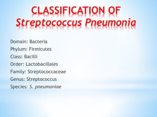
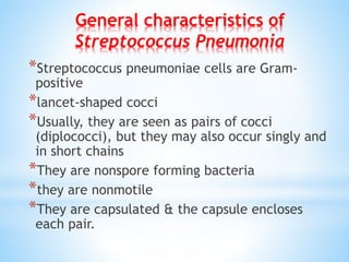
STREPTOCOCCUS PNEUMONIA
BY:
DR. Mohamed Taha Yassin Hassan
CLASSIFICATION OF
Streptococcus Pneumonia
Domain: Bacteria
Phylum: Firmicutes
Class: Bacilli
Order: Lactobacillales
Family: Streptococcaceae
Genus: Streptococcus
Species: S. pneumoniae
*Streptococcus pneumoniae cells are Gram-
positive
*lancet-shaped cocci
*Usually, they are seen as pairs of cocci
(diplococci), but they may also occur singly and
in short chains
*They are nonspore forming bacteria
*they are nonmotile
*They are capsulated & the capsule encloses
each pair.
General characteristics of
Streptococcus Pneumonia
STREPTOCOCCUS PNEUMONIA
*They grow only in enriched media.
*They are aerobes & facultative anaerobes.
*The optimum temperature being 37ºC & pH 7.8.
*Growth is improved by 5-10% CO2
Microscopy and Staining :
Perform Gram staining of the sample (sputum/CSF)
Gram staining shows Gram positive lanceolate shaped
diplococci
Electron microscope image of
Streptococcus Pneumonia
Colony morphology:
Colonies on blood agar plate are small (0.5 mm),
round, transluscent or mucoid with alpha-
hemolysis (A green discolouration of the agar
around the colonies).
Alpha-hemolytic property differentiates S.
pneumoniae from many species
alpha-hemolytic colonies are further
identified by the following confirmatory tests
Biochemical identification
*Optochin test (6 mm disc with 5µg).
*Inoculate blood agar plate with suspected alpha-hemolytic
isolates.
*Apply commercially available optochin discs on the streaked
blood agar plate
*Incubate plates at 37°C with 5-10% CO2 for 18-24 hours.
* Observe the zone of growth inhibition around the disc and
interpret as:
*A zone size > 14mm indicates susceptibility which is diagnostic of
Streptococcus pneumoniae.
*alpha-haemolytic colonies with zone of inhibition between 9 and
13 mm should be tested for bile solubility.
Bile solubility test:
*alpha-haemolytic colonies showing zone of inhibition around
optochin disc between 9 to 13 mm should be tested for bile
solubility.
*Prepare 1.0 ml of saline suspension of the organism from blood agar
plate.
*Bile solubility test: Streptococcus pneumonie colonies are lysed by
bile
*Inoculate 0.5 ml of the suspension into two tubes.
*Add an equal amount (0.5 ml) of 2% sodium deoxycholate in one
tube marked as test and 0.5ml of saline into the second tube
marked as control.
*Shake gently and incubate the tubes at 37°C for 2 hours
VIRULENCE FACTORS OF
STREPTOCOCCUS PNEUMONIA
1-Capsule:
is the major virulence factor of Streptococcus Pneumonia
Resistance to phagocytosis
2-Cell wall or(CWPS):
Inflammatory Factors
Activation of the alternative complement pathway,
resulting in anaphylatoxin production
During complement activation, the anaphylatoxins C3a
and C5a, which enhance vascular permeability, induce
mast cell degranulation, and recruit and activate
polymorphonuclear leukocytes (PMN) at the
inflammation site
3-Pneumococcal Proteins:
Include:
A-Pneumolysin:
stimulates the production of inflammatory cytokines like
tumor necrosis factor alpha and interleukin-1b by human
monocytes
inhibits the beating of cilia on human respiratory epithelial
cells
disrupts the monolayers of cultured epithelial cells from the
upper respiratory tract and from the alveoli
decreases the bactericidal activity and migration of
neutrophils
inhibits lymphocyte proliferation and Ab synthesis
B- Pneumococcal surface protein A. :
Pneumococcal surface protein A (PspA) is a surface protein with
structural and antigenic variability between different pneumococcal
strains.
required for full virulence of pneumococci
inhibition of complement activation
PspA enhances pneumococcal virulence
C- Autolysin:
Autolysin-negative mutants have been shown to be less virulent than
wild-type pneumococci
Release of pneumolysin and cell wall products
autolysin triggered by human lysozyme, a defense factor released
upon infection and inflammation.
Autolysin thus seems to take advantage of the (protective) function
of lysozyme.
D- Complement factor H-binding component:
Inhibition of complement activation
Inhibition of phagocytosis
E-Peptide permeases:
Enhancement of adhesion
F-IgA1 protease:
Counteracts mucosal defense mechanisms
4-Hydrogen peroxide:
Lung injury
STREPTOCOCCUS PNEUMONIA
PATHOGENESIS
the initiation of pneumonia requires escape from mucous defenses and
bacterial migration into the alveolus.
Firm adherence of bacteria to the alveolar epithelium and subsequent
replication and initiation of host damage
The pore-forming toxin pneumolysin and hydrogen peroxide released
in copious amounts by the bacteria disrupt the alveolar epithelium and
edema fluid accumulates in the alveolar space
Lipoteichoic acids, lipoproteins, and an array of proteins associated
with the bacterial surface further amplify interactions with the host
*Streptococcus pneumoniae bacteria (“pneumococcus”). These
bacteria can cause diseases include:
* pneumonia (infection of the lungs)
* ear infections
* sinus infections
* meningitis (infection of the covering around the brain and
spinal cord)
* bacteremia (blood stream infection). Pneumococcus bacteria
are spread through coughing, sneezing, and close contact with
an infected person.
*Symptoms of pneumococcal disease depend on the part of the
body that is infected. They can include fever, cough, shortness
of breath, chest pain, stiff neck, confusion and disorientation,
sensitivity to light, joint pain, chills, ear pain, sleeplessness,
and irritability. In severe cases, pneumococcal disease can
cause hearing loss, brain damage, and death.
TREATMENT:
*For penicillin sensitive strains Penicillin is drug of choice for serious
cases & Amoxycillin for milder ones.
*For penicillin resistant strains a third generation cephalosporin is
indicated.
*Vancomycin is to be reserved for life threatening illness with highly
resistant strains.
Albiger B, Dahlberg S, Sandgren A, Wartha F, Beiter K, Katsuragi
H, Akira S, Normark S, Henriques-Normark B. 2007. Toll-like
receptor 9 acts at an early stage in host defence against
pneumococcal infection. Cell Microbiol 9: 633–644
Bagnoli F, Moschioni M, Donati C, Dimitrovska V, Ferlenghi I,
Facciotti C, Muzzi A, Giusti F, Emolo C, Sinisi A, et al. 2008. A
second pilus type in Streptococcus pneumoniae is prevalent in
emerging serotypes and mediates adhesion to host cells. J
Bacteriol 190: 5480–5492
Bergmann S, Rohde M, Chhatwal GS, Hammerschmidt S. 2001. α-
Enolase of Streptococcus pneumoniae is a plasmin(ogen)-binding
protein displayed on the bacterial cell surface. Mol Microbiol 40:
1273–1287
Streptococcus pneumonia

Streptococcus pneumonia

  • 1.
  • 2.
    CLASSIFICATION OF Streptococcus Pneumonia Domain:Bacteria Phylum: Firmicutes Class: Bacilli Order: Lactobacillales Family: Streptococcaceae Genus: Streptococcus Species: S. pneumoniae
  • 3.
    *Streptococcus pneumoniae cellsare Gram- positive *lancet-shaped cocci *Usually, they are seen as pairs of cocci (diplococci), but they may also occur singly and in short chains *They are nonspore forming bacteria *they are nonmotile *They are capsulated & the capsule encloses each pair. General characteristics of Streptococcus Pneumonia
  • 4.
    STREPTOCOCCUS PNEUMONIA *They growonly in enriched media. *They are aerobes & facultative anaerobes. *The optimum temperature being 37ºC & pH 7.8. *Growth is improved by 5-10% CO2
  • 5.
    Microscopy and Staining: Perform Gram staining of the sample (sputum/CSF) Gram staining shows Gram positive lanceolate shaped diplococci
  • 7.
    Electron microscope imageof Streptococcus Pneumonia
  • 8.
    Colony morphology: Colonies onblood agar plate are small (0.5 mm), round, transluscent or mucoid with alpha- hemolysis (A green discolouration of the agar around the colonies). Alpha-hemolytic property differentiates S. pneumoniae from many species alpha-hemolytic colonies are further identified by the following confirmatory tests
  • 10.
    Biochemical identification *Optochin test(6 mm disc with 5µg). *Inoculate blood agar plate with suspected alpha-hemolytic isolates. *Apply commercially available optochin discs on the streaked blood agar plate *Incubate plates at 37°C with 5-10% CO2 for 18-24 hours. * Observe the zone of growth inhibition around the disc and interpret as: *A zone size > 14mm indicates susceptibility which is diagnostic of Streptococcus pneumoniae. *alpha-haemolytic colonies with zone of inhibition between 9 and 13 mm should be tested for bile solubility.
  • 12.
    Bile solubility test: *alpha-haemolyticcolonies showing zone of inhibition around optochin disc between 9 to 13 mm should be tested for bile solubility. *Prepare 1.0 ml of saline suspension of the organism from blood agar plate. *Bile solubility test: Streptococcus pneumonie colonies are lysed by bile *Inoculate 0.5 ml of the suspension into two tubes. *Add an equal amount (0.5 ml) of 2% sodium deoxycholate in one tube marked as test and 0.5ml of saline into the second tube marked as control. *Shake gently and incubate the tubes at 37°C for 2 hours
  • 15.
    VIRULENCE FACTORS OF STREPTOCOCCUSPNEUMONIA 1-Capsule: is the major virulence factor of Streptococcus Pneumonia Resistance to phagocytosis 2-Cell wall or(CWPS): Inflammatory Factors Activation of the alternative complement pathway, resulting in anaphylatoxin production During complement activation, the anaphylatoxins C3a and C5a, which enhance vascular permeability, induce mast cell degranulation, and recruit and activate polymorphonuclear leukocytes (PMN) at the inflammation site
  • 16.
    3-Pneumococcal Proteins: Include: A-Pneumolysin: stimulates theproduction of inflammatory cytokines like tumor necrosis factor alpha and interleukin-1b by human monocytes inhibits the beating of cilia on human respiratory epithelial cells disrupts the monolayers of cultured epithelial cells from the upper respiratory tract and from the alveoli decreases the bactericidal activity and migration of neutrophils inhibits lymphocyte proliferation and Ab synthesis
  • 17.
    B- Pneumococcal surfaceprotein A. : Pneumococcal surface protein A (PspA) is a surface protein with structural and antigenic variability between different pneumococcal strains. required for full virulence of pneumococci inhibition of complement activation PspA enhances pneumococcal virulence C- Autolysin: Autolysin-negative mutants have been shown to be less virulent than wild-type pneumococci Release of pneumolysin and cell wall products autolysin triggered by human lysozyme, a defense factor released upon infection and inflammation. Autolysin thus seems to take advantage of the (protective) function of lysozyme.
  • 18.
    D- Complement factorH-binding component: Inhibition of complement activation Inhibition of phagocytosis E-Peptide permeases: Enhancement of adhesion F-IgA1 protease: Counteracts mucosal defense mechanisms 4-Hydrogen peroxide: Lung injury
  • 19.
    STREPTOCOCCUS PNEUMONIA PATHOGENESIS the initiationof pneumonia requires escape from mucous defenses and bacterial migration into the alveolus. Firm adherence of bacteria to the alveolar epithelium and subsequent replication and initiation of host damage The pore-forming toxin pneumolysin and hydrogen peroxide released in copious amounts by the bacteria disrupt the alveolar epithelium and edema fluid accumulates in the alveolar space Lipoteichoic acids, lipoproteins, and an array of proteins associated with the bacterial surface further amplify interactions with the host
  • 21.
    *Streptococcus pneumoniae bacteria(“pneumococcus”). These bacteria can cause diseases include: * pneumonia (infection of the lungs) * ear infections * sinus infections * meningitis (infection of the covering around the brain and spinal cord) * bacteremia (blood stream infection). Pneumococcus bacteria are spread through coughing, sneezing, and close contact with an infected person. *Symptoms of pneumococcal disease depend on the part of the body that is infected. They can include fever, cough, shortness of breath, chest pain, stiff neck, confusion and disorientation, sensitivity to light, joint pain, chills, ear pain, sleeplessness, and irritability. In severe cases, pneumococcal disease can cause hearing loss, brain damage, and death.
  • 22.
    TREATMENT: *For penicillin sensitivestrains Penicillin is drug of choice for serious cases & Amoxycillin for milder ones. *For penicillin resistant strains a third generation cephalosporin is indicated. *Vancomycin is to be reserved for life threatening illness with highly resistant strains.
  • 23.
    Albiger B, DahlbergS, Sandgren A, Wartha F, Beiter K, Katsuragi H, Akira S, Normark S, Henriques-Normark B. 2007. Toll-like receptor 9 acts at an early stage in host defence against pneumococcal infection. Cell Microbiol 9: 633–644 Bagnoli F, Moschioni M, Donati C, Dimitrovska V, Ferlenghi I, Facciotti C, Muzzi A, Giusti F, Emolo C, Sinisi A, et al. 2008. A second pilus type in Streptococcus pneumoniae is prevalent in emerging serotypes and mediates adhesion to host cells. J Bacteriol 190: 5480–5492 Bergmann S, Rohde M, Chhatwal GS, Hammerschmidt S. 2001. α- Enolase of Streptococcus pneumoniae is a plasmin(ogen)-binding protein displayed on the bacterial cell surface. Mol Microbiol 40: 1273–1287